Haematology 2 - Paediatric haematology Flashcards
How should congenital leukaemia in Down’s syndrome be managed?
It will resolve spontaneously so it’s okay
Why may there be Howel-Jolly bodies on the blood film in sickle cell disease?
They are produced when there is splenic infarction
If not identified in a Guthrie spot, at what age does sickle cell disease tend to present?
6 months
In what age group might the hand-foot syndrome of sickle cell disease present?
<2 years
Why is there no risk of splenic sequestration in sickle cell disease once Howel-Jolly bodies have been identified on blood film?
Once there has been a splenic infarction (which will cause Howel Jolly bodies) you will get hyposplenism but there is no risk of sequestration
Recall 2 drugs that are required lifelong in all sickle cell disease patients?
Folic acid
Penicillin (for protection against encapsulated bacteria because of hyposplenism)
In sickle cell disease, when is the highest risk of stroke?
In childhood (actually less common in adults with sickle cell)
What is the main risk of blood transfusions in treating thalassaemia?
Iron overload
Recall some inherited causes of haemolytic anaemia
Spherocytosis
Elliptocytosis
PKU deficiency
G6PD deficiency
Sickle cell
What is the most common cause of acquired haemolytic anaemia in children?
E coli causing haemolytic uraemic syndrome
Which inherited defect of coagulation often presents with mucosal bleeding?
Von willebrand disease
How can you test for von willebrand disease?
Factor VIII assay
What is the treatment for von willebrand disease?
desmopressin
recombinant vWF - severe disease, usually type 3
Low purity factor VIII (rarely used)
In which haemoglobinopathy is there benefit to carotid doppler monitoring?
Sickle cell
Do doppler monitoring alongside exchange transfusion if there is turbulent carotid flow
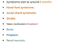
How do cell counts in children differ to adults?
In neonates?
- Higher Hb
- Higher WCC
- higher neutrophil count
- higher MCV
Neonates
- Higher Hb
- Higher WCC
- Higher Lymphocyte count
- higher neutrophil count
- higher MCV
How do enzymes in bloodd cells differ between neonates and adults?
Children have higher levels of G6PD in RBC
How does TTTS affect blood cell counts of foetus?
One will be anaemic, the other polycythaemic
When do Beta-globin disorders manifest in children?
3-6 months
Because of higher proportion of HbF - 2 alpha and 2 gamma
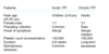
HbA: globin chains
2 alpha
2 beta
HbA2: globin chains
2 alpha
2 delta
Which genes are encoded on Chr 11?
- beta: found in HbA and HbA2
- delta: found in HbA2
- gamma: found in HbF
- Episolon…
Which genes are encoded on Chr16?
Alpha genes
α2 gene
α1 gene
ζ (zeta)
Which are the embryonic globin genes?
Episolon and zeta
(EZ!!!)
NORMAL FORMS OF HAEMOGLOBIN

What percentage of normal human haemoglobin should HbA2 comprise?
<3.5%
How do the haemoglobins in utero change over time?
- first 16 weeks: Haemoglobin Gower and Portland, comprise epsilon and zeta chains
- HbF predominates throughout foetal life
- HbA increases 32 weeks onwards
- At birth- HbA comprises 1/3 of Hb. Increases steadily with age
- prematurity- v low HbA levels
Causes of polycythaemia in the foetus
Twin-to-twin transfusion (twin bleeding into another twin)
Intrauterine hypoxia
Placental insufficiency- oxygen not getting to foetus → increased EPO production → Hb rises
Causes of anaemia in the foetus
Twin-to-twin transfusion
Foetal-to-maternal transfusion (baby bleeds into the mother’s circulation)
Parvovirus B19 infection (virus not cleared by immature immune system)
Haemorrhage from the cord or placenta
When do most mutations for leukamiea occur?
In utero
Leukamiea in down’s syndrome
Sometimes called transient abnormal myelopoiesis - as it is transient and spontaneously resolves within a few weeks, especially if they survive the first 4-6 weeks
*only invovles myeloid cells
*does not need treatment at this stage
NB: it can recur 1-2 years later in 25% of children so will need treatment then
Inherited haemolytic anaemias in children
Defects in:
- Red cell membrane
- Hereditary spherocytosis- defects in vertical interaction
- hereditary elliptocytosis- defects in spectrin protein (horizontal interaction)
- Haemoglobin molecule
- Haemoglobinopathy- abnormal structure of haemoglobin - SICKLE CELL DISEASE
- Haemoglobin quantity reduction - alpha and beta thalassamiea
- metabolic deficiencies
- G6PD deficiency
- Pyruvate kinase deficiency- rare
Difference in timing of presentation of defects in alpha vs beta globin genes vs gamma globin genes
- alpha- manifests early on as it’s part of HbF
- beta- only manifests in first year of life as HbF does not rely on beta
- gamma- only manifests in neonatal period as it’s only found in HbF
What infection can trigger G6PD deficiency?
Urinary tract infection
*infections in general can cause oxidative damage
Characteristic features of autoimmune haemolytic anaemia in children
- spherocytosis
- positive DAT test
Causes of acquired haemolytic anaemia in children
- autoimmune
- HUS
- haemolytic disease of the newborn
Triad of HUS
- MAHA
- thrombocytopaenia
- uraemia (renal failure)
*often triggered by E. Coli 0157:H7
Inherited defects of coagulation
- haemophilia A and B
- vWD
- TTP
Which type of leukamieas are common in children?
ALL is more common than AML
But you can still get AML!!
NB: in <1 yo, AML is MORE COMMON than ALL!!
How do you manage hyposplenism in children?
- Appropriate vaccinations
- Prophylactic penicillin
- Advice to parents regarding other risks:
- Malaria
- Dog bites
Definition of SCA vs SCD
Sickle cell disease
- encompasses homozygous and heterozygous forms
- homozygous- sickle cell anaemia
- heterozygous- SC sickle cell disease, sickle beta thalassamiea
Sickle cell anaemia
Sickle cell anaemia is an autosomal recessive condition that causes sickle (crescent) shaped red blood cells
→ red blood cells fragile and more easily destroyed
→ to a haemolytic anaemia
When does sickle cell anaemia manifest and why?
At 6 months
Because before this they have HbF.
At 6 months HbS production starts to increase
Aetiology of sickle cell disease
HbS forms as a result of a point mutation in codon 6 of the b-globin gene
This results in a change in the amino acid encoded, from glutamine to valine
**autosomal recessive
What is HbSC disease?
- Children inherit HbS from one parent and HbC from the other
- HbC is caused by a different point-mutation in b-globin
- NO HbA – because they have no normal b-globin genes
- Near normal total Hb level
- Symptoms:
- Causes a sickling disorder that is slightly milder than HbSS
- Fewer painful crises
- May develop proliferative retinopathy in adolescence
- Eyes should be regularly checked
- Prone to osteonecrosis of the hips and shoulders
What is HbSB disease?

What is the most common inherited disorder in childre in the UK?
Sickle cell disease
Epidemiology of sickle cell disease
More common in children of Afro-Caribbean origin and those found in the Middle East
How does the presentation of painful sickle cell crises differ between children and adults and why?
Red marrow in children is also present in long bones (in addition to axial skeleton)
Red marrow is more suscpetible to infarction than yellow marrow
This means that painful crises in children can affect hands and feet as well (whereas in adults it’s restricted to axial skeleton)
What type of haemolysis do you get in SCA?
Intravascular or extravascular (macrophages)
Findings will differ
presentation of haemolytic anaemia in SCA?
- reticulocytosis - expansion of bone marrow - leads to expansion of cheecks and skull (also in thalassamiea)
- extramedullary haematpoiesis - in liver–> hepatomegaly
What can cause acute anaemia in SCA?
- haemolytic crises
- aplastic crises- parvovirus B19
- sequestration crises
*aplastic crisis: reduced reticulocyte count
sequestration crisis: raised retiuclocyte count, can present with SOB
haemolyttic crisis: raised reticulocyte count, usually presents with pain and jaundice - VERY RARE compared to sequestration crises
What types of vaso-occlusion can you get in the first decade of life with SCD?

When does hand foot syndrome tend to present?
Tends to present in first 2 years
What is the most common cause of stroke in children?
Sickle cell disease
Who is more affected by vaso-occlusive crises of the spleen- younger or older children?
Younger>older
Splenomegaly vs hyposplenism in children
Young infants tend to present with splenomegaly due to acute splenic sequestration
Older children and adults tend to present with hyposplenism due to recurrent infarction
Treatment of splenic sequestration
blood transfusions and if recurrent splenic sequestration then consider splenectomy
What age groups is bacteramiea more common in? (Sickle cell disease)
in younger children - due to immature immune system and recurrent obstruction of splenic microvasculature
What infections are children w/ SCD more prone to?
- Pneumococcus (and other encapsulated bacteria)
- Parvovirus B19- aplastic anaemia
definitive diagnosis of sickle cell disease
Haemoglobin electrophoresis
What type of anamiea do you get in sikcle cell anamiea vs sickle beta thalassamiea?
SCA: normocytic
SB thalassamiea: microcytic due to reduced beta globin production
mode of inheritance of G6PD deficiency
x linked recessive
affects males more
how does g6pd deficiency cause haemolysis?
Causes buildup of glutathione - haemolysis
EPidemiology of G6PD deficiency
Common in areas where malaria is endemic -african and medeterrenean
Less common in caucasian population
Does G6PD defiiency cause acute or chornic haemolysis?
USually acute episodes of haemolysis following exposure to oxidative stress
eg
- Infections (EG UTIS!!!)
- Drugs:
- Antimalarials (primaquine)
- Antibiotics:
- sulphonamides
- ciprofloxacin
- nitrofurantoin
- Dapsone
- Vitamin K
- Naphthalene (this used to be in moth balls)
- Fava beans (broad beans)
**can rarely cause chronic haemolysis
What is teh most common cause of kernicterus?
G6PD deficiecny
Peripheral blood smear of G6PD deficiency
Steady state: NORMAL
In acute haemolysis:
- Bite cells
- Hemighost cells
- Irregularly contracted cells
- Nucleated RBC
- Methyl-violet staining shows HEINZ BODIES (sign of oxidative haemolysis; short-lasting as quickly removed by the spleen)
Is G6PD deficiency intravascular or extravascular haemolysis?
INTRAVASCULAR
*think enzyme is INSIDE the cell- hence intravascular
Is hereditary spherocytosis intravascular or extravascular haemolysis?
EXTRAVASCULAR
*think membrane is on the OUTSIDE of the cell; hence EXTRAvascular
Blood + urine findings of G6PD deficiency during an acute episode
Intravascular haemolysis hence:
- high LDH
- low haptoglobins
- high unconjugated bilirubin
Urine: haemoglobinuria
Mode of inheritance of haemophilia A and B
X linked recessive
Which factors are deficient in Haemophilia A and B?
Which disease presents similarly to Haemophilia A?
Clinical presentation of Haemophilias
Management
A: F8
B: F9
Haemophilia A presents similarly to vWD as VWF is needed to stabilise F8.
Clinical presentation
- Spontaneous, deep bleeding
- Haemarthroses (MOST COMMON)
- Bruises
- Soft tissue haematomas (e.g. muscle atrophy, shortened tendons)
- Ecchymoses are typical of coagulation disorders
- Other sites of bleeding (e.g. urinary tract, CNS, neck)
- Prolonged bleeding after surgery or dental extractions
- Bleeding following circumcision
Management: replacement of clotting factors for life
Haemophilia blood results

Mode of inheritance of VWD
Autosomal dominant- except Type 3
Types of VWD
Type 1: partial quant deficiency
Type 2: qual deficiency
Type 3: quant and qual deficiency
Clinical features of VWD
Mucocutaneous bleeding
Bruises
Post-traumatic bleeding
Menorrhagia
Which disease does VWD present similarly to and why?
Haemophilia A
Because VWD is needed to stabilise F8.
Lab results of VWD
- Platelet count: normal (except Type 2- low)
- Bleeding time: prolonged
- APTT: prolonged
- PT: normal
- F8: low
- Ristocetin: low
- (Platelet aggregation studies)
- Type 1: (quant)
- Addition of ristocetin- No aggregation
- Addition of pro-aggregative factors - Yes aggregation
- Type 2; (qual)
- Addition of ristocetin - no aggregation
- addition of pro-aggregative factors - no aggregation
- Type 1: (quant)
Differentials for VWD

Treatment of VWD
- desmopressin
- recombinant vWF (in severe vWD, usually type III)
- Factor VIII concentrates (rarely used)
Pathophysiology of ITP
IgG autoantibodies target glycoprotein IIb and IIIa on platelet surface
→ type II hypersensitivity reaction
→ platelets destroyed by reticuloendothelial system (spleen, liver, bone marrow)
Clinical features of ITP
- Petechiae
- do NOT blanch
- Haematomas
- Bruises
- subconjunctival haemorrhages
- Blood blisters in the mouth
Which type of ITP do children get?
Acute ITP
What is the difference between acute and chronic ITP
