Innate immunity Flashcards
What is innate immunity?
It is immunity present from birth- using strategy 1 (recognising molecular patterns).
Not enhanced by exposure- has no memory. Uses cellular and soluble components.
Is innate immunity fast?
Yes! Can respond within minutes to hours. Very effective and directs adaptive immunity
What are the pattern recognition strategies?
- PAMP (pathogen associated molecular patterns)
- DAMP (damage associated molecular patterns)
Both above are not antigen specific and they use pattern recognition receptors (PRR)
- They also detect ‘self’. Natural killer cells
Innate immunity physical barriers
Skin- mechanical barrier, acidic environment
Mucous membranes- mucus secretions trap microorganisms, cilia (respiratory) expel microorganisms
Physiological barriers innate immunity
- Body Temp
- low pH
- Chemical mediators (lysozymes, interferons, complement)
Phagocytic:cells ingest material (neutrophils)
Inflammatory:local vascular permeability increases
Which cells are involved in innate immunity?
Neutrophil- phagocytosis of microbe
Eosinophil- phagocytosis, granule release, defence against parasitic infection, help B cell response in GALT
Basophil- may act as APC
Monocyte/macrophage- phagocytosis, cytokine release act as APC
mast cell- granule release histamine
Nk cells- lysis of infected cell (cytotoxic t cells)
Dendritic cells- antigen capture and presentation
What is the function of a neutrophil
40-75% of leukocytes
Short lived
Migrate to tissues- first cells to be recruited to a site of tissue damage/ infection
What is the function of a macrophage?
Dispersed throughout tissues.
Signal infection by releasing soluble mediators (cytokines)
Characteristics of neutrophils?
- Multilobed nucleus
- Primary granules- site of the enzymes that are going to kill the phagocytosed pathogen
- Secondary granule- replenish teh primary granules and regulate toxins that are produced in the lysis
- Primary granules stain darker than secondary granules
- The granules fuse with the vacuole to form a phagolysosome
What do neutrophils need to do?
- Move from the cirulation into tissues where the infection is
- Bind to the pathogen
- Phagocytose the pathogen
- Kill the pathogen
Move towards–> bind —> phagocytose —> kill
How do neutrophils move into tissue?
Rolling –> activation—> adhesion
- Near the site of infection there will be some damage or activated macrophages. Initially, will have low affinity for selective binding.
- When it gets to endothelium, chemokines are released which bind to the local endothelium layer ( from macrophage)
- Chemokines are only present on the endothelium if there is an infection
- Neutrophils roll along the surface with low affinity interactions (binding to selectin)- integrin in a low affinity state
- Integrin activation when chemokine receptor on neutrophil binds to chemokine on endothelium surface- it is activated ta high energy state
- Integrin binds strongly to integrin ligand, immobilising the neutrophil.
- Cells migrate into the tissue and the cells follow a chemokine gradient to figure out where to go (chemotaxis)
What is diapedesis?
Movement across the endothelial layer
How does migration of neutrophils to tissues start?
Macrophage with microbes release signals of infection - CHEMOKINES
They are released to local endothelium to activate it so cells know where to leave the circulation and enter the tissue
What is opsonisation?
Coating of microorganisms with proteins to facilitate phagocytosis.
They bind to antigens AND phagocytes.
It makes it easier for neutrophils to recognise the pathogen
Give 2 examples of opsonins
Antibodies
Complement
(humoral immunity)- the soluble component
Describe how neutrophils bind to opsonins
- Antibody binds to receptor on the pathogen (the antigen)
- Complement binds to the cell SURFACE of the pathogen
- The bound antibody and complement can then bind to the neutrophil and activate it.
- The neutrophil will engulf the bacteria and lyse it.

How do neutrophils kill pathogens?
- OXYGEN INDEPENDENT- enzymes, lysozymes, hydrolytic enzymes, antimicrobial peptides
- OXYGEN DEPENDENT- respiratory bursts, superoxide anion, hydrogen peroxide, NO and reactive nitrogen intermediates
What are neutrophil extracellular traps? (NETs)
Activated neutrophils release granule proteins and chromatin to form extracellular fibres. It stops microorganisms from spreading everywhere and contains the site of infection.
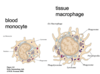
What is the difference between a monocyte and macrophage?
Monocytes leave circulation to mature into macrophages
Macrophages are bigger than monocytes
When in the blood, they are called monocytes. When in tissues, they are called macrophages.
They have lysosomes and are phagocytic- they have pattern recognition receptors.
Describe macrophage function

Signal infection by releasing soluble mediators (alarm cytokines)
This helps to recruit other cells (like neutrophils) and activate subsequent adaptive immune responses.
What do mast cells do?
They secrete histamine and other inflammatory mediators, including cytokines. There are different types:
- mucosal mast cells (lung)
- connective tissue mast cells (skin and peritoneal cavity, near blood vessels)
The recognise, phagocytose and kill bacteria. They can be activated by complement products (anaphylatoxins).
This leads to vasodilation and increased vascular permeability.
What do natural killer cells do?
They are large granulated lymphocytes: cytotoxic, lyse target cells and secrete the cytokine interferon-gamma. They kill self
•5-10% peripheral blood lymphocytes
•no antigen-specific receptor, but express both activating and inhibitory receptors. 50/50 - this is why there are two mechanisms for cell recognition
- have receptors which bind to antibody-coated cells (Antibody DependentCell-mediatedCytotoxicity)
- important in defence against tumour cells and viral infections (esp. herpes)
How do NK cells recognise target cells?
There are 2 types of target cell recognition:
- Missing self recognition
- Induced self recognition

What is missing self recognition?
Healthy cells present MHC Class I. Receptors on NK cells can bind to this and they send inhibitory signals to stop lysis of the cell. When the cell is infected MHC class I expression is downregulated- they go ‘missing’. Inhibitory signal does not exist


