Inflammatory Dermatosis and Blistering Diseases--Westra Flashcards
Urticaria
Affects Dermis
Dermal edema
immunologic (IgE and histamine), mast cell–mediated condition that may be allergic or nonallergic.
Transient Wheals
Pruritic>(painful)
Ask about new exposures/medications
Angioedema
severe Urticaria
Intense dermal and subcutaneous swelling
Burning/Painful>pruritic
Laryngeal Involvement = Emergency
Typically a medication reaction
Urticaria and Angioedema–how would you begin to discover the cause?
Ask questions! Take a thorough history.
Medications, food, travel, physical stimuli
pruritic
itching
Urticaria and Angioedema: Immune causes
A. Type 1 IgE mediated
Foods: shellfish*, fish*, peanuts*, tree nuts*, eggs, milk, soy, wheat. (* seen in adults) (seen in kids)
Latex
Stinging insects including hymenoptera (bees, wasps, hornets), fire ants, bedbugs, fleas, mites.
Medications: penicillin, cephalosporin, sulfa
Aeroallergens: dust mites, pollens, molds, animal dander
B. Autoimmune disease: Hashimoto’s immune thyroiditis (production of anti-thyroid antibodies), SLE, vasculitis.
(there may be thyroid anti-autobodies in the absence of thyroid dysfunction)
C. Infection: viral (cytomegalovirus, Epstein-Barr, HIV, hepatitis A, B & C); parasitic, fungal or bacterial.
Urticaria and Angioedema: Non-immune causes
A. Physical urticaria
B. Direct mast cell de-granulation
C. Foods containing high levels of histamine
Small urticarial papules on red skin (axon reflex erythema) occurring on the neck within 30 minutes of vigorous exercise.

Cholinergic Uticaria
as it appeared 5 minutes after stroking the skin with a wooden stick. The patient had experienced generalized pruritus for several months with no spontaneously occurring urticaria.

Dermographism
Urticaria and Angioedema Laboratory Evaluation
Rule 1: don’t get carried away! Expensive lab evaluations usually have poor yields.
Punch biopsy to exclude vasculitis if lesion persists > 48 hrs or looks atypical
Urticaria and Angioedema Tx
Avoid agitating cause!
First choice: Second generation, non-sedating H1-blockers
Second choice: (if symptoms not controlled, add) first generation, sedating H1 blockers
…
Last choice: add oral corticosteroid. Prednisone 40 mg →5 mg (course tapered over 10-14 days)
Reaction pattern of blood vessels in the dermis

Erythema multiforme
erythematous iris-shaped papular and vesiculobullous lesions
an immune-mediated dermatologic disease of young adults and young children in which target lesions appear on the hands, feet, and the extensor surfaces of the limbs
EM minor
erythema multiforme minor
mild form involving 1 mucosal site. major cause is herpes simplex infection with onset of EM rash at day 10
EM major
erythema multiforme major
**severe with extensive skin and mucous membrane involvement. **
Stevens-Johnson Syndrome
Usually due to drugs (sulfa, PCN, dilantin) and after mycoplasma pneumoniae infection
Stevens Johnson Syndrome
and
Toxic Epidermal Necrolysis
SJS: a hypersensitivity reaction commonly caused by drugs; infection can also be a trigger
Skin Tenderness and Erythema of Skin and Mucosa
Extensive Cutaneous and Mucosa Epidermal Necrosis and Sloughing
Potentially Life Threatening
How do you differentiate between:
Erythema Multiforme
Stevens-Johnson Syndrome (SJS)
Toxic Epidermal Necrolysis
EM = 1 mucosal membrane
SJS = EM major (2+ mucosal membrane an <10% epidermal detachment)
TEN = maximal variant of SJS (2+ mucosal membrane and 30% epidermal detachment)
Fixed Drug Eruption
a localized, sharply demarcated erythematous patch that can itch, burn or be asymptomatic
Panniculitis
Major focus of inflammation: subcutaneous tissue
Described as lobular or septal depending on where disease process begins
Accurate diagnosis by skin biopsy
Panniculitis:
Erythema Nodosum
Erythematous tender nodules
Typically ANTERIOR shins
Young women most common
Triggers
Infection (strep, TB, fungal)
Meds (OCP, Sulfa, NSAIDS)
Autoimmune (IBD, Sarcoid)
Treatment: rest, ice, pain control
Panniculitis:
Erythema Induratum
Tender red nodules
Lobular panniculitis and vasculitis
Middle aged, usually female
Posterior legs>ant
Chronic, recurrent subcutaneous nodules and plaques with ulceration
Associated with TB
6 Ps

LICHEN PLANUS (6 Ps)
- Planar (flat topped)
- Purple
- Polygonal
- Pruritic
- Papules
- Plaques
Erythematous to violaceous polygonal papules especially on the flexor areas such as wrists and ankles
Subacute cutaneous lupus
A chronic multisystem autoimmune disease that predominantly affects the skin and joints, although any body system can be involved.
The most common presentation is a butterfly rash on the face, low-grade fever, and nondeforming arthritis
Psoriasis (General)
common chronic skin disorder characterized by excessive proliferation of keratinocytes, resulting in the formation of thickened, scaly plaques, and inflammation, resulting in erythema and often pruritus
Risk factors: 30% genetic, stress, medications, infections, drugs (beta blockers, antimalarials)
Post strptococcal infection
Psoriasis:
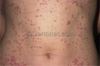
Guttate Type
Post strptococcal infection
children or young adults
eruptive trunkal dermatosis
What is this?

Psoriasis: Pustular type
Generalized: Potentially life threating; Small pustules becoming generalized with fever
Localized: Hand and foot form involves the palms and soles. May be termed Pustular Psoriasis of Barber
Hallmark of hives?
wheals
Butterfly rash across the nose and the cheeks.
Photosensitive pattern of erythema accross upper chest and extensor areas.
Triggers: Sunlight and Medications (anti-seizure, antibiotics, BP Rx)
Treat with NSAIDs, antimalarials, corticosteroids
Systemic Lupus Erythematosus (SLE)
Hallmark is development of Wickham’s Striae
Chronic, inflammatory, autoimmune disease
Association with Hepatitis C (HCV)
Location: wrists, shins, mucous membranes, Wickham’s stria (lacy, reticular, white lines)
lichen planus
Group of vesicles on a red base which rapidly become purulent and crusted
Two types
Herpes Simplex
“dew drop on a rose petal”
Incubation: average 14 days
fever, chills, malaise, 2-3 days before onset of rash
Varicella – Zoster virus (Chickenpox)
Primary infection usually occurs in childhood with lesions on the lips or face
HSV 1
Disease of adults involving the genital area, primary infection extensive, painful vesiculations and necrosis
Recurrent, lifelong disease with no cure
HSV 2
Follows nerve root – single dermatome.
Prodrome: pain along nerve root up to 5 days prior to rash
Recurrence of varicella
Herpes Zoster (Shingles)
Treatment options: Acyclovir, Prednisone,
IV Acyclovir
Bullae (bull’s eye filled with fluid), blisters occurring in the axillary, groin, fold areas most commonly.
Superficial vesicles progress to rapidly enlarging, flaccid bullae with sharp margins and no surrounding erythema. When the bullae rupture, yellow crusts with oozing result.
Bullous Impetigo
Caused by toxin producing Staph
Treat with:
Hygenic Measures
Topical Antibiotics
Oral Antibiotics
Begins as a single red macule or papule that quickly becomes a vesicle. The vesicle ruptures easily to form an erosion, and the contents dry to form characteristic honey-colored crusts that may be pruritic.
Nonbullous Impetigo
70% of cases of impetigo are nonbullous
Erythema, small pustules, fringe of white scale.
Cutaneous and mucosal (thrush, vulval vaginitis)
Yeast-Candida Albicans
Common in skin folds and on mucous membranes.
Bright beefy red dermatitis surrounded by satellite micropustules
or
Infections can produce superficial blisters or pustules
Fungal Infections
Candida Albicans
Dermatophytes
Autoimmune
Tense bullae (grouping) on normal or erythematous skin
Age: 60-80 years
Diagnosis based on histological exam.
Bullous pempigoid
Treat with:
Prednisone is the cornerstone of treatment
Topical cortisone for mild cases
Autoimmune condition typified by clusters of erythematous papules, excoriations and vesicles that arise as a consequence of gluten sensitivity.
Most patients are between 20 and 40 years of age, but the condition may occur at any age.
Pruritic and distributed symmetrically along extensor surfaces
Dermatitis herpetiformis
Associated with Celiac Disease
Autoimmune disease that affects the skin and mucous membranes. The predominant skin lesions are flaccid blisters.
The blisters are located on the head, trunk and intertriginous areas.
40 and 60 years of age.
Associated with Nikolsky sign (top layers of the skin slip away from the lower layers when slightly rubbed and may create a blister) and has a mortality rate of 5 to 15 %
Pemphigus vulgaris
skin hyperpigmentation and urine discoloration (increased uro and coproporphyrins in the urine)
Blistering of the skin occurs on sun-exposed areas, especially the hands and forearms and occasionally face.
Patients may also exhibit hypertrichosis (growth of hair) of the forehead and cheeks.
Risk factors for the disease include Hepatits C, hemochromatosis and alcoholism.
Porphyria Cutanea Tarda
Results from a deficiency in a heme-synthesizing enzyme.
history findings and associated shoulder Disorders:
Diabetes or thyroid disorders
Adhesive capsulitis
