Immunology: mechanism of self defense Flashcards
Innate immunity definition
the basic resistance to disease that a species possesses - the first line of defense against infection
characteristics of the innate immune response
- Broad-Spectrum (non-specific)
- no memory or lasting protective immunity
- limited repertoire of recognition molecules
Not equipped to fight something major – just general traffic, maintaining integrity
Antigen
Foreign Invader, Intruder
Antibody
Your Fire-power
Leukocytes
Your Soldiers
Phagocytes
Eaters - We eat our enemies
Macrophages
Big Eaters
Opsonization
Coating (coating surface of antigen, so possibly dealt w/later in various ways)
1st, 2nd, and 3rd line of defense
- First line of defence
- Innate resistance
- Second line of defence
- Inflammation
- Third line of defence
- Adaptive (acquired) immunity. Very Specialized soldiers/specific reactions
What is our 1st line of defense?
- physical and mechanical barriers
- biochemical barriers
- anatomic barriers
- physiologic barriers
- chemical factors
- Endocytic and phagocytic barriers
- biochemical barriers
Why do smokers develop respiratory infections, chronic bronchitis?
Failure in 1st line of defense: loss of ciliary action –> inflamed and chronically inhabited by bacteria
Physical and mechanical barriers
1st line of defense
- skin
- lining of GI, GU, respiratory tracts
- sloughing off of cells
- coughing & sneezing
- flushing
- vomiting
- mucus & cilia
Biochemical barriers
- Synthesized and secreted saliva, tears, earwax, sweat, and sebum
- sebaceous glands in skin secrete antibacterial and antifungal FAs and lactic acid
- perspiration, tears, saliva contain lysozome, which attacks cell walls of gram-positive bacteria (= acidic skin surface)
- Normal Flora – bacteria that live w/us, prevent other invaders. Our health depends on them in addition to our own genetic material *
What causes an “opportunistic infection”?
infection - taking advantage of weakened immune system
biochemical barriers and opportunistic infections
Antibiotics: subsequent vaginal infection, etc – b/c normal flora has been changed along w/ridding body of problem. in vaginal infections, protective lactobacillus is diminished leading to growth of other bacteria and yeast
Antimicrobial Peptides
- Biochemical barriers: proteins secreted by epithelial surfaces of body.
- 2 classes: Cathelicidins & Defensins
- class division based on structure
- high local concentrations. Toxic to several bacteria, fungi, viruses
Cathelicidins
- small cationic peptides that possess broad-spectrum antimicrobial activity
- linear alpha-helical shape
- only one known to function in humans
- stored in neutrophils, mast cells, variety of epithelial cells
Defensins
- Defensins are a family of small cationic, antibiotic peptides that contain six cysteines in disulfide linkage
- abundant in phagocytes and small intestinal mucosa
- about 50 different defensins ID’d thus far

Defensin structure
- triple-stranded ß-sheet structures
- 3 intrachain disulfide bonds
- Carboxy end and NH2 end
- Subdivided depending on how many cystein residues are connected in formation of disulfide linkages
- into alpha (at least 6 in humans) and beta (at least 10, but maybe up to 40) types
alpha vs beta defensins
- alpha
- often require activation by proteolytic enzymes
- esp in granules of neutrophils, paneth cells lining sm intestine
beta defensins: variety of epithelial cells lining respiratory, urinary, intestinal tracts, & skin.
- beta defensins
- synthesized in active forms
- Have antibacterial properties AND
- may also help protect epithelial cells from HIV!
- Both classes can activate cells of innate and adaptive immunity
how antimicrobial peptides kill bacteria
bacteria have cholesterol-free cell membranes - may allow cathelicidins to insert selves into and disrupt membranes
Defensins have similar chemical charge to cathelicidins and may work in same way
Physiologic Barriers
1st line of defense
- Temperature
- pH
- Oxygen Tension
1st line of defense: Temperature
- normal body temperature inhibits growth of most microorganisms.
- Elevated body temperature (fever) can have a direct effect on pathogenic microorganisms.
1st line of defense: pH
low pH of stomach, skin, & vagina (inhibits microbial growth)
1st line of defense: Oxygen Tension
particularly damaging to group of bacteria that prefer anaerobic environment. **e.g., diabetes –> poor circulation –> infection by anaerobes –> poor wound healing
Why chronic wound clinics use hyperbaric O2 chambers –> several hundred percent oxygen concentration to tissues
1st line of defense: chemical factors
a number
- Fatty acids, lactic acid
- Pepsin (digestive enzyme which hydrolyzes proteins)
- Lysozyme
1st line of defense: endocytosis
endocytic & phagocytic barriers
Lectins: definition
- proteins which specifically bind (or crosslink) carbohydrates
- involved in a variety of recognition processes
- exhibit considerable structural diversity.

- like flags sticking out of cell membranes*
- 2nd line of defense*
Types of lectins
- *Collectins**: Collectins are a family of collagenous calcium-dependent defence Lectins.(respiratory tract)
- *Selectins**: an important family of Lectins!
antimicrobial lectins: intestinal epithelium, against gram+ bacteria
Lectin forms & shapes
a variety!

Lectins & inflammation
- Release of Mediators into blood stream
- Release of Lectin like substances from the endothelial cells (b/c they sense inflammation)
- Lectin like substances slowing down the circulating leukocytes (expressed like little flags (lectin flag), slow leukocytes down – to come fight inflammation)
- Leukocytes get attached to the blood vessel wall near the site of inflammation
- Leukocytes leave the vessel and go to the site
Selectin: A Modified Lectin
- During an inflammatory response, some of the inflammatory mediators (such as histamine and thrombin) cause endothelial cells to mobilize P-selectin from stores inside the cell to the cell surface.
- P-Selectin has low affinity for a protein on the surface of leukocytes by the name of P-selectin glycoprotein ligand-1 (PSGL-1)
Putting it together: you step on a nail. How do leukocytes know to come and participate/attack the invader?
Antigens invade. Area hot/red/inflamed. Inflammation causes endothelial cells to produce selectin flags (P-selectins). P-selectin-leukocyte interaction brings leukocytes
What is the selectin roll?
- Leukocytes are able to “roll” along the blood vessel wall during their adhesion cascade!
- As the leukocyte rolls, the distal Lectin-like domain of the Selectin binds to certain carbohydrate groups presented on proteins on the leukocyte (PSGL-1)
- Selectin attachment slows the leukocytes and allows it to leave the blood vessel and enter the site of infection.
- The “rolling action” is because of Selectin’s low affinity. (they’re able to attach, but then they let them go fight infection)
innate immunity: 2 lines of defense
natural barriers & inflammation
cells involved in first line of defense
epithelial
Cells involved in 2nd line of defense
inflammation:
mast cells, granulocyte (neutrophils, eosinophils, basophils), monocytes/macrophages, natural Killer cells, PLTs, endothelial cells
Cells involved in adaptive immunity
T lymphocytes, B lymphocytes, macrophages, dendritic cells
Peptides involved in 1st line of defense/barriers
defensins, cathelicidins, collectins, lactoferrin, bacterial toxins
Peptides involved in inflammatory response
complement, clotting factors, kinins
Peptides involved in adaptive immunity
antibodies, complement
Where does inflammation occur?
tissue that has a blood supply
Cardinal Signals of inflammation
**redness: **vasodilation & increased BF
**heat: **” “
swelling: exudate (fluid & cells) accumulate
**pain: **PGs, bradykinin, pressure of exudate
Inflammation: 3 characteristic changes in microcirculation (arterioles, capillaries, venules)
- Vasodilation
- increased vascular permeability
- WBC adherence to inner walls of vessels and their migration through vessel walls to site of injury (diapedesis)
Describe Acute Inflammatory Response
- (usually) Cellular injury > mast cell degranulation / activation of 3 plasma systems / release of subcellular components from damaged cells.
- Interdependent systems: e.g., mast cell degran can result in other two
- leads to dvpt of microscopic changes in inflamed site AND characteristic clinical manifestations

What causes edema?
cap permeaility, leakage of plasma (also > viscous blood in microcirculation)
What causes warmth and redness of inflammation?
increased blood flow and increase in RBCs
What stimulates endothelial cells lining capillaries to retract during inflammation and what is the effect?
biochemical mediators (e.g., histamine, bradykinins, leukotrienes, PGs)
Effect: spaces at junctions btwn cells, allowing leukocytes and plsma to enter surrounding tissue
Inflammation: what do the cells and chemicals do together once in the tissues
- prevent infection/further damage
- contaminate microorganisms through
- influx of fluid to dilute toxins produced by bacteria and released by dying cells
- influx & activation of plasma protein systems that help destroy and contain bacteria (e.g., complement, clotting)
- influx of “eater” cells (neutrophils, macrophages) to destroy cell debris & infectious agents
- LImit & control inflammatory process (prevent spread)
* through influx of plasma protein systems (e.g., clotting), plasma enzymes, cells (e.g. eosinophils)
3. Interact with components of adaptive immune system to elicit specific response
- through influx of macrophages and lymphocytes
4. Prepare area of injury for healing - through removal of bacterial products, dead cells, other products of inflammation
- and initiation of mechanisms of healing & repair
Role of lymph nodes in adaptive immunity
microbial Ags in lymphatic fluid activate B & T lymphocytes
Phases of inflammation and repair
- cell injury
- Acute inflammation
- healing or chronic inflammation
- healing or granuloma formation
- healing
Type of cell does most work in inflammatory response
leukocyte
Types of leukocytes
neutrophils, monocytes, eosinophils, lymphocytes, macrophages, mast cells, and basophils.
What is an inflammatory mediator?
molecules inside and outside your body that play a role in inflammation.
endotoxin
- an “exogenous” inflammatory mediator, called endotoxin, or lipopolysaccharide (LPS).
- present in the outer covering of some types of bacteria, and signals to immune cells that there are bacteria present, causing an increase in inflammation.
Selectin Roll image (good to know)
PMN: neutrophil
Rolling: L-selectin, P-selectin, E-selectin
Margination: Integrin alpha4beta1 (VLA4) & alpha4beta7, VCAM-1
Diapedesis: integrin aLB2 (LFA-1), integrin aMB2 (MAC-1), PCAM-1, ICAM-1, ICAM-2, PCAM-1

Neutrophil Adhesion and Chemotaxis in inflammatory response (image)
Selectin mediates neutrophil attraction & attachment
Rolling & sticking initially mediated by selectin then integrins
CAM molecules help enter tissue, spread, extravasate

How does the body recognize inflammatory mediators?
Toll-like receptors: on surface of our cells. Can sense presence of several different microbial stimuli

How are macrophages activated during inflammation?
by various immune system molecules (such as cytokines, including interferons, or IFNs), endotoxin, or by other microbial signals.
chemical message! not selectin like leukocytes
How do active macrophages differ from non-active?
- develop different properties, e.g., capacity to stop dividing and phagocytosis. “Make war not love”
- vigorous production of inflammatory molecules such as TNF and interleukins, clotting factors, prostaglandins, free radicals, nitric oxide, and tissue remodelling enzymes (i.e. enzymes promoting bone resorbtion) and growth factors
- promote the growth of new blood vessels. NOT just destruction
Macrophage overview (image)
Monocytic cells give rise to resident and recruited macrophages w/functional heterogeneity
Clockwise from top left
- 1st of defense against intracellular pathogens by
- generating an inflammatory and respiratory burst and
- initiating antigen presentation to activate adaptive immunity; (unlike neutrophils)
- clearing immune complexes and downregulating inflammatory responses
- promoting wound healing via elaboration of growth factors.(primary to do so)
- A subset, e.g. splenic red pulp macrophages and osteoclasts, become highly specialized in their location and function.
- splenic red pulp macrophages: clear and recycle senescent red blood cells
- osteoclasts: critical to remodeling bone throughout adult life

What are the 1st cells to go fight bacteria?
neutrophils!
Role of neutrophils (aka PMN) in defense
- buy time devouring organisms (esp bacteria) waiting for healing & inflammation to begin
- degrade organisms through activation of proteases, of other antibiotic molecules, and generation of toxic oxygen radicals
- remove debris if sterile
- phagocytize bacteria if nonsterile
- die early - leftover is pus
How do neutrophils kill?
carry molecular weapons called free radicals that are poisonous to bacteria. Typically, these poisons are hidden in specialized storage sacks kept separate from body tissues.
Meaning of “band” or “left shift”
Immature neutrophils. Sign of bacterial infection.
Life of a neutrophil
myeloid lineage. Short lived.
bands, neutrophils, recruitment & rolling….

angry neutrophils
Golgi complex is making more vesicles- so electron microscopy looks denser w/black vesicles (similar to macrophages)
abnormal neutrophil function
uncommon, impair ability respond to life-threatening infections
noninfectious attraction of neutrophils
major role in rheumatic conditions, noninfectious stimuli e.g. complement activation & lipid inflammatory mediators
Lymphocytes: 2 broad classes
T cell and B cell
Where do lymphocytes begin and mature?
Begin in bone marrow
B mature in marrow
T mature in thymus
Role of PLTs in inflammation
we know they stimulate blood clotting
- In inflammation
- shed certain molecules that directly activate inflammation inside blood vessels
- produce variety of inflammatory mediators, e.g., GFs, adhesion molecules, cytokines
so site is stickier for other cells, signals sent

When is platelet action unwanted in inflammation?
Clots - e.g., during CAD. Why we give PLT inhibitors (aspirin, heparin)
Overview of innate immunity and inflammation

3 plasma protein systems essential to effective inflammatory response
complement, clotting, kinin
all contain inactive enzymes (proenzymes) which are sequentially activated. Substrate of activated enzyme becomes next component in series
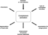
3 pathways to complement activation
classical, lectin, alternative
Complement Cascade Image

Complement: interaction w/other components of inflammatory response
all of them! Team players.
How do complement proteins get to target area
Quickly, freely, through bloodstream.
React directly w/antigens
What can the complement system do when it is activated?
- Trigger inflammation
- Attract eater cells such as macrophages to the area
- Coat intruders so that eater cells are more likely to devour them
- Kill intruders : direct hole in chest

What does complement need to blast holes into lipid bilayer of target membrane?
must develop membrane attack complex (MAC)

Complement Cascade, another image

How does the classical pathway start for the complement cascade?
usually started through antigen/antibody complexes through component C1 (consists of C1q, two C1r, C1s molecules)
C1q must simultaneously bind to 2 Ab molecules

Important common point to all complement pathways
Get to C3 convertase - then you have a common path

What begins the lectin pathway in the complement cascade?
Mannose-binding lectin (MBL) binding to oligosaccharides on certain virions/infected cells. (or 2 mannose-rich pathogen associated molecules on surface of bacterium)
Lectins bring leukocytes in during inflammation, even if no bacteria present - so complement can get started w/o Ag/Ab

How does alternative pathway to complement cascade begin?
Component of complement (C3) spontaneously breaks down as a result of inflammation.
activated by many agents, e.g., gram-negative bacterial and fungal cell wall polysaccharides, which bind and stabilize C3b, which is produced by normal breakdown of C3 in blood
Relationship between number of pathways begun and magnitude of response
Just one molecule of C42b can cleave up to 1000 molecules of C3 and C3b. Many C3b molecules bind to microbial surface > multiple holes in the membrane

The complement snowball
Classical complement pathway (image)

Test question: C3a and C3b - uses in and around complement cascade
C3b: opsonization; C3a (and C5a, to limited extent C4a): recruiter of mast cell (histamine): leaky blood vessels, attract phagocytes. (“anaphylatoxins - induce mast cell degranulation)
complement helps mediate direct destruction of enemy (cytolysis)
*carboxypeptidase inactivates C3a and C5a to stop anaphylatoxic activities

5 processes begun by bacterial activation of complement
chemotaxis, degranulation of mast cells, enhancement of humoral immune responses, opsonization for phagocytes, lysis of susceptible microbes
complement pathway: cleaving components into fragments during activation
name components & effect
components: 2b, 4a, Ba, C3a, C5a
smaller fragments often have biologic activities & serve as chemotactic factors and anaphylatoxins
Larger activated fragments usually converted to active enzymes (bar above names in book image) and form complexes w/additional components in the cascade
Characteristics of MBL as part of complement cascade
MBL contains 2 associated enzymes, MASP-1 and MASP-2, and functions in a manner similar to C1.
Main substance in coagulation system
fibrin, an insoluble protein
Complement cascade: similar function of C1 and MBL
each activate complement components C4 and C2
Role of C3b in alternative complement pathway and following steps
C3b produced by normal breakdown of blood. Bound and stabilized by many agents, e.g., bacterial polysaccharides.
C3b forms site of binding of factor B (fB), which is activated by factor D (fD) into Bb and the small fragment Ba. Properdin (P) helps stabilize the complex.

C3 and C5 convertases in complement pathways
- both produced by all pathways
- enzymatically active complexes that activate C3 and C5, respectively
- C3b produced by C3 convertase can function as an opsonin
- C5b initiates assemblage of Membrane attach complex (MAC), which results in multiple C9 molecules forming a pore in the bacterial membrane
- C5a is the major chemotactic factor for neutrophils

What is C1?
a macromolecular complex consisting of C1q and two molecules each of C1r and C1s
begins classic complement pathway w/Ag-Ab immune complex. A conformational change in C1 results in enzymatically active molecules whose substrates are C4 and C2
Other ways to activate classical pathway
e.g. heparin, DNA, RNA, C-reactive protein (increased during inflammation)
chemotactic factor
biochemical substance that attracts leukocytes to the site of inflammation
C5a is the major CF for neutrophils
Purpose of coagulation system
- Prevents the spread of infection
- Keeps microorganisms and foreign bodies at the site of greatest inflammatory cell activity
- Forms a clot that stops bleeding
- Provides a framework for repair and healing
Action of coagulation system
Forms a fibrinous meshwork at an injured or inflamed site
2 coagulation pathways

Two ways to activate clotting
intrinsic & extrinsic pathways

How is the intrinsic pathway initiated?
by the activation of Hageman factor (XII) into XIIa
(activated factors are enzymes and are indicated by lowercase a)

sequential activation of the intrinsic pathway components results in the formation of a complex of ….
IXa, VIIIa, and X

Extrinsic pathway is activated by
exposure of tissue factor (TF) during tissue damage.
TF complexes w/factor VII, which is activated (VIIa), and forms a complex with factor X (TF, VIIa, X)
similarities between intrinsic and extrinsic pathways
- dependent on calcium
- form on phospholipid membranes that are rich in phosphatidylserine
- have “tenase” activity (can activate factor X into Xa)
Intrinsic/extrinsic common pathway
Factor X begins a common pathway in which Xa complexes w/Va and prothrombin (PT), with calcium and phospholipid membranes, to form an active prothrombinase (activates prothrombin into thrombin).

Thrombin and fibrin
an enzyme that cuts high-molecular-weight fibrinogen into fibrin molecules. Fibrin polymerizes to form a clot.
fibrin is end product of coagulation cascade!

What substances can activate the clotting system?
substances released during tissue injury and infection, including collagen, proteinases, kallikrein, plasmin, bacterial products such as endotoxins
activation of extrinsic vs intrinsic pathway
- Extrinsic is tissue factor pathway, activated by TF (aka tissue thromboplastin) released by damaged endothelial cells in blood vessels. Reacts w/factor VII (VIIa)
- Intrinsic pathway - contact activation pathway. Activated when vessel wall is damaged and hageman factor (XII) in plasma contacts negatively charged subendothelial substances.
- Factors converge at factor X
clotting system and inflammation
as w/complement, fragments produced by cascade enhance the inflammatory response.
2 low molecular weight fibrinopeptides, A and B, are released from fibrinogen when fibrin is produced. Both, esp B, are chemotactic for neutrophils and increase vascular permeability by enhancing the effects of bradykinin
Function of kinin system
to activate and assist inflammatory cells
- Primary kinin is bradykinin
- Causes dilation of blood vessels, pain, smooth muscle contraction, vascular permeability, and leukocyte chemotaxis
What activates the kinin pathway?
Factor XIIa from the clotting system (the hagemon factor!)
XIIa functions as an enzyme (prekallikrein activator) to convert prekallikrein intokallikrein. Enzymatically active kallikrein converts kininogen into bradykinin.

Hageman factor
The mastermind! XIIa. Connects all 3 pathways
know how all three activate each other

Kinin system & inflammation
- in a sense, once you have bradykiinin, you have vasodilation, vascular permeability, pain.
- Sometimes want these things, sometimes not. Inhibit bradykinin – prevening vasodilatation & pain

PAMPs
essential polysaccharides and polynucleotides that differ little from one pathogen to another but are not found in the host.
many recognized by receptors in innate immunity
Cellular mediators of inflammation
Cellular components
- Granulocytes, platelets, monocytes, and lymphocytes
Cell surface receptors
- Pattern recognition receptors (PRRs)
- Pathogen-associated molecular patterns (PAMPs)
- Toll-like receptors
- Complement receptors
- Scavenger receptors (nonspecific receptors)
Mast Cells
Cellular bags of granules located in the loose connective tissues close to blood vessels
Skin, digestive lining, and respiratory tract
Mast cell activation
- Physical injury, chemical agents, immunologic processes, and toll-like receptors
Mast cell chemical release
Two ways:
Degranulation and synthesis of lipid-derived chemical mediators
(Degranulation: release “bags” of granules on site of inflammation)
Mast cell degranulation: what is “in the bag”?
Histamine & chemotactic factors
H1 receptor
- Proinflammatory
- Present in smooth muscle cells of the bronchi
- Why histamine plays a role in the devpt of asthma. Prevent mast cell degranulation through drugs called “mast cell stabilizers”. Brings inflammation in bronchus down. We used to think asthma was bronchospasm, but bronchospasm tied to inflammation
H2 receptor
- Anti-inflammatory
- Present on parietal cells of the stomach mucosa
- Induces the secretion of gastric acid
- Some pts want you to block these – even if anti-inflammatory, b/c they have ulcers, etc. H2 blockers – not benefiting from their anti-inflammatory, but need to stop acid/ulceration. OTC dosage of H2 blockers is half “real” dosage.
action of histamine
- Vasoactive amine that causes temporary, rapid constriction of the large blood vessels and the dilation of the postcapillary venules (does both!)
- Retraction of endothelial cells lining the capillaries
- there are 2 histamine receptors (one pro and other antiinflammatory)
Action of mast cell’s chemotactic factors
- Chemotactic factors – named after who they attract
- Neutrophil chemotactic factor
- Attracts neutrophils
- Eosinophil chemotactic factor of anaphylaxis (ECF-A)
- Attracts eosinophils
4 mediators synthesized by mast cells
Lipid derived
- Leukotrienes
- Product of arachidonic acid from mast cell membranes
- Similar effects to histamine in later stages (used w/asthma)
- Prostaglandins
- Similar effects to leukotrienes; they also induce pain. PG inhibitors address pain (ibuprofen)
- Platelet-activating factor
- Similar effect to leukotrienes and platelet activation
Growth Factors & cytokines

Margination (pavementing)
Adherence of leukocytes to endothelial cells
Diapedesis
Emigration of cells through the endothelial junctions
Types of phagocytes
monocytes & macrophages, eosinophils, NK cells, PLTs
Monocytes and macrophages
- Monocytes are produced in the bone marrow, enter the circulation, and migrate to the inflammatory site, where they develop into macrophages
- Macrophages typically arrive at the inflammatory site 3 to 7 days after neutrophils
- Macrophage activation results in increased size, plasma membrane area, glucose metabolism, number of lysosomes, and secretory products
Eosinophils
- Mildly phagocytic - main job is not to eat
- Duties
- Defense against parasites and regulation of vascular mediators
- Eosinophils high on CBC: 1) drug allergy or 2) parasitic infection. More likely #1 in U.S., 3) lab error
- Defense against parasites and regulation of vascular mediators
NK Cells
Function is to recognize and eliminate cells infected with viruses and some function in eliminating cancer cells. We’d prob have cancer more frequently & younger w/o them. As we age, NK cells lose surveillance ability
Platelets
Activation results in degranulation and interaction with components of the coagulation system – need both to make a good clot
Important cytokines
interleukins, interferons, TNF-alpha
Interleukins
- Produced primarily by macrophages and lymphocytes in response to a pathogen or stimulation by other products of inflammation
- Many types
- Examples
- IL-1 is a proinflammatory cytokine
- IL-10 is an anti-inflammatory cytokine
- Must be able to move to inflammatory state and back out of inflammatory state
Interferons: who do they fight and how?
- Protects against viral infections
- Produced and released by virally infected host cells in response to viral double-stranded RNA
- Types
- IFN-alpha and IFN-beta
- Induce production of antiviral proteins
- IFN-gamma
- Increases microbiocidal activity of macrophages
- IFN-alpha and IFN-beta
- Important in drug therapy

TNF: who does it fight and how?
- Secreted by macrophages in response to PAMP and toll-like receptor recognition (also in mast cells)
- enhances endothelial cell adhesion molecule expression
- Systemic effects:
- Induces fever by acting as an endogenous pyrogen
- Increases synthesis of inflammatory serum proteins
- Causes muscle wasting (cachexia) and intravascular thrombosis
- probably responsible for shock fatalities caused by gram- bacteria
some attempts to use w/septic pts
Steps to inflammation, agents

Transudative vs exudative fluid
3 components to r/o exudative: protein, LDH, total LDH (LDH is 2 of clinical parameters)
Systemic manifestations of inflammation
fever, leukocytosis, increased plasma protein synthesis
What causes fever?
Caused by exogenous and endogenous pyrogens
Act directly on the hypothalamus
What are the plasma proteins that are increased in systemic inflammation?
Acute-phase reactants: C-reactive protein, fibrinogen, haptoglobin, amyloid, ceruloplasmin, etc.
Role of plasmin in protein cascades
- regulates clot formation: degrades fibrin and fibrinogen
- can activate complement cascade through C1, C3, C5
- can activate kinin cascade by activating Hageman factor (factor XII) and producing prekallikrein activator
4 effects of Hageman factor (XII)
- Activation of clotting cascade through factor XI
- Control of clotting through conversion of plasminogen proactivator to plasminogen activator, resulting in the generation of plasmin
- Activation of the kinin system by activated Hageman factor (prekallikrein activator)
- Activation of C1 in the complement cascade

C1-esterase inhibitor (C1-inh)
inhibits:
complement activation
- through reactivity w/all 3 pathways
- C1, MASP-2, C3b
clotting & kinin
- e.g., kallikrein, activated hageman factor XIIa
*genetic defect w/C1-inh: hereditary angioedema
Role of eosinophils
parasites or regulate vascular mediators released from mast cells.
Mast cells produce ECF-A, which attracts eosinophils to site of inflammation. There, they help control.
Role of basophils
assoc w/allergies and asthma, but primary role unknown
macrophage and T-cell
T-cells or cells activated by TLRs produce inflammatory cytokines which increase bacteriacidal activity of macrophages
macrophages have cell surface receptors for these cytokines
