Alterations in Immunity and Inflammation Flashcards
4 types of inappropriate immune response
allergy, autoimmunity, alloimmunity, immune deficiency
allergy
exaggerated response against environmental Ags
autoimmunity
misdirected against host’s own cells
alloimmunity
directed against beneficial foreign tissues, such as transfusions or transplants
a.k.a. isoimmunity
immune deficiency
insufficient to protect host
2 classifications for hypersensitivity
- source of Ag (allergy, autoimmunity, alloimmunity)
- mechanism of Dz (Types I, II, III, IV)
mechanism of onset not understood
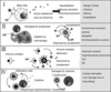
Hypersensitivity Types
- Type I: immunoglobulin E mediated
- Type II: Tissue specific
- Type III: Immune complex mediated
- Type IV: cell-mediated/delayed
artificial. Rarely just one.
Sensitization to an antigen
when adequate # of Abs or T cells to cause noticeable reaction on reexposure
Immediate vs delayed hypersensitivity
Immediate: minutes to hours
Delayed: several hours to days
Type I: name
IgE-mediated reaction
Type I: rate of development
immediate
Type I: Class of Ab involved
IgE
Type I: Principal effector cells
Mast
release histamine –> vasodilation, mucous secretion, bronchoconstriction, itch
Type I: complement participation?
No
Type I: example d/os
allergic rhinitis, urticaria, asthma, anaphylaxis
Type II: name
Tissue-specific reaction
Type II: rate of dvpt
Immediate
Type II: Ab
IgG, IgM
dissolved by complement

Type II: effector cells
Macrophages in tissues
(damaged by lymphocytes, uptaken by macrophages)
Ab directed against cell surface Ags mediates cell destruction via complement activation or ADCC
Type II: complement participation
frequently
Type II: example d/os
autoimmune thrombocytopenic purpura, Graves dz, autoimmune hemolytic anemia,
Type III: Name
Immune complex-mediated reaction
Type III: Rate of dvpt
immediate
Type III: Ab
IgG, IgM
Ag-Ab complex deposited in various tissues induce complement activation and an ensuing inflammatory response mediated by massive infiltration of neutrophils

Type III: effector cells
neutrophils
Type III: complement participation
Yes
Type III: example d/os
SLE, RA, hemolytic anemia, hypersensitivity pneumonia
Type IV: name
cell-mediated reaction
Type IV: Rate of dvpt
delayed
Type IV: Ab
None
Type IV: effector cells
lymphocytes, macrophages
sensitized Th1 cells release cytokines that activate macrophages or Tc cells which mediate cell damage

Type IV: complement?
No
Type IV: examples of d/os
contact sensitivity/dermatitis to poison ivy and metals (jewelry), liver damage d/t drug allergy
Mechanism of Type I IgE mediated reactions
Type I IGE mediated reaction: initial exposure, ___ cells activate ___ cells.
And what is the result?
- Antigen Presenting dendritic cells activate Th2 cells
- Th2 produce cytokines IL-3, IL-4, IL-5 and GM-CSF (granulocyte macrophage colony stimulating factor)
- IL-3, IL-5, and GM-CSF attract and promote survival of eosinophils
- other cytokines, e.g., IL-4, induce B cells to class-switch to IgE producing plasma cells
- IgE coats surface of mast cell by binding w/IgE specific Fc receptors on mast cell’s PM (sensitization)
Type I IgE mediated reaction: subsequent exposure
- cross-links the surface bound IgE and activates signals from teh cytoplasmic portion of the IgE Fc receptors
- this initiates 2 parallell and interdependent processes
- mast cell degranulation and discharge of preformed mediatory (e.g., histamine, eosinophil chemotactic factor of anaphylaxis)
- production of newly formed mediators such as arachidonic metabolites (leukotrienes, PGs)
- this initiates 2 parallell and interdependent processes
Type I IgE mediated reactions: 2 phases
- Initial phase: vasodilation, vascular leakage, and, depending on location, smooth muscle spasm or glandular secretions (5-30 min post-exposure)
- Late phase: 2-8hrs later w/o additional exposure. more intense infiltration of tissues w/eosinophils, neutrophils, basophils, monocytes, Th cells, and tissue destruction in form of mucosal epithelial damage
IgE mediated reactions: what happens when mast cells are activated?
degranulation of preformed mediators (primary mediators) and synthesis of newly formed (de novo) mediators (secondary mediators)
what characterizes a Type II hypersensitivity reaction?
specific cell or tissue targeted. MHC locus antigens (HLAs) or other tissue specific Ags
some TSAs only on certain cells
IgE mediated reactions: primary mediators
Granule contents: histamine, proteases, chemotactic factors (ECF, NCF)
IgE mediated reactions: secondary mediators
membrane phospolipids lead to arachidonic acid (leukotrienes and PGs) and PAF (plt activating factor)
Mechanisms of Type II reactions
- destruction via Ab/Ag binding
- phagocytosis by macrophages in tissue
- neutrophil mediated immune destruction
- ADCC
- Modulaton or blocking the normal function of receptors by antireceptor Ab
Type II reaction: Ab/Ag destruction
complement mediated
Type II reaction: Phagocytosis
opsonization (IgG and C3b) & eating by macrophages
e.g., Abs against PLT-specific Ags or RBC Ags of Rh system coat and they’re removed in spleen
Type II: neutrophil mediated damage
Ag deposition -> Ab binds _> complement cascade -> C3a & C5a –> neutrophil chemotaxis -> neutrophil adhereance (bia Fc portion of Ab or C3bR) and degranulation (enzymes, ROS) into healty tissue!
Type II reaction: ADCC
- Ab dependent cell mediated cytotoxicity
- apoptosis of target cells is induced
- FcR on NK cell recognizes Ab on target cell
- by granzymes and perforin produced by NK cells and interactions of FasL - FasR)
Type II: antireceptor Ab
e.g., hyperthyroidism of Graves dz - binds and activates TSH
blocks normal function of receptors. Not destroy, just malfunction
Myasthenia gravis - what type of hypersensitivity and what does it do?
Type II
acetylcholine receptor Abs block acetylcholine from attaching to receptors on motor end plates of skeletal muscle, thereby impairing neuromuscular transmission and causing muscle weakness
autoimmune hemolytic anemia: what type and why?
Type II
d/t alloimmune reaction to ABO mismatched transfused blood cells - Ag/Ab reaction w/complement cascade
What characterizes Type III
- Ag-Ab immune complexes formed in circulation and deposited later in vessel walls or extravascular tissues
- Not organ specific and Sx not to do w/particular antigenic target.
- harmful effects d/t complement activation - esp through chemotactic factors for neutrophils
Primary difference from type II and Type III
Type II Ab binds to Ag on cell surface
Type III: Ab binds to soluble Ag that was rleased into blood or body fluids and complex is then deposited in tissues.
Why are neutrophils usually unsuccessful at ingesting immune complexes in Type III?
bound to large areas of tissue - deposit bad stuff into tissue instead of phagolysosomes
Type III: how do immune complexes form?
in blood from circulating Ag and Ab
Type III: What happens to small and large immune complexes?
Removed successfully from circulation, not deposited
What happens to intermediate sized immune complexes (type III)
deposited in certain target tissues in which circulation is slow or filtration of blood occurs. They activate the complement cascade through C1 and generate fragments, e.g., C5a, C3b
C5a brings neutrophils which attach to IgG and C3b in immune complexes. These fail at phagocytizing and instead destroy healthy tissue
Complement & Type III
Only type in which complement levels may be depleted
Serum Sickness
Type III. foreign serum introduced. Typically affect blood vessels, joints, kidneys. +fever, LAD, rash, pain
Raynaud phenomenon
Type III. Form of serum sickenss. Temperature dependent deposition of immune complexes
Arthus reaction
Localized immune complex mediated inflammatory response. Type III.
Repeated exposure to Ag reacts w/preformed Ab and forms immune complexes on walls of local blood vessels.
Sx 1 hour of exposure and peak 6-12
Characterize Type IV
No Ab. Delayed.
Mechanism of Type IV
- Ags from target cells stimulate T cells to differentiate into
- Tc cells (direct cytotoxic) and
- Th1 cells (delayed hypersensitivity)
- produce lymphokines (esp IFN-gamma) that activate macrophage through specific receptors (IFN-gammaR).
- Macrophage can attach to targets and release enzymes and ROS responsible for most tissue destruction
- produce lymphokines (esp IFN-gamma) that activate macrophage through specific receptors (IFN-gammaR).
ABO: surface of erythrocytes blood group O
core H antigenic carbohydrate - not Abs against it, so universal donor

O blood serum, Abs against
IgM against A and B carbohydrates

A blood type, what type of Ag?
Some H modified into A Ags by addition of N-acetylgalactosamine (NAGA)

Serum Abs of Type A blood
IgM against B antigen

B blood group, antigens
Some H modified to B antigens by addition of galactose (Gal)

B blood group, Abs against
IgM Abs against A antigen
AB blood group, antigens
Some Ags modified into both A and B

AB blood group, antibodies
no Abs to A or B antigens – universal recipient
Brutons agammaglobulinemia:
- agammaglobulinemia = antibodies are almost totally absent
- most severe B lymphocyte deficiency
- X linked, mutation in the gene for Bruton’s tyrosine kinase (Btk).
- enzyme is involved in intracellular signaling from several B cell receptors (IgM B antigen receptors, IL-5 receptor & IL-6.
- Ineffective signaling – arrest of development in bursal-equivalent tissue (bone marrow) of early cells in the B cell maturation process.
- Few circulating B cells, IgM and IgA are almost absent
DiGeorge’s
- Congenial thymic aplasia or hypoplasia: Lack/ partial lack of thymus
- Decreased T cells and functioning
- Usually due to deletions on chromosome 22
- May have partial or complete absence of the parathyroid: decreased blood Ca levels, structural defects in heart & aorta, abnormal facial features- underdeveloped chin, low-set ears, shortened structure of upper lip
Reticular Dysgenesis
- Combined B & T cell deficiency
- Failure of blood cells to develop- a common stem cell for all WBCs is absent so B/T and phagocytic cells never develop
- Baby usually dies in utero or after birth
ADA deficiency: Adenosine Deaminase (ADA)
- Enzyme involved in purine metabolism
- Autosomal recessive
- Accumulation of toxic levels or purine metabolites which affects rapidly dividing cells like lymphocytes
- Development of B, T & NK cells arrested very early, very few lymphocytic cells found in blood
TB SKIN TEST (TST):
aka PPD, purified protein derivative; the first type IV hypersensitivity reaction described (1890s); intradermal injection of the tuberculin antigen –> in a sensitized individual (someone previously exposed to the Ag), the site becomes infiltrated with T cells and macrophages, leading to a clear hard center (induration) and a reddish surrounding area (erythema); DELAYED hypersensitivity RXN –> reaches peak intensity at 24-72 hours; measurement of the induration diameter guides test interpretation
Atopy:
genetic predisposition to developing type I allergies; 40% chance if one parent has an allergy; atopic individuals tend to produce ↑ IgE, have ↑ Fc receptors on their mast cells, and more responsive skin / airways