Immunology Flashcards

1
Q

Define innate immunity and recall the host-microbe interactions

A

Innate immunity:

  • First line (and second line) of defence against invading pathogens
    • First line: skin, mucous membranes, secretions of skin and mucous membranes
    • Second line: phagocytic leukocytes, antimicrobial proteins, inflammatory response
  • Can activate acquired immune system
  • Ability to differentiate between host and pathogen (e.g. sentinel cells in stratum spinosum)

Host-microbe interactions – (bacterial) infection pyramid:

2
Q

List the main functions of the innate immune system (5 points)

A

Functions of innate immune system:

  1. Anatomic and physiologic barriers
  2. Phagocytosis
  3. Activation of acquired immune system
  4. Immune cell recruitment
  5. Completement activation
3
Q

List the anatomic and physiologic barriers of the innate immune system

A

Anatomic and physiologic barriers of innate immune system:

_S_avage _M_ermaids _S_mash _T_urtles”

  • Skin – physical barrier; tight junctions in epithelium; desquamation to remove adhered pathogens
  • Mucous membranes – resp. and GI tracts; slimy surface traps pathogens, ciliary clearance; defensins
  • Secretions – stomach acid, digestive enzymes, tears (lysozyme), mucous, sweat
  • Temperature – Normal body temperature and/or fever inhibits growth of some pathogens.
4
Q

Describe the function of defensins in the innate immune system

A

Defensins:

  • Contained in mucous membranes of all animal and plant cells
  • Short, +ve charge (cationic) proteins, with hydrophobic and amphiphilic domains
  • Abundant in neutrophils (to kill phagocytosed pathogens)
  • Broad spectrum of antimicrobial activity (Gram +ve/-ve bacteria, fungi, parasites, enveloped viruses).
  • Difficult for microbes to acquire resistance to defensins.
5
Q

What is the overall host response to injury or infection?

A

Host response to injury or infection – the inflammatory response.

Inflammatory response has two main immunological components:

  1. Innate, non-adaptive response (non-specific)
  2. Acquired, adaptive response (specific response)
6
Q

Outline the stages of the innate immune response against injury or infection

A

Stages of the innate immune response:

  • Occurs immediately on injury or infection
  • Comprises vascular and cellular changes
  • PRRs on sentinel cells detect PAMPs on pathogens – triggers cytokine release (IL-1, TNF-α):
    • vasodilatation
    • expression of adhesion molecules on cell surfaces
  • Leukocytes roll on, adhere to and migrate through activated vascular endothelium towards pathogen
  • Phagocyte (leukocyte) and NK cell (lymphocyte) activity
  • Complement system activation (end result is cell lysis via MAC complex)
  • Activation of acquired immune response
7
Q

Describe and explain the role of pathogen associated molecular patterns (PAMPs) and pattern recognition receptors (PRRs)

A

Recognizing a pathogen – PAMPs and PRRs

Pathogen associated molecular patterns (PAMPs):

  • Microorganism surface components
    • Peptidoglycan cell wall
    • Flagella of bacteria
    • Lipopolysaccharide (LPS) on Gram -ve bacteria
    • Teichoic acids on Gram +ve bacteria
    • Molecules in the cell walls of fungi (zymosan, glucan, chitin)
  • Short bacterial DNA sequences (CpG motif – twenty times less common in vertebrate DNA than in bacterial DNA)

PAMPs are specific to pathogens (pathogen-associated) and enables the immune system to distinguish self from non-self via host pattern recognition receptors.

Patten recognition receptors (PRRs)

  • Soluble receptors in cytoplasm:
    • NOD-like receptors (NLRs)
    • RIG-I-like receptors (RLRs)
  • Membrane-bound receptors:
    • Toll like receptors (TLRs)
    • C-type lectin receptors (CLRs)
8
Q

Recall the 6 main outcomes TLR stimulation

A
9
Q

Which TLRs recognize bacterial lipids?

A

11 families in mammals (TLR 1–11):

TLR 1, 2, 4, and 6 recognize bacterial lipids

10
Q

Which TLRs reconize viral RNA?

A

TLR 3, 7, and 8 recognize viral RNA

11
Q

Which TLRs recognize bacterial DNA?

A

11 families in mammals (TLR 1–11):

TLR 9 recognizes bacterial DNA

12
Q

Which TLRs recognize bacterial and parasite proteins?

A

11 families in mammals (TLR 1–11):

TLR 5 and 10 recognize bacterial and parasite proteins

13
Q

Which TLRs are present on the cell surface?

Which TLRs are intracellular?

A

Cell surface: TLR 1, 2, 4, 5, 6, and 10

Intracellular: TLR 3, 7, 8, 9, and 11

14
Q

Describe the stages involved in phagocytosis (7 points)

A

Stages of phagocytosis:

  1. Binding of opsonins (complement or antibody) to pathogen (or antigen presenting cell)
  2. Phagocyte recognizes opsonin +/- PAMPs of pathogen
  3. Cell membrane of phagocyte extends and envelops opsonized target
  4. Pinches off in cell cytoplasm to form a phagosome
  5. Phagosome fuses with lysosome to form phagolysosome
  6. Lysosomal enzymes digests contents
  7. Release of digestion products from cell

TLR-mediated signalling pathways lead to the translocation of transcription factors that activate the elements of immune response

15
Q

List 3 types of phagocyte

A

Phagocyte:

  1. Monocytes
    • Dendritic cell
    • Macrophage (Mφ)
  2. Neutrophil
  3. Mast cell
16
Q

List the 3 distinctive pathways of the complement system

A

Distinct pathways of the complement system (3 points):

  1. Classical pathway (Ag-Ab)
  2. Lectin pathway (microbial carbohydrates: MBL, MASP-1, -2)
  3. Alternative pathway (spontaneous, activating surfaces)

All meet at level of C3 convertase which reacts with C5 to produce membrane attack complex (MAC)

17
Q

Describe the action of natural killer cell (NK cell)

A

NK cells cause lysis of targets cells via induction of apoptotic or necrotic pathways

Action of NK cell depends on balance of activating and inhibitory signals via receptors on NK cell:

  • Activating receptors recognize molecules that are expressed on the surface on cancer cells and infected cells, and ‘switch on’ the NK cell
  • Inhibitory receptors on the surface of NK cell recognize cognate MHC I (found on normal healthy cells), and this ‘switches off’ the NK cells, prevent it from killing.
18
Q

Recall the mechanism of action of NK cell (4 points)

A

Cancer cells and infected cells often lose their cell surface MHC I, leaving them vulnerable to NK cell killing (since MHC I normally inhibits NK cells).

Mechanism of action of NK cell (4 points):

  1. NK cell releases cytotoxic granules containing perforin and granzymes
  2. Perforin creates pore in cell membrane of target cell (e.g. pathogen, tumor)
  3. Granzymes enter cell cytoplasm
  4. Granzymes induce apoptosis
19
Q

Define acquired immunity

A

Acquired, adaptive immune response (specific response):

  • Immunity that an organism develops during lifetime – after exposure to antigens
  • Involves activity of lymphocytes
  • Includes 3rd line of defence (1st and 2nd line involved in the innate, non-specific immune response)
20
Q

Recall the different types of passive vs active acquired immunity

A

Acquired immunity may be acquired either actively or passively:

21
Q

List the 4 key attributes of acquired immune response

A

Acquired immune response exhibits 4 immunologic attributes:

  1. Specificity
  2. Diversity
  3. Memory
  4. Self/non-self recognition
22
Q

Explain why acquired immunity elicits a specific, later response than innate immunity

A

The acquired immune response is slower-acting, longer-lasting, and more specific than the innate immune response:

  • The acquired immune response occurs in two phases: the induction phase and effector phase
  • The acquired immune response first requires signalling from the innate immune response to function
    • Antigen presenting cells (APCs) of the innate immune system, e.g. macrophages and dendritic cells, display antigens via MHC molecules to complementary naïve T lymphocytes e.g. CD4+ (MHC II) or CD8+ (MHC I), of the acquired immune system.
  • In response, the T lymphocytes need time to differentiate and proliferate into TH cells or Tc cells (i.e. the induction phase).

Therefore, the process of responding to the APCs of the innate immune system takes time because it involves protein synthesis and cell division, but lasts much longer and has greater specificity.

23
Q

Which lymphocytes are involved in cell-mediated immunity?

Which lymphocytes are involved in antibody-mediated immunity?

A

T lymphocytes – involved in cell-mediated immunity (i.e. cellular immunity)

B lymphocytes – involved in antibody-mediated immunity (i.e. humoral immunity)

24
Q

Describe the key features and functions T lymphocytes:

  1. Expression of cell surface receptors and secretion of proteins
  2. Structure of cell surface receptors and secretion of proteins
  3. Diversity of cell surface receptors and secretion of proteins
A

T lymphocytes (matured in the thymus):

  • T cell receptor (TCR) – protein complex which detects antigen bound to MHC I or MHC II:
    • Class I MHC proteins – found on virtually all body cells (including cancer cells)
    • Class II MHC proteins – found on certain cells in the immune response (APCs)
  • T lymphocyte types (diversity):
    • Helper T cell (TH) – secrete cytokines to help B cells and Tc cells to divide
    • Cytotoxic T cells (T­c , or killer T cells) – kill infected body cells
    • Memory T cells (MT) – remain in the body in case of subsequent exposure to antigen.
25
Q

Described the key features and functions B lymphocytes:

  1. Expression of cell surface receptors and secretion of proteins
  2. Structure of cell surface receptors and secretion of proteins
  3. Diversity of cell surface receptors and secretion of proteins
A

B lymphocytes (matured in the bone marrow):

  • B cell receptor (BCR) – antibodies (i.e. immunoglobulins, Ig) can detect extracellular antigens
  • IgD is the antibody specific to B lymphocytes (B cell activation, can’t cross placenta)
  • Behave directly as APCs, and differentiate into plasma cells (release Ab) and memory B cells (MB).
  • B lymphocyte types (diversity):
    • B cell which differentiates into plasma cells (release antibodies)
    • B cell which differentiates into memory B cells (MB cells) which remain in the body in case of subsequent antigen exposure.
26
Q

Explain the role of B lymphocytes in humoral immunity

A

Antibody-mediated (humoral) immunity:

  • Target extracellular microorganisms (bacteria and viruses circulating in blood)
  • TH cells stimulate B cells that have engulfed and presented pathogen-derived antigens.
  • B cells differentiate into plasma cells that secrete antibodies.
27
Q

Identify the immunoglobulin classes (IgG, IgA, IgM, IgD, IgE) and their functions

A
28
Q

Explain the nature of immunoglobulin-antigen binding

A

Immunoglobulin (antibody) nature:

  • 2 variable ‘arms’ regions specifically for antigen binding (antigen-binding fragment, Fab)
  • 1 constant ‘tail’ region specifically for cells of immune system (constant fragment, Fc).

‘Y-shape’ Ig

29
Q

Differentiate between the primary and secondary phases of the humoral immune response (8 step explanation)

A
  1. First ever exposure to antigen (e.g. pathogen): innate immune system recognizes antigen as foreign (sentinel cell phagocytoses pathogen, presents antigen protein fragments – now considered APC).
  2. APC interacts with CD4+ and CD8+ T lymphocytes via the APC’s MHC I and MHC II surface proteins, respectively. Induction phase begins:
  3. CD4+ and MHC II interaction initiates TH cell proliferation and B cell proliferation (humoral response)
  4. CD8+ and MHC I interaction initiates proliferation of Tc cell proliferation (cellular response)
  5. Predominant antibody type in primary response to first ever antigen exposure: IgM (pentameric)
  6. Both interactions also produce memory T cells and memory B cells ready for subsequent exposure
  7. Subsequent exposure to the same antigen initiates a more rapid secondary response (since there is no need for innate immune response) through activation of memory B cells (act directly as APC) and T cells into effector cells.
  8. Predominant antibody type in secondary response: IgG (monomeric)
30
Q

Explain how immunoglobulins (antibodies) protect against infectious agents, including:

  1. Neutralisation
  2. Agglutination
  3. Opsonisation
  4. Activation of complement
  5. Antibody-dependent cell-mediated cytotoxicity
A

Neutralization – antibodies block the activity of the pathogen by saturating pathogen’s cell surface antigens.

Agglutination – multiple pathogens are aggregated by antibody molecules (macrophages then phagocytose).

Opsonization – pathogens bound by antibodies are more efficiently engulfed by phagocytes (free Fc region).

Activation of complement – antibodies bound to pathogens activate the complement cascade (free C1 protein binds to Fc region of antibody)

Antibody-dependent cell-mediated cytotoxicity – abnormal body cells that are bound by antibodies are recognized by NK cells and are subsequently lysed (free Fc region).

31
Q

Describe the three complement pathways (classical, alternative and lectin)

A

Classical pathway:

  • Ag-Ab complex on pathogen surface (C1q,r,s)

MB-Lectin pathway:

  • Mannose-binding lectin binds mannose (sugar monomer) on pathogen surface

Alternative pathway:

  • Spontaneous hydrolysis of C3-H2O
32
Q

Describe the extracellular (exogenous) antigen processing pathway by APCs (4 points)

A

Extracellular (exogenous) antigen processing by APC:

  1. Exogenous antigen is ingested by phagocytosis (trapped inside intracellular phagosome)
  2. Phagosome combines with lysosome to form phagolysosome (contains lysozymes)
  3. Lysozymes and acidic environment of phagolysosome degrade antigen into small peptides
  4. Small peptides are then presented with class II MHC molecules on cell membrane of APC.
33
Q

Describe the intracellular (endogenous) antigen processing pathway by APCs (4 points)

A

Intracellular (endogenous) antigen processing by APC (4 points):

  1. Endogenous antigen is produced inside the cell itself (e.g., in a virus-infected cell)
  2. Antigen is degraded within the cell cytoplasm into small peptides
  3. Small peptides move into endoplasmic reticulum and bind to class I MHC molecules
  4. Peptide-class MHC I complexes then move through Golgi apparatus to cell membrane of APC.
34
Q

Define major hisocompatibility complex (MHC)

A

Major histocompatibility complex (MHC) molecules:

  • Major (i.e. essential)
  • Histo- (relating to tissues)
  • -Compatibility (genetic variation so different shape protein; leads to non-self rejection of tissues).

Two main classes of MHC:

  1. Class I MHC
  2. Class II MHC
35
Q

Explain the role of MHC molecules in adaptive immunity

A

Class I MHC presents antigen and interacts with complementary CD8+ T lymphocytes

  • MHC I-TCR complex is stabilized by CD8+ peptide (co-receptor) to allow for IL-1, -2 signalling
  • Initiates cell-mediated immune response (proliferation of Tc ­­cells)

Class II MHC presents antigen and interacts with complementary CD4+ T lymphocytes

  • MHC II-TCR complex is stabilized by CD4+ peptide (co-receptor) to allow for IL-1, -2 signalling
  • Initiates cell-mediated (proliferation of TH cells, Treg cells) and antibody-mediated immune response (proliferation of B cells then differentiation into plasma cells)
36
Q

Explain how helper, cytotoxic and regulatory T cells contribute to cell-mediated immunity

A

Cell-mediated immunity (immune response towards infected cells, cancer cells and transplant cells):

  • Helper T cells – secrete cytokines and help B cells and Tc cells to divide
  • Cytotoxic T cells – kill infected body cells by perforin and granzyme action
  • Regulatory T cells – restrain and inhibit the cell-mediated response to prevent excessive reaction and autoimmunity
37
Q

Explain how helper T cells regulate the immune system

A

Helper T cells (TH):

  • Bind to other white blood cells that have previously encountered an antigen and release cytokines:
    • Stimulate proliferation of other T cells
    • Stimulate B cells that have already become bound to antigen
  • Without TH cells there is no immune response
38
Q

Explain the function of cytotoxic T cells

A

Cytotoxic T cell (Tc):

  • Destroys infected body cells
    • binds to target cell
    • secretes perforin protein (punctures cell membrane) and granzymes (induces apoptosis).
39
Q

Describe the role of regulatory T cells

A

Two main types of regulatory T cells:

  1. Inducible regulatory T cells (iTreg) are derived from T helper cells precursor (TH0)
  2. Naturally occurring regulatory T cells (nTreg) are matured in the thymus

Regulatory T cells restrain and inhibit the development of the immune response, preventing autoimmunity and excessive immune activation.

40
Q

Recall the overall picture of the innate and adpative immune responses

A
41
Q

List the 3 basic layers of the mucosa

A

Mucosa made up of 3 basic layers:

  1. Epithelial lining (innermost, luminal aspect)
  2. Lamina propria (cellular connective tissue deep to epithelial layer, e.g. lymphocytes)
  3. Muscularis mucosae (deepest aspect of mucosa; enables peristalsis of mucosa)
42
Q

Summarize the lymphatic system

A

The lyphatic system:

  1. The immune system is organised into several specialized tissues which are collectively termed as lymphoid or immune tissues
  2. Tissues that have evolved to a high degree of specificity of function are termed as lymphoid organs
  3. Organs of the immune system have been divided into:
    • Primary lymphoid tissue
    • Secondary lymphoid tissue
43
Q

List the 2 primary (central) lymphoid tissues

A

Primary (central) lymphoid tissues:

  1. Bone marrow
  2. Thymus
44
Q

List the 3 secondary (peripheral) lymphoid tissues

A

Secondary (peripheral) lymphoid tissues:

  1. Lymph nodes
  2. Spleen
  3. MALT
45
Q

Describe the structure of lymph nodes (5 points)

A

Structure of lymph node (5 points):

Surrounded by a fibrous capsule from which trabeculae penetrates the nodes

  1. Outer cortex – accumulation of lymphocytes (primary lymphoid follicles) within which germinal centres (secondary follicles) develop during antigenic stimulation. Follicle also contain dendritic macrophages.
  2. Inner medulla – lymphocytes, plasma cells and macrophages are arranged as elongated branching bands (medullary cords).
  3. Bursa dependent areas – the cortical follicles and medullary cords that contain B-lymphocytes.
  4. Thymus dependent area – between the cortical follicles and medullary cords there is an ill-defined intermediate zone (paracortical area) which contains T lymphocytes.
46
Q

List the key functions of lymph nodes (4 points)

A

Functions of lymph node:

  1. Filter for lymph, each group draining specific part of the body.
  2. Phagocytose foreign materials including microorganisms.
  3. Help in proliferation and circulation of T cells and B cells.
  4. They enlarge during local antigenic stimulation.
47
Q

Describe the structure of the spleen

A

Structure of the spleen

Largest lymphoid organ which has a capsule from which trabeculae descend, dividing the organ into several interconnected compartments. The spleen contains two main tissue types: white pulp and red pulp.

  1. White pulp – lymphatic tissue (material which is part of the immune system) mainly made up of white blood cells, constitute 75% of the organ.
  2. Red pulp – made up of blood-filled cavities (venous sinuses) and splenic cords. Splenic cords are special tissues which contain different types of red and white blood cells.
48
Q

List the key functions of the spleen (4 points)

A

Key functions of the spleen:

  1. Filtering and clearing of infectious organisms.
  2. Serves as a ‘graveyard’ for affected blood cells􀍘
  3. Reserve tank/settling bed for blood & systemic filter to trap circulating blood-borne foreign particles.
  4. The immunological function of spleen is primarily directed against blood-borne antigens.
49
Q

Identify the 4 types of mucosa-associated lymphoid tissue (MALT)

A

Mucosa-associated lymphoid tissue (MALT) includes 4 types:

  1. Gut-associated lymphoid tissue (GALT)
    • Peyer’s patches (PP)
    • Mesenteric lymph nodes (MLN)
    • Appendix
    • Solitary lymphoid nodes
  2. Nasopharyngeal-associated lymphoid tissue (NALT)
    • Salivary glands
    • Tonsils
  3. Bronchus-associated lymphoid tissue (BALT)
  4. Urogenital
50
Q

Describe the structure-to-function relationship of MALT

A

Structure of MALT:

  • MALT contains lymphoid as well as phagocytic cells
  • Both B and T cells are present
  • Predominant immunoglobulin produced in the mucosa is secretory IgA
  • Other immunoglobulin classes: IgG, IgM and IgE also formed locally.

Function of MALT:

  • The systemic immune system – largely sterile environment. Vigorous response to microbial invasion.
  • In contrast, mucosal immune system – constant exposure to ingested and inhaled foreign antigens and microbes (therefore MALT functions in a non-sterile environment with high antigen load)
  • Human gut hosts an enormous number of commensal microorganisms (1 x 10¹⁴)
  • Constant exposure to food matter (potential to cause pathogenicity)
51
Q

Describe the 5 innate immune components of MALT (specifically, GALT)

A

Innate immunity of MALT with reference to GALT (5 components)

  1. Mucosal barriers:
    • Glycocalyx – goblet cells produce thick mucus with IgA to trap antigens
    • Tight junctions – proteins: claudin, occludin (connections just below microvilli)
  2. Proteolytic enzymes:
    • Pepsin (stomach), small intestine (trypsin, pancreatic protease)
    • Breakdown polypeptides into oligopeptides (< 8–10 aa poor immunogens)
  3. Antimicrobial molecules:
    • Lactoferrin (binds iron to inhibit bacterial growth), lysozyme (cleaves +ve cell wall)
    • Defensins – produced in crypt lumen (contains Paneth cells):
      • Human defensin 5 and 6; Human β defensin 1 and 2)
  4. Intestinal commensals (vital for certain drug and dietary nutrient metabolism – Vit K and B):
    • Indigenous microflora promotes formation of Peyer’s patches
    • Expansion of intraepithelial lymphocytes populations and IgA production
    • Expansion of germinal centre reactions involving T cells and B cells
  5. Immune cells (IgA – may be considered an innate component)
52
Q

The acquired immune system of MALT (specifically, GALT) can be broken down into 4 main components:

  • Unique epithelium for antigen uptake
  • Unique lymphocyte/APC repertoire​
  • IgA dominated humoral response
  • The need to minimize injury to the mucosal tissue while providing protection

​Described the unique epithelium for antigen uptake of GALT

A

Unique epithelium for antigen uptake:

  • Mucosal epithelium is an easy portal of entry; specific defence mechanisms required for specialized protection of gut
  • Peyer’s patch samples intestinal antigens to stimulate production of its lymphocytic family to populate the epithelial lining and isolated lymphoid follicles.
  • Epithelium overlying Peyer’s patch composed of M cells:
    • Lack microvilli
    • No glycocalyx coating
    • Interacts directly with antigens in the gut (portal of entry into GALT via transcytosis)
    • Certain Gram -ve pathogens can invade M cells (salmonella, shigella)
  • Dendritic cells can extend dendrites out onto luminal aspect of gut (between lateral junction of enterocytes, not M cells) to sample for antigens and present to T and B lymphocytes.
53
Q

The acquired immune system of MALT (specifically, GALT) can be broken down into 4 main components:

  • Unique epithelium for antigen uptake
  • Unique lymphocyte/APC repertoire​
  • IgA dominated humoral response
  • The need to minimize injury to the mucosal tissue while providing protection

​Described the unique lymphocyte/ APC repertoire​ of GALT

A

Unique lymphocyte/APC repertoire:

  • Intraepithelial lymphocytes, IEL (CD8+ cytotoxic T lymphocytes)
    • Limited repertoire of TCR (compared to peripheral T lymphocytes), so can recognize a limited number of antigens to prevent indiscriminate inflammation of gut
    • Epithelial cells of gut undergo changes due to stress from infection, damage and toxic peptides (e.g. patient in ICU) and express self-stress antigens: MIC-A, MIC-B.
    • NKG2D receptors on IEL bind to MIC-A, B antigens of epithelium to induce apoptosis – a local reaction to a system stress (e.g. breakdown of gut cells in patients on ICU).
  • Lamina propria lymphocytes (CD4+ T helper lymphocytes, macrophages, neutrophils, B cells)
    • Limited proliferative capacity in lamina propria
    • Still acts as helper cell to mount a response for B cells and Tc cells action
  • Mast cells (release IgE in those sensitive to antigens, e.g. corn; results in GI upset)
  • Dendritic cells
    • Primarily protects colonic (large bowel) epithelial integrity by secreting IL-22.
54
Q

The acquired immune system of MALT (specifically, GALT) can be broken down into 4 main components:

  • Unique epithelium for antigen uptake
  • Unique lymphocyte/APC repertoire​
  • IgA-dominated humoral response
  • The need to minimize injury to the mucosal tissue while providing protection

​Describe the IgA-dominated humoral response​ of GALT

A

IgA-dominated humoral response:

  • Dimer antibody transported via endocytosis through epithelial cells (submucosa to lumen)
  • Three mechanisms employed by IgA for mucosal immunity:
    • Secreted IgA on gut surface can neutralize pathogens and toxins
    • IgA is able to neutralize antigens already internalized in epithelial endosomes
    • IgA can export toxins from lamina propria while being secreted out into lumen
55
Q

The acquired immune system of MALT (specifically, GALT) can be broken down into 4 main components:

  • Unique epithelium for antigen uptake
  • Unique lymphocyte/APC repertoire​
  • IgA-dominated humoral response
  • The need to minimize injury to the mucosal tissue while providing protection

​Describe the need to minimize injury to the mucosal tissue while providing protection to GALT

A

The need to minimize injury to the mucosal tissue while providing protection:

  • IgA is unable to activate complement pathways via classical or alternative pathways
  • IgA induced IL-1 R antagonist from monocytes (downregulate mounted immune response)
  • Treg cells (from CD4+ cell activation) secrete IL-10 to restrain lamina propria immune cells.
56
Q

Summarize the structure-to-function relationship of MALT (specifically, GALT)

A
57
Q

Define autoimmunity, autoantigens and autoantibodies

A

Autoimmunity – disease involving the breakdown of mechanisms responsible for self-tolerance and induction of specific adaptive immune response against components of the self.

Autoantigen – any antigen that stimulates antibodies in the organism that produced it.

Autoantibody – antibody formed in response to an agent produced by the organism itself.

58
Q

Classify autoimmune diseases into organ-specific and non-organ specific (systemic) and provide examples

A
59
Q

Described the 3 main phases of autoimmunity development

A

Main phases of autoimmunity development (3 points):

  1. Initiation phase – genetic and environment triggers; subclinical phase.
  2. Propagation phase – clinical presentation, self-perpetuating inflammation and tissue damage
  3. Resolution phase – activation of inhibitory pathways; relapse and remission of disease.
60
Q

List the proposed theories (mechanisms) leading to autoimmunity (7 points)

A

Proposed theories (mechanisms) of autoimmunity:

  1. Forbidden clone theory
  2. Altered antigen
  3. Sequestered antigen
  4. Immunologic deficiency
  5. Genetic influence
  6. Molecular mimicry
  7. Hygiene hypothesis.
61
Q

Describe the forbidden clone theory for autoimmune development

A

Forbidden clone theory:

  • Lymphocytes mutate during maturation producing “forbidden patterns”
  • Forbidden patterns mount immune response to host cells.
62
Q

Described the altered antigen theory for autoimmune development

A

Altered antigen theory:

  • Environmental factors alter host cells to render them non-recognisable as self and therefore mount antibody response:
    • Chemical (e.g. drug)
    • Biological (e.g. virus)
    • Physical (e.g. radiation).
63
Q

Described the sequestered antigen theory for autoimmune development

A

Sequestered antigen theory:

  • Specific areas of body are immune privileged (e.g. eyes, testes, foetus)
  • No contact with lymphocytes during development
  • Damage causes exposure in later life
  • Host cells recognize immune privileged tissues as foreign (e.g. sympathetic ophthalmia).
64
Q

Described the immunologic deficiency theory for autoimmune development

A

Immunologic dificiency theory:

  • Mutation or loss of immunological regulatory functions, i.e. Treg cells (either general or specific loss) so that self behaves as foreign antigen
  • Defects in Treg cell activity are associated with multiple sclerosis.
65
Q

Described the geneteic influence theory for autoimmune development

A

Genetic influence theory:

  • Certain immune disorders predominate in females and families
  • Genetic links have occurred between diseases and inherited human leukocytes antigen (HLA)
    • HLA gene complex on chromosome 6 encodes proteins that make up the MHC
  • Predisposition to autoimmune disease due to combined effects from multiple genes coding for:
    • Cytokines, inhibitory T cell surface molecule, complement, TLR, apoptosis
    • Single gene mutation:
      • Fas (death receptor) – failure of apoptosis of self-reactive B and T cells results in autoimmune lymphoproliferative syndrome.
66
Q

Described the molecular mimicry theory for autoimmune development

A

Molecular mimicry theory:

  • Structural similarity between viral T cell epitopes and self-peptides lead to induction of autoaggressive T cell response.
67
Q

Describe the hygeine hypothesis for autoimmune development

A

Hygiene hypothesis theory:

  • Lack of exposure to pathogens in early childhood leads to defective development of self-tolerance
  • Increased susceptibility to allergy
  • Increased asthma rates (especially in relatively sanitary conditions of western world).
68
Q

Describe the role of hormones in autoimmune disease

A

Hormones and autoimmune disease:

  • Oestrogen may trigger AI disease by influencing gene expression and altered immune response:
    • Largest difference between genders is between menarche and menopause
    • Disorders exacerbated during pregnancy (significant hormonal changes)
69
Q

Explain the establishment of self-tolerance (2 main types) in autoimmunity

A

T and B lymphocyte selection pressures result in the establishment of self-tolerance.

There are two main types of self-tolerance:

Central tolerance – discriminates self from non-self:

  • Deletion of autoreactive lymphocytes during maturation in thymus and bone marrow
    • Induction of apoptosis
    • Weakly autoreactive B cells do not respond to receptor stimulation
    • Weakly self-recognising T cells differentiate into natural regulatory T cells (nTreg cells).

Peripheral tolerance – prevents over-reactivity

  • Develops after mature lymphocytes enter the peripheral tissues and lymph nodes
    • Treg cells suppress circulating lymphocytes that react to self
    • Apoptosis following activation
    • Anergy (cellular inactivation due to weak activation with no co-stimulus)
70
Q

Describe the aetiology and pathophysiology of myasthenia gravis

A

Myasthenia gravis:

Autoantibodies block nicotinic acetylcholine receptors at the neuromuscular junction in skeletal muscle

  • Aetiology: certain genotypes (HLA allele – DR3); thymic follicular hyperplasia (express AChR and may trigger autoantibody synthesis).
  • Signs and symptoms:
    • Muscle weakness that increases with exercises and improves on rest
    • Dropping eye lids
    • Diplopia
    • Oropharyngeal weakness (successive swallows become more difficult without rest)
    • SOB
71
Q

Discuss the aetiology and pathophysiology of​ systemic lupus erythematosus (SLE)

A

Systemic lupus erythematosus (SLE):

Chronic multi-system disorder, most commonly affecting women during their reproductive years, characterised by presence of anti-nuclear antibodies (ANA). Immune complexes deposit in blood vessels around body, especially nephrons of kidney, digits of extremities (type 3 hypersensitivity reactions).

  • Aetiology: poorly understood, familial aggregation (HLA allele – DR3), drugs (e.g. sulfasalazine, carbamazepine), Epstein-Barr virus, hormonal (oestrogen)
  • Signs and symptoms:
    • Malar “butterfly” rash on face
    • Photosensitive rash
    • Nephrosis
    • Arthritis
    • Raynaud’s phenomenon
    • Thrombosis
72
Q

Discuss the aetiology and pathophysiology of​ pernicious anaemia

A

Pernicious anaemia

  • Aetiology:
    • Vitamin B12 is required for formation of red blood cells
    • Vitamin B12 is absorbed in terminal ileum (requires intrinsic factor, IF)
    • IF is secreted from parietal cells in stomach
    • Gastric anti-parietal cell antibodies cause atrophy – loss of IF
    • Body unable to absorb vit B12 if there is no IF, resulting in macrocytic anaemia
  • Signs and symptoms:
    • Glossitis
    • Oral ulceration
    • Neuropsychiatric disturbance (mood, memory, cognition)
    • Peripheral neuropathy (paraesthesia)
73
Q

Discuss the aetiology and pathophysiology of rheumatic fever

A

Rheumatic fever:

Disease of the developing world: autoimmune process following infection with group A streptococci

  • Jones criteria: carditis, arthritis, chorea, erythema marginatum & subcutaneous nodules
  • Chronic destruction of cardiac valves and resultant cardiac failure
74
Q

Discuss the aetiology and pathophysiology of Sjögren syndrome​

A

Sjögren syndrome:

Lymphocytic infiltration of exocrine glands and subsequent destruction

  • Aetiology:
    • Genetic factors (HLA)
    • Environmental (EBV, hep C, human T cell leukaemia virus)
    • Hormonal (e.g. oestrogen)
75
Q

Compare the key autoimmune mechanisms of pemphigus and pemphigoid (2 points)

A

Pemphigus and pemphigoid (type 2 hypersensitivity “cytotoxic” reactions):

  • Pemphigus – antibodies against desmoglein (desmoglein 1 and 3)
  • Pemphigoid – antibodies against hemidesmosome (BPAG 1 and BPAG2)
76
Q

Describe the pathogenesis of bullous pemphigoid (5 points)

A

Bullous pemphigoid pathogenesis:

  1. Fab (variable region) of IgG auto-antibodies bind to BPAG-1 and BPAG-2 of hemidesmosomes
  2. Fc (constant region) of IgG then binds to complement protein C1 → initiates complement cascade → C3a, C4a, and C5a (mast cell chemotactic factors)
  3. Mast cells arrive and degranulate → TNF-​α, LKT, cytokines (WBC chemoactic factors)
  4. WBCs (i.e. neutrophils, eosinophils, T cells, macrophages) arrive and release proteolytic enzymes against BPAG-1 and BPAG-2 antigens of hemidesmosomes
  5. Proteolysis occurs and bullous pemphigoid progresses:
    • Subepidermal bullous (blister) beneath subepidermal cells of stratum basale
    • Negative Nikolsky sign (as desmosomes between cells of stratum spinosum remain unaffected and so retain integrity)
77
Q

Describe the pathogenesis of pemphigus vulgaris (5 points)

A

Pemphigus vulgaris pathogenesis:

  1. Fab (variable region) of IgG auto-antibodies bind to desmoglein proteins of desmosomes (epidermis = types 1 and 3; mucosa = only type 3)
  2. Triggers apoptosis which results in the release of protease enzymes by the affected cells
  3. Protease enzymes breakdown desmogein proteins of desmosomes, resulting in acantholysis (cells detach from each other)
  4. Since hemidesmosomes are not affected, the basal cells of the stratum basale remain anchored to the underlying basement membrane, but separate from the more superficial cells of the stratum spinosum → tombstoning effect
  5. Intraepidermal bullae (blisters) form throughout:
    • Positive Nikolsky sign (as desmosomes between cells of stratum spinosum have undergone acantholysis)
    • Presence of Tzanck cells in areas of acantholysis
78
Q

Recall the difference between mucocutaneous and mucosal pemphigus vulgaris

A
79
Q

Describe mucous membrane pemphigoid

A

Mucous membrane pemphigoid (cicatricial pemphigoid):

  1. Most significant complication is eye involvement
  2. Similar to pemphigus vulgaris, but not as severe, and healing results in scar formation (hence ‘cicatricial’ pemphigoid)
  3. Oral mucosal lesions in mucous membrane pemphigoid appear as vesicles or bullae that, unlike the lesions of pemphigus vulgaris, may be intact because the epithelium is not as friable
  4. Although epithelium is not as friable, there is still a positive Nikolsky sign
  5. Oral lesions frequently limited to the gingiva.
  6. The gingival lesions have been called desquamative gingivitis
  7. The appearance ranges from erythema to ulceration and involves both the marginal and attached gingiva
80
Q

Recall the pathogenesis and clinical features of lichen planus

A
81
Q

Describe the autoimmune response in periodontal disease

A

Periodontal disease – loss of the normal supporting tissues of the teeth and a humoral and cellular immune response to bacterial antigens of dental plaque which accumulates at the dentogingival junction.

Observations of autoimmune response in periodontal disease:

  • Genetic predisposition (HLA)
  • Higher incidence of periodontal disease in patients with diabetes
  • Autoantibodies against ECM found in serum of patient with aggressive periodontitis
  • Autoimmune mechanisms prominent in aetiology of gingival lesions.
82
Q

Define allergy, allergen and hapten

A

Allergy – a chronic condition involving an abnormal reaction to an ordinarily harmless substance, i.e. allergen

Allergen – a substance capable of inducing hypersensitivity or an allergic reaction, e.g. latex

Hapten – a small molecule that elicits an immune response only when attached to a large carrier molecule (usually a protein). The carrier molecule does not elicit a response by itself.

83
Q

Describe the type I hypersensitivity reaction and provide clinical examples

A

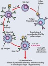
Type I hypersensitivity (“anaphylactic”) reaction:

  • Antigen presented to CD4+ Th2 cells
  • B cells recruited and IgE antibodies formed
  • IgE binds to granular cells (e.g. mast cells and basophils) – sensitization
  • Re-exposure to the same antigen leads to release of mediators from granular cells – degradation
    • Mediators: histamine, leukotriene (LKT), platelet-activating factor (PAF), prostaglandin (PG)
  • Clinical examples:
    • Anaphylaxis
    • Urticaria
    • Allergic asthma
    • Drug allergy (e.g. penicillin)
    • Allergic rhinitis (i.e. hey fever)
84
Q

Describe the type II hypersensitivity reaction and provide clinical examples

A

Type II hypersensitivity (“cytotoxic”) reaction

IgG or IgM antibody binds to cellular antigen, leading to activation of complement. IgG can also mediate antibody-dependant cell-mediated cytotoxicity.

Complement dependant cytotoxicity:

  • Transfusion reaction – rapid destruction of the donor red blood cells by host antibodies
  • Autoimmune haemolytic anaemia
  • Erythroblastosis fetalis
  • Goodpasture’s syndrome

Antibody-dependant cell-mediated cytotoxicity:

  • Transplant rejection
  • Immune reaction against neoplasms
  • Immune reaction against parasites
85
Q

Describe the type III hypersensitivity reaction and provide clinical examples

A

Type III hypersensitivity (“immune complex”) reaction:

Phase 1 – soluble immune complexes are formed by the combining of antigens and antibodies

Phase 2 – Ab-Ag complexes deposit in small vessels where blood flow is reduced (e.g. joints, kidneys):

  • Inflammation
  • Complement binding
  • Platelet aggregation

Examples:

  • Systemic lupus erythematosus
  • Rheumatoid arthritis
  • Acute glomerulonephritis – commonly occurs after streptococcus infection
  • Farmer’s lung – inflammation of alveolar walls à increased collagen and decreased lung compliance
86
Q

Describe the type IV hypersensitivity reaction and provide clinical examples

A

Type IV hypersensitivity (“delayed”) reaction:

  • Cell-mediated response (T lymphocytes)
  • MHC II-antigen complex elicits macrophage response
    • Proliferation of T lymphocytes (T and B cells)
    • Inflammatory reaction
  • Reaction takes several days to develop (cell signalling, and protein synthesis takes time)

Two main phases of type 4 hypersensitivity:

  1. Sensitization: macrophage (Mφ) + CD4+ T lymphocyte → memory CD4+ T lymphocyte (MT)
  2. Re-exposure: (Mφ) + MT → effector CD4+ Th1 cell (IL-2, IFN-γ) → leukocytes, CD8+, Mφ

Examples:

  • Allergic contact dermatitis
  • Autoimmune myocarditis
  • Mantoux test
87
Q

Describe the type V hypersensitivity reaction and provide clinical examples

A

Type V hypersensitivity (“autoimmune”) reaction:

  • Autoimmune disease – antibodies bind to specific cell target receptors
  • Type V not classically used as part of the Gell and Coomb’s classification

Examples:

  • Graves’ diseases
  • Myasthenia gravis
88
Q

Recall the Gell and Coombs classification of hypersentivity reactions (Type I–Type IV)

A
89
Q

Recall the mnemonic for the types of hypersensitivity and the respective antibody/cell

A

Mnemonic and respective antibody/cell:

Type I–Type IV

  • ACID:
    • Anaphylaxis
    • Cytotoxic
    • Immune complex
    • Delayed
  • EGG(T):
    • IgE
    • IgG
    • IgG
    • T lymphocyte
90
Q

Define pathogenicity

A

The ability of an organism to cause disease

91
Q

Define virulence

A

The extent of pathology (harm) caused by the organism

92
Q

Describe the regulation of bacterial virulence

A

Two major regulatory control mechanisms to control expression of virulence genes (2 points):

  1. Sigma factors:
    • Protein synthesis of bacterial RNA polymerases control initiation of transcription
    • Regulate prokaryotic gene expression in response to microenvironment.
  2. Two component system:
    • Sensor protein embedded in bacterial membrane “senses” different physiological conditions of bacterial cell
    • Response regulator which binds to the promotor region of a gene to activate or repress transcription.
93
Q

List 7 mechanisms which microbes employ to overcome host immunity

A

Mechanisms which microbes employ to overcome host immunity – i.e. mechanisms of pathogenicity:

Corona Virus Vaccine Could Enrage Rebellious Antivaxxers”

  1. Changing out protein expression
  2. Viral antigenic drift and shift
  3. Viral interference with antigen presentation
  4. Counteract complement
  5. Extra cell wall protection
  6. Render phagocytosis ineffective
  7. Avoid phagocytosis
94
Q

Mechanisms of pathogenicity:

  • Changing out protein expression
  • Viral antigenic drift and shift
  • Viral interference with antigen presentation
  • ​Counteract complement
  • Extra cell wall protection
  • Render phagocytosis ineffective
  • Avoid phagocytosis

Describe changing out protein expression

A

Changing out protein expression (viruses):

  • Virus proteins may up-regulate host cell proteins that inhibit apoptosis
  • Ability of virus to mutate genotype (and hence phenotype by changing out protein expression) to escape host immune response

Viruses are obligate intracellular parasites, and can manipulate the host cell in which it is residing to benefit its own survival.

95
Q

Mechanisms of pathogenicity:

  • Changing out protein expression
  • Viral antigenic drift and shift
  • Viral interference with antigen presentation
  • ​Counteract complement
  • Extra cell wall protection
  • Render phagocytosis ineffective
  • Avoid phagocytosis

Describe viral antigenic drift

A

Viral antigenic drift:

  • Evolutionary/variation mechanism by which viruses mutate the genetic code for antibody-binding site morphology
  • Creation of new virus, not effectively targeted by pre-existing antibodies
  • Influenza A, B and C- glycoproteins differ between flu subtypes (e.g., H1N1, H3N2, H5N1)
  • Seasonal variation of flu strains (and hence vaccination)
96
Q

Mechanisms of pathogenicity:

  • Changing out protein expression
  • Viral antigenic drift and shift
  • Viral interference with antigen presentation
  • ​Counteract complement
  • Extra cell wall protection
  • Render phagocytosis ineffective
  • Avoid phagocytosis

Describe viral antigenic shift

A

Viral antigenic shift:

  • The combination of ≥2 viral strains to form new virus with combination of both surface antigens
    • Occurs only in influenza A
    • Results in major reorganisation of strains
    • Responsible for jumping between species and pandemics

One species passes viral strain to intermediate host animal, a human also passes on a separate strain to the same animal.

Genes from both strains combine to form new strain which can jump from intermediate host to humans.

97
Q

Mechanisms of pathogenicity:

  • Changing out protein expression
  • Viral antigenic drift and shift
  • Viral interference with antigen presentation
  • ​Counteract complement
  • Extra cell wall protection
  • Render phagocytosis ineffective
  • Avoid phagocytosis

Describe viral interference with antigen presentation

A

Viral interference with antigen presentation:

  • Viruses can prevent proteasomal fragmentation (needed so fragment can be presented on MHC)
  • Block synthesis of MHC molecule itself
  • Redirection of endogenous MHC-antigen complexes to cytosol for destruction → MHC doesn’t even make it to the cell surface of APC
  • Exclusion of APC presentation
98
Q

Mechanisms of pathogenicity:

  • Changing out protein expression
  • Viral antigenic drift and shift
  • Viral interference with antigen presentation
  • ​Counteract complement
  • Extra cell wall protection
  • Render phagocytosis ineffective
  • Avoid phagocytosis

Describe how microbes counteract complement

A

Intracellular bacteria survive and replicate inside host cells.

They enter host cells via receptors or phago-/endocytosis. Whilst intracellular they escape antibodies and avoid subsequent complement mechanisms of host immune system.

Intracellular lifestyles of bacteria:

  • Facultative intracellular bacteria – capable of living either inside or outside of host cells:
    • Listeria monocytogenes, Salmonella typhi, legionella.
  • Obligatory intrasellar bacteria – only survive inside host cells as reliant on intracellular host resources:
    • Chlamydia, rickettsia, Coxiella, Mycobacterium tuberculosis.

Viruses are by nature obligate intracellular parasites and so naturally avoid the complement system once inside the host cell.

Viruses can interfere with the host cell to prevent the host cell itself from activating complement (viral interference with antigen presentation)

99
Q

Mechanisms of pathogenicity:

  • Changing out protein expression
  • Viral antigenic drift and shift
  • Viral interference with antigen presentation
  • ​Counteract complement
  • Extra cell wall protection
  • Render phagocytosis ineffective
  • Avoid phagocytosis

Describe the extra cell wall protection of microbes

A

Extra cell wall protection:

Two major groups of bacteria based on cell wall structure:

  • Gram-positive
    • Cell wall components: peptidoglycan and teichoic acid
    • Common microbes: Staphylococcus aureus, Staphylococcus epidermidis, streptococci
  • Gram-negative
    • Cell wall component: LPS/endotoxin
    • Primary receptor for endotoxin is CD14 (found on macrophages)
    • Common microbes: E. coli, P. aeruginosa, meningococci

Bacterial capsule for extra cell wall protection:

  • Coat of polysaccharide (sugar) “slime” – glycocalyx (esp. Gram -ve)
  • External to cell membrane and cell wall
  • Protection against desiccation
  • Enhances ability to adhere to host
  • Protection from phagocytosis:
    • does not all allow opsonization by antibodies → frustrated phagocytosis
  • Virulence factor

Notorious species: Streptococcus pneumoniae, Neisseria meningitidis, Pseudomonas aeruginosa

100
Q

Mechanisms of pathogenicity:

  • Changing out protein expression
  • Viral antigenic drift and shift
  • Viral interference with antigen presentation
  • ​Counteract complement
  • Extra cell wall protection
  • Render phagocytosis ineffective
  • Avoid phagocytosis

Describe how microbes can avoid phagocytosis and employ mechanisms to render phagocytosis ineffective

A

Avoid phagocytosis:

  • Intracellular lifestyle of bacteria and viruses – able to survive, replicate and avoid clearance by phagocytes in ECM

Render phagocytosis ineffective:

  • Bacterial capsule prevents opsonization → “frustrated phagocytosis”
101
Q

Describe how viruses can escape the host immune system by mutation mechanisms (pathogenicity) (6 points):

A

Viral escape – mutation mechanisms (pathogenicity) (6 points):

Ebola Epidemic Should Be Carefully Identified”

  1. Evasion of NK cell immunity – encode decoy analogue of MHC molecules to veto the NK killing process
  2. Evasion of antibody and complement – antigenic drift and antigenic shift
  3. Sequestration – infection of host cells to store genetic information – virus remains in latent state
  4. Blockade of antigen presentation – disruption of MHC synthesis and APC presentation
  5. Cytokine evasion – encode mimics of normal cytokines (virokines) which inhibit normal immune cells
  6. Inhibition of apoptosis by viral proteins – up regulate proteins to inhibit apoptotic cellular events
102
Q

Recall the differences between antigenic drift and antigenic shift

A

Antigenic dift vs antigenic shift

103
Q

Define primary (inherited) immunodeficiency (PID) and list 6 causes of PID

A

Primary immunodeficiency disorder (PID) refers to a heterogeneous group of disorders characterized by poor or absent function in one or more components of the immune system.

Causes of primary immunodeficiency (6 points):

Peter Pan Should Challenge Pirates Carefully”

  1. Pure” T cell disorders
  2. Pure” B cell disorders
  3. Severe combined immunodeficiency (SCID)
  4. Combined immunodeficiencies
  5. Phagocyte deficiencies
  6. Complement deficiencies
104
Q

Describe “pure” T cell disorder in primary immunodeficiency

A

“Pure” T cell disorders – affecting T cell arm of acquired immunity:

  • Complete insufficiency of T cell function can result from hereditary conditions such as severe combined immunodeficiency (JAK3-deficient SCID: T-, B+):
    • JAK3 (Janus Kinase 3) deficiency is an autosomal recessive form of SCID.
    • It is characterized by lack of circulating T and NK cells but normal number of B lymphocytes.
105
Q

Describe “pure” B cell disorder in primary immunodeficiency

A

“Pure” B cell disorder – most common type of primary immunodeficiency (~50% of PID cases)

Heterogeneous group of disorders (>20) characterised by increased susceptibility to respiratory tract infections with bacteria, particularly Streptococcus pneumoniae and Haemophilus influenzae.

Clinical examples of “Pure” B cell disorders:

  • XLA (X-linked agammaglobuminaemia, or Bruton’s syndrome)
    • Inability to mature B cells
    • Complete or incomplete lack of antibodies.
  • Common variable immunodeficiency (ID):
    • Gene deletion of surface proteins and cytokine receptors
    • Lack of IgG, IgM, IgA.
  • Selective IgA deficiency:
    • Undetectable IgA
    • Most common genetic primary immunodeficiency (PID).
106
Q

Explain severed combined immunodeficiency (SCID)

A

SCID (more aggressive form of CID) is sometimes referred to as ‘bubble boy disease’:

  • 1 in 100,000 live births
  • Can be X-linked or autosomal recessive
  • Multiple variants of genetic mutations responsible
  • Early diagnosis imperative
  • Without treatment, fatal within first year
  • Treated with stem cell (BM) transplant, gene therapy, enzyme replacement therapy.
  • Bubble boy disease

Do not give live vaccines.

Hx and exam: recurrent infections, failure to thrive (poor weight gain), chronic diarrhoea; absence of lymphoid tissue, diffuse erythematous rash, microcephaly, skeletal abnormalities.

Risk factors for SCID: FHx of infant death, FHx SCID (consanguinity).

107
Q

Describe combined immunodeficiency (CID)

A

Combined immunodeficiency (CID)

Overview of combined immunodeficiency:

  • Categorized into presence/absence of T cells and B cells (T-/B+) or (T-/B-)
  • May also have normal T cell count
  • If associated with neurological findings, may be life-threatening

Clinical presentation:

  • < 12 months
  • Chronic diarrhoea
  • Failure to thrive
  • Severe, recurrent infections with opportunistic pathogens
  • Skin rashes

Clinical examples:

  • SCID (more aggressive form of CID)
  • DiGeorge syndrome
  • Wiskott-Aldrich syndrome
  • Ataxia-telangiectasia
108
Q

Explain DiGeorge syndrome

A

DiGeorge syndrome:

  • Deletion in chromosome 22 (segment 22q11.2)
  • Neurological, immunological, endocrinological, or cognitive defects
  • Treatment depends clinical manifestations present in the individual patients
  • Symptomatic management

Hx and exam: cyanosis, heart failure, facial features (bulbous nose tip and prominent ears), cleft lip/palate, growth failure, seizure/tetany, learning disorder, frequent infection, hypocalcaemia, hypoplastic thalamus.

109
Q

Explain Wiskott-Aldrich syndrome

A

Wiskott-Aldrich syndrome:

  • X-linked condition characterised by thrombocytopenia
  • Small platelet size is consistent feature
  • Eczema, recurrent infections and easy bruising
  • Autoimmunity complicates up to 70% of cases
  • Increased risk of haematological malignancies
110
Q

Give 3 examples of phagocyte deficiency in PID

A

Examples of phagocyte deficiency in PID:

  1. Chronic granulomatous disease – severe infection, abscess, granuloma formation
  2. Hyper IgE syndrome – chronic dermatitis, recurrent severe lung infection, bone fragility, failure to lose primary teeth
  3. Leukocyte adhesion deficiency – recurrent severe bacterial infections, poor wound healing
111
Q

Recall the complement deficiencies and their respective clinical manifestations in PID

A

Specific complement deficiency results in specific clinical manifestations

112
Q

Define secondary (acquired) immunodeficiency and list 6 example causes

A

Secondary immunodeficiency is an acquired state in which the immune system’s ability to fight infectious disease and cancer is compromised or entirely absent.

It can result from various immunosuppressive processes/agents:

  1. Malnutrition
  2. Aging
  3. Infectious disease
  4. Malignant
  5. Medications
  6. Environmental toxins – mercury and other heavy metals, pesticides, and petrochemicals
113
Q

Describe the aetiology and pathophysiology of human immunodeficiency virus (HIV)

A

HIV aetiology and pathophysiology

Aetiology:

  • Retrovirus – infects and replicates in CD4+ T cells and macrophages
  • Transmitted via blood, blood products, sexual fluids and breast milk
  • 0.1% per contact for heterosexual transmission (increases with concurrent ulcerative STDs, high HIV viral load and lack of ART)

Pathophysiology:

  • HIV infection follows a 7-step mechanism:
    • Fusion, entry, reverse transcription, integration, synthesis, budding, maturation
  • Provirus resides in host cell nucleus and infected cells remain quiescent
  • Uses host cell to replicate itself
  • High level of viral replication within few weeks
  • Decline in CD4+ T cells
  • Progression to AIDS and death if not treated
114
Q

List the 4 stages of HIV presentation

A

HIV stages of presentation (4 points):

  1. Acute seroconversion
  2. Asymptomatic clinical latency
  3. Symptomatic period (milder immune dysfunction)
  4. Severe immunodeficiency and AIDS
115
Q

Recall the mechanism of HIV infection (7 points)

A

Mechanism of HIV infection (7 points):

  1. Fusion to host cell
  2. Entry of HIV contents: RNA, reverse transcriptase and integrase
  3. Reverse transcription of viral RNA to create viral DNA
  4. Integration of viral DNA into host DNA via integrase enzyme
  5. Synthesis of viral proteins by host using HIV RNA as genomic RNA
  6. Budding of immature HIV virions containing new RNA and proteins
  7. Maturation of virus by protease releasing individual HIV proteins
116
Q

Describe the process and manifestation of HIV seroconversion

A

Seroconversion is the period during which the specific HIV antibodies develop and become detectable in the blood.

Manifestation of HIV seroconversion:

117
Q

Define AIDS and list some examples of AIDS defining illnesses

A

Acquired immunodeficiency syndrome (AIDS) – CD4 count <200 or AIDS defining illnesses:

  • Candidiasis of oesophagus
  • Candidiasis of upper airways
  • Cytomegalovirus
  • CMV retinitis
  • Encephalopathy
  • Multifocal leukoencephalopathy
  • Chronic herpes simplex
  • Kaposi’s sarcoma
  • Lymphoma
  • Mycobacterium tuberculosis
  • Cerebral toxoplasmosis
  • HIV wasting syndrome
  • Cervical cancer
  • Pneumocystis jirovecii pneumonia.
118
Q

Describe HIV diagnosis and management

A

HIV diagnosis:

  • Fourth generation HIV test (ELISA) – tests for p24 antigen as well as antibodies, so are better suited for detecting recent infection (reduces risk of false negative result during 12-week window period)
  • High levels of p24 are present in the blood during the short period between HIV infection and seroconversion, before fading away
  • Test routinely used in the UK.

HIV management (4 points):

  1. Antiretroviral drugs
  2. Counselling (pre- and post- testing)
  3. Vaccinations
  4. Micronutrient supplementation