Image questions Flashcards

1
Q

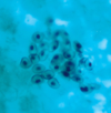
50 yom with chest pain for 24 hours: (a fib/ventricular tachycardia/ventricular bigeminy)

A

ventricular bigeminy: a ventricular premature beat follows each normal beat

2
Q

55 yom with 4 hours of crushing chest pain. (anterior wall MI/inferior wall MI/anteroseptal MI/RV MI/L lateral wall MI)

A

inferior wall MI.

The electrocardiogram shows the early hyperacute injury phase of inferior wall myocardial infarction. This is reflected by the marked slope elevation of the ST segments, and the increased amplitude of the T waves in Standard leads II and III, and lead aVF. Reciprocal ST segment depression occurs in Standard leads I, lead AVL and leads V 4 to V6.

3
Q

lipid deposition and smooth muscle cell hyperplasia (accumulation in intima)

A

atherosclerosis

4
Q

two findings

A

atherosclerosis and thrombus

5
Q
A

dissecting aorta

6
Q

medium sized arteries, smoking history

A

Buerger disease (thromboangiitis obliterans)

7
Q

what vessel does syphilitic aneurysm typically affect

A

ascending aorta (vasa vasorum)

8
Q

xanthomas in which disease

A

familial cholesterolemia (foamy macrophages, ApoE involved, lacks LDL receptor)

9
Q

filarial worm infestation causing obstruction of what

A

lymph nodes (elephantiasis)

10
Q

biopsy of skin lesion in 25yow patient with proteinuria, recent hx of Hep B infection, P-ANCA, elevated erythrocyte sed rate

A

polyaderitis nodosa (medium sized and smaller muscular arteries, fibrinoid necrosis)

11
Q

AIDS patient with multiple purple-colored skin nodules on hands and feet. What virus is implicated

A

HHV 8 (Kaposi sarcoma)

12
Q

70yow with throbbing unilateral headache and vision problems, mandibular pain

A

Giant cell arteritis

13
Q
A

cavernous hemangioma

14
Q

thoracic aorta with aldehyde fuchsin stain

A

cystic medial necrosis (dissecting aneurysm). focal loss of elastic and muscle fibers in the aortic media leads to cystic spaces filled with pools of metachromatic myxoid material

15
Q
A

aneurysm

16
Q

coronary artery section in pt with familial hypercholesterolemia. what’s in it?

A

platelets and RBCs + fibrin = thrombus (at an early age)

17
Q

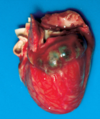
fever, skin rash, mucosal inflammation, lymph node enlargement. usually self limited but may involve coronary arteries and lead to aneurysm
A

Kawasaki

18
Q

renal biopsy. (papillary necrosis/polyarteritis nodosa/monckeberg medial sclerosis/hyaline ateriolosclerosis)

A

hyaline ateriolosclerosis

19
Q
A

varicose veins

20
Q

excessive collagen deposition in the skin, subintimal fibromuscular thickening, renal biopsy. what is the disease

A

scleroderma

21
Q

infant heart. (cardiac amyloidosis/endocardial fibroelastosis/hypoplastic left heart syndrome)

A

endocardial fibroelastosis (fibroelastotic thickening of the endocardium of the left ventricle, may also affect valves)

22
Q

left main coronary artery 2 days after an MI. what histologic changes are present?

A

two days after MI: heart muscle will show myofiber necrosis, edema, and focal hemorrhage. this artery shows necrosis of cardiac myocytes and infiltrates of neutrophils

23
Q

35 yom with MI, L main coronary artery two days later. what underlying disease does the patient have?

A

familial hypercholesterolemia

24
Q

ruptured myocardial wall causes what

A

cardiac tamponade

25
Q

this infarct was caused by thrombotic occlusion of which coronary artery

A

LAD (most common) supplies anterior wall and anterior septum of LV

26
Q

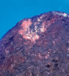
image shows hemopericardium. myocardial rupture and hemorrhage into the pericardial sac is most commonly seen when after an MI
A

between 1 to 4 days after MI

27
Q

this heart shows mural thrombosis in the aneursym. what is a likely complication?

A

stroke

28
Q

RV hypertrophy and dilation secondary to pulmonary HTN

A

cor pulmonale (MCC is COPD)

29
Q

presence of sterile vegetations on apparently normal cardiac valves in assoc with cancer or some other wasting disease

A

nonbacterial thrombotic endocarditis/marantic endocarditis

30
Q

78yom with hx of recurrent syncope. aortic valve disease (bacterial endocarditis/calcific aortic stenosis/hypertrophic subaortic stenosis/marantic endocarditis)

A

calcific aortic stenosis

31
Q

the boxcar nuclei in the myocardium on the right indicate what?

A

hypertension

32
Q

sudden death in young athletes

A

hypertrophic cardiomyopathy–cardiac hypertrophy is out of proportion to the hemodynamic load placed on the heart, autosomal dominant

33
Q

most common type of cardiomyopathy

A

dilated cardiomyopathy

34
Q

heart with CHF shows biventricular dilation and hypokinesis, patchy interstitial mononuclear inflam infiltrate composed of T lymphocytes and macrophages

A

viral myocarditis

35
Q

most common primary tumor of the heart

A

cardiac myxoma, most arise in LA

36
Q

chronic fibrosing disease of the pericardium that compresses heart and restricts inflow (results from an exuberant healing response)

A

constrictive pericarditis

37
Q

higher risk for cerebral embolism

A

mitral valve prolapse

38
Q

mitral valve. (bacterial endocarditis/carcinoid heart disease/Libman-Sacks endocarditis/marantic endocarditis/mitral valve prolapse)

A

bacterial endocarditis

39
Q

72 yow dies of CHF. aortic valve at autopsy

A

chronic rheumatic valvulitis (fusion of commissures!)

40
Q

alcoholic patient, 40yom currant jelly sputum, diffuse pulmonary consolidation. (atypical/broncho/interstitial/lobar) PNA

A

lobar PNA

41
Q

interstitial PNA in infants and immunocompromised pts. alveolar cells are large, show basophilic nuclear inclusions, peripheral halo and multiple cytoplasmic basophilic inclusions

A

CMV

42
Q

pt undergoing chemo. cough, fever, multiple areas of consolidation and a large cavity in the RUL

A

invasive aspergillosis

43
Q

38 yow with SLE being treated with corticosteroids who breeds pigeons. granulomas and budding yeast forms, stain positively for polysaccharides (actinomycosis/coccidiomycosis/cryptococcosis/histoplasmosis)

A

cryptococcosis–pigeon droppings

44
Q

pt with AIDS, CXR shows bilat and diffuse infiltrates

A

pneumocystis jiroveci

45
Q

(atelectasis/bronchiectasis/bronchoPNA/lobar PNA/pulmonary fibrosis)

A

pulmonary fibrosis

46
Q

alveoli are filled with granular, proteinaceous, eosinophilic material, PAS positive, assoc with compromised immunity, leukemia, lymphoma

A

alveolar proteinosis aka lipoproteinosis

47
Q

COPD in a young woman, long history of SOB, chronic cough, sputum production, wheezing

A

alpha 1 AT deficiency

48
Q

status asthmaticus. what histo is present

A

smooth muscle hyperplasia and basement membrane thickening

49
Q

numerous small, fibrotic nodules in both lungs

A

silicosis

50
Q

worked on ships. pulmonary fibrosis and iron stains of lung tissue show numerous ferruginous bodies

A

asbestosis (diffuse interstitial fibrosis)

51
Q

25 yo black woman with 3 month hx of cough and SOB on exertion. elevated ACE

A

sarcoidosis

52
Q

rare pneumonitis that occurs in Sjogren and HIV

A

lymphocytic interstitial PNA (LIP)

53
Q

23yom with nasal obstruction, serosanguinous discharge, cough, bloody sputum. CXR shows cavitated lesions and multiple nodules over both lung fields. Obliteration of maxillary sinuses. Hematuria, C-ANCA

A

Wegener granulomatosis–aseptic necrotizing granulomatous inflammation and vasculitis

54
Q

pulmonary eosinophilic granuloma due to what disease

A

langerhans cell histiocytosis

55
Q

patient responds favorably to antiestrogen and antiprogesterone tx. rare interstitial lung disease that occurs in women of childbearing disease

A

lymphangioleiomyomatosis

56
Q

22yom with 6 months of increasing SOB and rusty sputum. linear deposits of IgG and C3 are detected in the alveolar basement membrane by immunofluorescence

A

Goodpasture

57
Q

28 yom with 6 days of fever and SOB. pt does not respond to standard antibiotics. (lipid/pneumococcal/pneumocystis/eosinophilic/usual interstitial) PNA

A

eosinophilic PNA

58
Q

53yom with increasing SOB on exertion, dry cough that has developed over a period of a few years. patchy scarring with extensive areas of honeycomb cystic change

A

usual interstitial PNA

59
Q

55yow with sudden onset fever, dry cough, SOB. seen for flu like symptoms 6 weeks ago. biopsy shows inflamed airways with plugs of fibrous tissue

A

cryptogenic organizing PNA (bronchiolitis obliterans organizing PNA)

60
Q

45yow with severe kyphoscoliosis with fatigue, SOB on exertion, fainting, bloody sputum. CXR shows enlargement of the central pulmonary arteries

A

pulmonary HTN

61
Q

popcorn pattern of calcification

A

pulmonary hamartoma

62
Q

what type of cancer in the lung

A

squamous cell carcinoma

63
Q

history of smoking, peripheral subpleural mass

A

adenocarcinoma

64
Q

lung cancer, stains for keratin and chromogranin are negative

A

large cell undifferentiated

65
Q

primary pulmonary adenocarcinoma originating from stem cells in the terminal bonchioles

A

bronchioalverolar carcinoma

66
Q

“oat cell” carcinoma, exhibits neuroendocrine features, Cushing Syndrome

A

small cell carcinoma (carcinoid tumor is also neuroendocrine BUT no Cushing syndrome)

67
Q

neuroendocrine granules within tumor cells in the lung

A

carcinoid tumor

68
Q

the heart of a patient who died in uremia displays what type of pericarditis

A

fibrinous pericarditis