HISTOLOGY - WBC Disorder and Pulm Flashcards
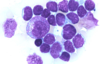
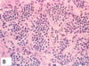
ALL and AML
- Scant basophilic cytoplasm
- FINE Nuclear Chromatin w/Convolutions
(Note: Unable to differentiate ALL from AML with this Histology)

Positive TdT Staining indicative of
ALL Specifically

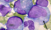
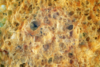
Erythroid Precursors in
MyeloDysplastic Syndrome (MDS)
[Nuclear BIMS - Budding / irregularities / multinucleation / separation of lobes]

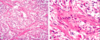
ProMyelocytes with AUER RODS –indicative of
aPL - Acute PROmyelocytic Leukemia
(type of AML)

Positive [MyeloPerOxidase Stain] with Dark Brown consolidation indicating
AML

Positive [AnBe Stain - Alpha napthyl Butyrate esterase] indicating
AML
(monocytic lineage)
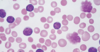
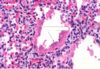
Smudge Cells
in [CLL-SLL]

[small lymphocytes w/scant cytoplasm]
see in a lymph node of [CLL-SLL]

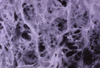
Hairy Cell Leukemia


Floret Cell
in
ATL - [Adult T-cell Leukemia/Lymphoma]

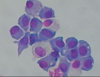
[Benign Reactive Follicle]
in
Follicular Lymphoma


Follicular Lymphoma
Abnormal Brown Stain in center of lymph node


Follicular Lymphoma

Mantle Cell Lymphoma
Cyclin D1 Overexpression

Burkitt Lymphoma
Starry Sky Appearance


DLBL [Diffuse Large B-cell Lymphoma]
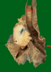
Spleen with Fish Flush Appearance

DLBL - [Diffuse Large B-cell Lymphoma]
Multiple Nucleoli (dark structures) with
[Vesicular Chromatin - bubbly appearance]

PTCLn (Peripheral T-Cell Lymphoma - not otherwise specified)
Heterogenous Polymorphic appearance

[Hodgkins Lymphoma: Nodular Sclerosis]
Lacunar Cells

[RSO cell - Reed Sternberg OwlEye]
Hodgkins Lymphoma

[RSO cell - Reed Sternberg OwlEye] -
Mononuclear variant
in Hodgkins Lymphoma

[RSO cell - Reed Sternberg OwlEye] -
Lacunar variant
in Hodgkins Lymphoma

Popcorn RSO Variant cells
in
Hodgkins Lymphoma: Lymphocyte Predominant

[Dark Lytic Lesions] of the Skull
indicating
Multiple Myeloma

Mott Cell in Bone Marrow Biopsy
indicating
Multiple Myeloma

CML - Chronic Myelogenous Leukemia
Peripheral Blood Smear showing more WBC than usual

CML - Chronic Myelogenous Leukemia
Peripheral Blood Smear showing INC Basophils

CML - Chronic Myelogenous Leukemia
Bone Marrow showing [Abnormal 100% Cellularity-_mainly Granulocytes_] with no Fat Cells

Tear Drop Cells
indicating
[CIPM- Chronic Idiopathic Primary Myelofibrosis]

Essential Thrombocytosis

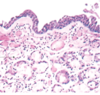
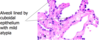
Primary Bronchus

TERTIARY BRONCHUS AND BRONCHIOLE

Bronchogenic Cyst

Cyst of Varying Sizes in the Lung
Pulmonary Sequestration
Pink, Granular Edema Fluid within alveolar spaces

ARDS
(as long as no cardiac failure is present)

Early Exudative Phase of ARDS
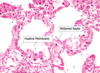
ARDS – late proliferative and fibrosis stages.
Hyaline Membrane
(Fibrin + Necrotic Cells)

INC [submucosal glands/goblet cells] and [Reid Index > 50%] in
Chronic Bronchitis

Centriacinar EmPhysema
Panacinar EmPhysema
Irregular EmPhysema

Cilia with Missing Dynein Arms in

Kartagener Syndrome

Idiopathic Pulmonary Fibrosis


Cobblestone Pleura
indicating
IPF (Interstitial Pulmonary Fibrosis)

Usual Interstitial PNA
(AKA [Interstitial Pulmonary Fibrosis] and [Cryptogenic Fibrosing Alveolitis]


Anthracosis
(from Coal Dust Pneumoconiosis)


Progressive Fibrosis

- (from Coal Dust Pneumoconiosis)*
- 3rd stage - Black Lung*

Silica Crystals under polarized light
indicating
Silicosis
Type of Pneumoconiosis


Asbestos Bodies
indicating
Asbestosis

[Pleural Plaques w/calcification]
indicating
Asbestos


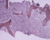
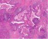
Granulomas
in
Bronchial Epithelium


Sarcoidosis


Sarcoidosis


Signet Ring
indicating
Bronchiectasis


Rim of Fibrosis (around lower lobes of lung)
indicating
Interstitial Pulmonary Fibrosis / UIP


Meniscus
indicative of
Pleural Effusion


[Layering while in Lateral Decubitus]
indicative of
Pleural Effusion
Loculations
indicating
Pleural Effusion

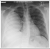
Black Area + No Lung Markings
indicating
PTX (pneumothorax)

PTX (pneumothorax)
SQuamous Cell Carcinoma


Intercellular Bridges
indicating
SQuamous Cell Carcinoma


Keratinization
indicating
SQuamous Cell Carcinoma
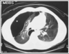
Irregular [Spiculated Mass] with entrapped anthracosis
indicating
Adenocarcinoma


Well demarcated nodule in
[AAH-Atypical Adenomatous Hyperplasia]
(which progresses to Adenocarcinoma)

[AiS BAC]

Adenocarcinoma in-Situ / BronchioloAlveolar Carcinoma

Thyroid Tx Factor 1
indicating
Adenocarcinoma

SubTypes of Adenocarcinoma
a. Acinar adenocarcinoma
b. Papillary adenocarcinoma
c. Solid adenocarcinoma
d. Mucinous adenocarcinoma

[Large Cell Undifferentiated Carcinoma]
- No squamous or adeno differentiation by morphology*
- OR by immunohistochemistry*

Azzopardi Effect-staining on vessel walls
indicating
[SOC- Small Oat cell Carcinoma]

[SOC- Small Oat cell Carcinoma]


[Polypoidal endobronchial mass] + [no necrosis]
indicating
Bronchical Carcinoid
Bland Cell Nest separated by vasculature
indicating
Bronchial Carcinoma

SQamous cell Carcinoma

ADenoCarcinoma

[SOLC - Small Oat cell Lung Carcinoma] (blue)

LCUC - Large Cell Undifferentiated Carcinoma
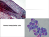
Normal Mesothelial Cells
in Pleura cavity

Mesothelioma
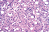

Mesothelioma
on EM and Stain


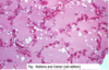
Metastatic signet ring ADenoCarcinoma
Engorged Alveolar capillaries
in
Pulmonary Edema

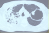
Pulmonary Embolus


Pulmonary Embolus
being shown in DVT

Saddle Pulmonary Embolus

Medial Hypertrophy
indicating
Pulmonary HTN

Plexiform Lesions
indicating
Pulmonary HTN
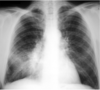
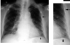
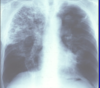
PNA

Streptococcus pneumoniae
most common cause of [CA PNA]

Palasading Epithelium
indicating
TB Lung Granuloma

[Multinucleated Giant Cells]
indicating
TB Lung Granuloma
[Amorphic Central Necrosis]
indicating
TB Lung Granuloma

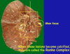
Ghon Focus
indicating
TB
[Cavitary NonTB Mycobacteria]
in the setting of Emphysema

[Bronchiectasis NonTB Mycobacteria]

