Histology of the Reproductive Tract Flashcards
Describe the gross structure and function of the ovaries
Ovaries are atached to the posterior face of the broad ligament. The broad ligament is a wide double folded peritoneum that envelops the uterus, fallopian tubes and ovaries.
The ovaries are anchored to the uterus via the **ligament of ovary **and to the pelvic wall via the **suspensory ligament **
The fallapian tubes are intricately related to teh ovaries.
The ovaries are responsible for:
- Producing gametes
- Producing hormones
- Responding to circulating hormones
Ovarian function declines with age and this is termed menopause
Describe the histological structure of the ovaries
An ovary is divided into an:
-
Inner medulla
* loose connective tissue and blood vessels - Outer cortex
- location of ovarian follicles and oocytes
- cortical stroma is composed of highly cellular connective tissue and smooth muscle cells
The surface of the ovaries are irregular due to the constant rupture and repair that occurs associated with the release of occytes in ovulation
Surface is simple epithelium (either squamous or cuboidal) continuous with mesothelium - 70% of ovarian tumours are from this epithelium
Directly beneath the epithelium is the tunica albuginea - a dense supportive connective tissue
How many oocytes are present in a female ovary over the course of time?
There are approximately 5 million / ovary in embryo and this reduces to 0.5 million by the time of birth - this is a female’s full compliment as she is unable to acquire more eggs than this.
Most of the oocytes present at birth die over the time of a persons life; a few thousand go through most of a maturation cycle.
Around 500 oocytes are ovulated into the fallopian tubes over the course of a females life
Characterise primordial oocytes
Primordial oocytes are the smallest and earliest form of oocyte
They are lined by squamous follicular cells with themselves are contained within a common basal lamina
These oocytes are arrested in prophase of meiosis 1 (incomplete division)
Characterise a primary oocyte
The oocyte is now surrounded by the zona pellucida (inner aspect of follicular cells)
**The oocyte itself enlarges **
Follicular cells become cuboidal and multilayered granulosa cells / stratum granulosum
Surrounding stromal cells start to form stratum interna and externa
Characterise a secondary follicle
As the stratum **granulosum **thickens a fluid filled cavity, known as theantrum, forms within the follicle and almost encloses the oocyte contained within.
The oocyte is suspended within the antrum by a stalk of granulosa cells called the cumulus oophorus
This circular layer of granulosa cells around the oocyte form the corona radiata after release
Describe the events that lead to ovulation
Under an LH surge, the primary oocyte completes its first meiotic division to form a secondary oocyte
Associated with this is the rupturing of the follicle containing the oocyte - releasing the secondary oocytes into the body cavity to enter fallopian tube where it can survive for a maximal 24 hours if not fertilised
What happens to a follicle once it releases its oocyte
Once a follicle ovulates it’s oocyte; it forms a corpus luteum
This occurs as the result of stromal, granulosa and thecal cells invading the cavity left by the oocyte and differentiating into luteal cells
**The **corpus luteum appears yellow due to lipid deposition, becomes vascularised and produced progesterone and estrogen to prepare and then maintain the endometrium
The deposited lipids are utilised on the synthesis of the hormones mentioned above
Corpus luteum survives for 14 days. If there is no fertilisation, it involutes with time -> transitioning through a non-functional stage of **corpus albicans **
Characterise the histological structure of fallopian tubes
Fallopian tubes have:
External serosa (mesothelium + thin connective tissue layer)
Smooth muscle muscularis
**Mucosa **(epithelium + connective tissue)
The lumen of the fallopian tubes is lined by cilliated epithelium that assists in moving the oocyte towards the uterus. Fluid secreted from the epithelium and/or drawn from the peritoneum is used by the cilia to create a current, in conjunction with smooth muscle peristalsis, that moves oocyte/blastocyst towards uterus
Fertilisation usually occurs in the ampulla and the zygote travells in the isthmus for 4 days - during which time the fallopian tube provides nutrients for development in the secreted fluid
Characterise the tissues of the uterine wall
The uterine wall of the uterus can be divided into three tissue layers: perimetrium, endometrium and myometrium
The perimetrium covers most of the external surface of the external surface.
Myometrium
Is composed of three layers of smooth muscle around the outside of the endometrium: the inner and outer layer is longitudinal muscle but the middle layer is circular and very vascular.
These smooth muscle cells are enlarged and in increased numbers during preganacy - remains after first pregnancy permanently as a thickened wall
Endometrium
Epithelial mucosa mix of ciliated and secretory columnar cells in simple epithelium
**Secretory glands** (no ciliated cells) penetrate into
lamina propria (dense connective tissue)
Mucosa supplied by helical arteries
Functional layer sloughed off each menstrual cycle
Basal layer regenerates functional layer
Growth of the endometrium is driven by estrogen initially and breakdown by later decrease of estrogen and progesterone
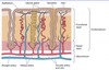
Compare the histological profile of the endometrium over the course fo the menstrual cycle
Over the course of the proliferative stages/secretory stages of mentrual cycle, the endometrium undergoes changes:
- Epithelial, stromal (lamina propria) and vascular cells proliferate vigorously
- Secretion accumulates (mucoid with glycogen)
In mid-proliferative phase glands of the endometrium become dilated and hypertrophied
In mid-secretory phase the glands have substance within them ready for implantation to occur

Discuss the histology of the cervix
The **endocervix **is composed of simple columnar epithelium and it **highly glandular **like the body of the uterus.
Secretion is serous and copious at ovulation (conducive to sperm migration); thick and plug like at other times (non-conducive to sperm migration)
The ectocervix is composed of **stratified squamous epithelium **and is non-glandular like the skin/vagina
There is an abrupt transition between the simple columnar and stratified squamous epithlium - although the position of transition cahnges with reproductive state of female and stage of menstrual cycle.
HPV infects female in this region -> stratified epithelium sheds cells that are inspected by Pap smears for tumourous changes
Describe the histology of the vagina
The **vagina **is a **fibromuscular tube **
It is lined by non-keratinised stratified squamous epithelium (undergoes cyclic changes with menstrual cycle) and forms mucosa with partly erectile lamina propria
It’s smooth muscle layers comprise of a thin inner and thick outer layer (continuous with muscle of uterus - but no middle layer)
No glands are present - the vagina is lubricated by cervical glands or vestibule glands
The most superficial cells of the epithelium retain nuclei which predisposes them to malignancy
Characterise the structure of breasts
Breasts vary with age, menstrual cycle or reproductive status
They contain multiple mammary glands embedded in dense connective tissue and abundant adipose tissue (majority of breast volume is fat)
Multiple mammary glands each open onto the nipple through independent and seperate openings
Mammary glands consist of several important structures:
Terminal duct lobular units (TDLUs)
- 15-20 lobular structures per mammary gland which contain acini that actually produce milk and interlobar connecting ducts to organise milk distribution
**Lactiferous ducts **
- Branching network that delivers milk from lobules to the nipple for secretion
Mammary glands are modified sweat glands
What differntiates the breast structure of males and females?
Until puberty, male and female mammary glands are similar
At puberty, male glands regress due to testosterone
At puberty, female glands grow due to estrogen and progesterone = extension of glands and breast enlarges (largely adipose tissue)
Note: mammary glands remain inactive until pregnancy as they are under negative influence of stromal cells
Discuss the events and histology associated with inactive glands
Inactive Glands
- are responsive to the menstrual cycle and under go growth and collapse with successive rounds of the menstrual cycles
- in luteal phase -> become more columnar, some secretions appear and fluid accumulates in stroma connective tissue
- immediately prior and during mestration the gland involute
Histologically:
Inactive gland relatively sparse, cuboidal/ columnar epithelial cells, surrounding
myoepithelial cells in extensive dense connective tissue
- flattened myoepithelial cells contract the ducts to release milk
What changes in breast structure occur in pregnancy?
Terminal ductules elongate and branch
Epithelial and myopeithelial cells
proliferate from progenitor cells
By term, have large cuboidal epithelial
cells containing lipid and secretory product
in lumen
Breast is enlarged as a result of growth
Characterise the profile of milk secreted in lactation
Colostrum - a premilk substance
Human milk is mixture of low lipid, low carbohydrate and high protein
It is high in IgA (from plasma cells that have invaded intralobular tissue) which assists in passive immunity
The production of milk is stimulated by the hormone prolactin
Mature milk contains less Ig and higher amounts of CHO and lipids
What mechanisms regulate lactation?
Suckling initiates a reflex that inhibits prolactin release-inhibiting hormone -> leading to the increased release of prolactin for milk production.
Suckling also releases oxytocin which acts to contract myoepithelial cells of mammary glands to squeeze milk out of TDLUs and lactiferous sinuses
What happens to mammary glands following menopause?
Following menopause, mammary glands
involute
Secretory cells disappear leaving only ductal systems
These ductal systems tend to be the site of breast cancer
Describe the structure of the testis
What is the pathway of sperm in ejaculation
Structures of the testis are surrounded by thick tunica albuginea connective tissue
This tunica albuginea is organised in septa that divied the testis into approximately 250 incomplete compartments
In each of these compartments is a seminiferous tubule (site of spermatogenesis) that connects to the epididymis via first the rete testis and then the efferent ductules.
The epididymis leads to the vans deferens which then enters the urethra

Describe the histology of the seminiferous tubule
Each lobule of the testis contains 1 - 4 highly convoluted seminiferous tubules (~50 cm long)
They are lined by stratified epithelium where spermatogenesis occurs:
- Spermatogonia are stem cells that produce spermatocytes to undergo meiosis and mature into sperm -> this process occurs as they migrate towards the tubule lumen
- Sertoli cells anchor, support and provide nutrients to spermatids so they can mature and develop in the lumen
Tunica/lamina propria surrounds the seminferous tubules and contains:
- Myoid cells = contractile smooth muscle like cells that propel sperm fluid and debris along tubule
- Leydig cells = large testosterone secreting cells
The final section of the seminiferous tubule is the straight tubuli recti which is lined solely by sertoli cells before final cuboidal epithelium. It deposits tubule contents into the rete testis - an interconnected set of channels lined by ciliated cuboidal cells which communicate with epididymis via efferent ductules

Describe the structure and function of the epididymis and efferent tubules
The epididymis is embryologically derived from the mesonepheric wolffian duct
The efferent ductules (derived from mesonepheric tubules) connect the epididymis to the rete testis
The epididymis is lined with pseudostratified
columnar epithelium surrounded by smooth muscle
It is 4-6 m long and highly coiled; consisting of a head, body and tail (efferent ductules connect to head)

What is the function of the epididymis?
Sperm mature as the move along epididymis
This includes decapacitation, the inhibition of
the ability of the sperm to fertilise an egg
(reversed in vagina) mediated by inhibitory factors
secreted by epididymis
Epididymis also absorbs most testicular
fluid around sperm and **clean up debris **
