Histological Sections Flashcards
(12 cards)
1
Q
Identify the overall image and label the different stuctures:
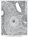
A
- Cell Soma
- Cell body
- Proximal Dendrites
- Nucleus
- Nucleolus
- Nissl bodies
- Mitochondria
- Globus cisternae

2
Q
Identify the overall image and label the different stuctures:

A
- Nissl Bodies/ Rough ER (Light microscope)
- Nucleus
- Nuclelous
- Nissl Bodies

3
Q
Identify the overall image and label the different stuctures:

A
- Nissl Bodies/Rough ER (Light Microscope)
- Nissl Bodies
- Dendrites
- Axon
- Axon hillock

4
Q
Identify the overall image and label the different stuctures:

A
- Nissl Bodies/Rough ER (Electron Microscopy)
- Rough ER
- Mitochondria

5
Q
Identify the overall image and label the different stuctures:

A
- Nissl Bodies/Rough ER (Electron Microscopy)
- Mitochondria
- Microtubule
- Neurofilament
- Globus Cisternae
- Rough Endoplasmic Recticulum (ER)
- Free Ribosomes

6
Q
Identify the overall image and label the different stuctures:

A
- Transverse section of a spinal motor dendrite
- Mitochondria
- Nissl Bodies
- Microtubules
- Neurofilament
- small spines
- synaptic termninals
- Axon terminals

7
Q
Identify the overall image and label the different stuctures:

A
- Longitudinal section of Dendrite
- Nissl Bodies
- Mitochondria
- Microtubules

8
Q
Identify the overall image and label the different stuctures:

A
- Longitudinal Section of an Axon
- Axon Terminal
- Mitochondria
- Microtubles
- Ribosomes
- Endoplasmic Reticulum
- Axon Terminals
9
Q
Identify the overall image and label the different stuctures:

A
- Transverse Section of a myleininated Axon in PNS
- Mylein Sheath
- Mitochondria
- Microtubules
- Nuerofilaments

10
Q
Identify the overall image and label the different stuctures:

A
- Longitudinal Section of myelinated axon
- Node of Ranvier
- Microtubules
- Myelin
- Pockects of oligodendrocyte cytoplasm

11
Q
Identify the overall image and label the different stuctures:

A
- Synapse
- axon terminal
- synaptic vesicles
- postsynaptic element
- dendrite
- synaptic cleft
- Post synaptic density (PSD)
- microtubules
- neurofilaments

12
Q
Identify the overall image and label the different stuctures:

A
- Myelinating oligodendrocyte (Electron Microscope)
- Oligodendrocyte
- Axon 1
- Axon2

