Histo - Female Flashcards
What are the components of the ovaries?
Basics:
- 2 ovaries present
- each has hilum
- suspended from mesenteries
- has cortex & medulla
Histo:
- Covered w/ germinal epithelium
- simple cuboidal lining
- continuous w/ mesothelium
- NOT germinal
- Tunica albuginea = deep to germinal epithelium

What is this?

Ovary Cortex and Medulla
Cortex:
- ovarian follicles
- stroma w/ CT
Medulla:
- loose CT & blood vessels
- blood vessels enter from hilum

What are Ovarian Follicles?
Basics:
- follicle = oocyte + cells surrounding it
- enlarge as they develop –> increasing the cell layers that surround them
Cells surrounding oocyte:
- Follicular cells or granulosa cells (w/in BL)
- Thecal cells (outside BL)
Stages:
- Primordial
- Primary
- unilaminar
- multilaminar
- Secondary
- Tertiary
What are the different stages of follicle development in the ovary?
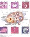
Follicular Development
- Primordial follicles
- Primary follicles
- unilaminar
- multilaminar
- Secondary follicle aka Antral
- antrum forms
- Tertiary follicle aka Graafian follicle
- antrum expands & becomes mature

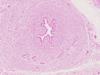
What is this?

Primordial - Follicle in Ovary
Primordial
- single layer of flattened follicular cells around the oocyte
- BL surrounds it
- becomes primary oocyte
Location
- outer cortex
What is this?

Primary Follicles
Unilaminar
- has primary oocyte
- has single layer of cuboidal follicular cells
- Zone pellucida forms
- BL around oocyte
Multilaminar
- has primary oocyte
- has multiple layers of follicular cells (granulosa cells)
- Theca begins to organize
- Zone pellucida

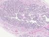
What is this?

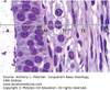
Secondary & Tertiary Follicles - in Ovary
Secondary follicle:
- start of antrum = callexner bodies
- contains follicular fluid made by follicluar (granulosa) cells
- can have complete antrum… but do not protrude from ovary
- smaller
- multiple layers of granulosa cells
- theca organized
- zona pelucida
Tertiary (Mature or Graafian) follicle:
- secondary oocyte
- zona pellucida
- corona radiata
- antrum complete = continuous + large
- protrudes from surface of ovary when read to ovulate
- 1 or 2 per cycle undergo ovulation (others becocme atretic)
- granulosa cell
- theca organized

What is this?

Follicular Structures

What is this?
Theca Interna & Externa
Theca Interna
- Steroid secreting cells
- Vacuolated
Theca Externa
- Fibroblasts
What is the difference between primary & secondary oocyte?
Primary oocyte
- meiosis arrested in prophase I
- meiosis I = completed prior to ovulation
- meiosis II = starts & arrests in metaphase II
- now = secondary oocyte in mature follicle
Secondary oocyte
- ovulates
- if fertilized, meiosis II = complete
- second polar body
What is this?
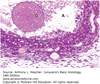
Atresia
- Degeneration
- can happen at any stage of follicle development
- Apoptosis of granulosa cells
- Autolysis of oocyte
- Macrophages do clean up
What phase is Oogenesis suspended in during childhood?
Meiosis I (Prophase I)

What is the Hormonal Regulation of Ovarian Function?
Hormonal Regulation of Ovaries
- Hypothalamus secretes GnRH
- stimulates anterior pituitary (AP)
- FSH & LH released
- stimulate follicular development
- Maturing ovarian follicles
- secrete inhibin (inhibits FSH production)
- low levels of estrogen (initially inhibits both the hypothalamus & AP)
- Estrogen (low levels)
- assists w/ dev of vesicular follicle
- Vesicular follicle
- produces a large threshold amount of estrogen
- stimulates the hypothalamus & AP
- produces a large threshold amount of estrogen
-
LH surge from the AP
- induces ovulation
-
Corpus luteum forms
- due to influence of LH
- Corpus luteum secretes large amounts of progesterone, estrogen, inhibin
- inhibits hypothalamus & AP

What happens during Ovulation?
Ovulation
- LH surge = causes ovulation
- Graafian follicle ruptures
- Causes oocyte w/ surrounding cells, blood & follicular fluid to leave the ovary
- if contacts peritoneum = cause mid-cycle lower abd pain
If secondary oocyte in meiosis II metaphase is fertilized…
- meiosis II = completes
- LH causes follicle to become a corpus luteum
- becomes corpus albicans
What is this?

Corpus Luteum
After ovulation:
- follicle involutes
-
theca interna cells —> theca lutein cells
- darker staining cells than granulosa lutein cells
-
follicular cells —> granulosa lutein cells
- ligher staining cells than theca lutein cells
- theca externa contracts
- granulosa cells collapse
- theca interna cells INVADE into granulosa cells
If NO fertilization:
- involutes w/in 14 days into a corpus albicans
If fertilization:
- involutes w/in 6 months to corpus albicans

What are the 2 Types of Corpora Lutea?
Corpus Luteum of Menstruation
- persists for part of 1 cycle
- phagocytosed by macrophages
- forms a corpus albicans
Corpus Luteum of Pregnancy
- uterine mucosa cannot menstruate (would lose embryo)
- corpus luteum of pregnancy = maintain by HCG
- 4-5 months until placenta makes progesterone & estrogen
- then becomes a corpus albicans
What is this?

Corpus Albicans
- Scar tissue
- Macrophages phagocytose debris
-
Hemosiderin in macrophages
- brown color
What are the Parts of the Uterine Tubes?
-
Fimbriae & infundibullum
- catches oovum
- large open space w/ folds
-
Ampulla
- where fertilzation takes place
-
Isthmus
- narrowing near the uterus
-
Intramural segment
- opens to uterus

What is this?

Mucosa of the Uterine Tube Wall
Layers of Oviduct
-
Mucosa
- simple columnar epithelium
- ciliated cell
- partially responsible for movement of ovum (mostly occurs via tubal peristalsis)
- secretory non-ciliated (aka Peg Cells)
- produce tubal fluids rich in K+, Cl-, and Ig’s
- nutrition; helps move egg along
- ciliated cell
- lamina propria
- simple columnar epithelium
- Muscle
-
Serosa
- simple squamous

What is this?

Oviduct Fimbriae
What is this?
Oviduct Isthmus
- blood vessels
- peg cells & ciliated cells in epithelium
What is this?
Infundibulum of Oviduct
- lots of mucosal folds
- little bit of muscle on outter edge

What is this?

Uterus
Endometrium
-
mucosa (lined by simple columnar epithelium; some ciliated)
- stratum basalis
- stratum functionalis (shed during menstruation)
Myometrium
- 3 layers of smooth muscle
Serosa or Adventitia
- continuous w/ perimetrium
Body & Fundus
- cervix

What is the Arterial Supply to the Endometrium?
Straight arteries
- supply stratum basale
Spiral arteries
- extend further
- supply stratum functionalis
- supply a capillary bed w/ vascular lacunae

What are the 3 Phases of the Uterus?

Uterus - Endometrium
Proliferative Phase
- Estrogen
- Straight glands = increase in length
- Glycogen = increases
- Endometrium = increases thickness
Secretory Phase
- Progesterone
- Glands becomes tortuous
- Coiled arteries = extend
- Veins = distend
Menstrual Phase
- Stratum functionalis = shed

What are the important characteristics of Myometrium?
Basics:
- Smooth muscle fibers + CT
- CT has venous plexi and lymphatics
When things go wrong:
- Leiomyoma
- aka fibroids
- common benign tumor of SM
During pregnancy:
- Hyperplasia of SM cells
- Increased collage from cells
- Contracts during parturition
- after, cells shrink & may apoptose
What phases of the uterus are these?

Proliferative Phase (Left) & Secretory Phase (Right)
Proliferative Phase:
- Estrogen
- Glycogen increases
- Straight glands
Secretory Phase:
- Progesterone
- Glands become tortuous
- Coiled arteries extend

What are the 2 parts of the Cervix?
Ectocervix
- SSNK epithelium
- Lots of glycogen (light staining)
- Cyclic changes under influence of estrogen/progesteron
Endocervix
- Simple columnar epithelium/mucous secreting
- Btw uterus & vaginal cavities
- Provides lubrication + protective barrier
When things go wrong…
-
Nabothian cysts:
- SSNK epithelium covers mucous secreting epithelium at external os
- epithelium can change –> lead to cancer
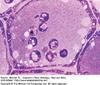
What is this?

Pap Smear
Basics:
- cells scraped from exocervix/external os
Stain:
- stained w/ hematoxylin, orange G, & eosin
- surface cells = pink/orange
- subsurface cells = blue/green
When things go wrong…
-
Cervical carcinoma if…
- High # of blue/green cells
- Cells w/ atypical nuclei
What is this? What are the 3 layers?

Vagina
Mucosa:
-
SSNK epithelium
- washed out due to loss of glycogen/presence of lipids
- glycogen maximal at ovulation
- high estrogen
- Lactobacilli break down glycogen & produce lactic acid
- acidifies environment
- prevents bacterial & yeast infections
- Lamina propria
Fibromuscular layer
- Muscularis externa
Adventitia

What are Bartholin’s ( Vestibular) Glands?
Bartholin’s ( Vestibular) Glands
-
Analogous to bulbouretral glands in males
- Glands that open into the vestibule
- space surrounded w/in the labia minora
- part of external genitalia
- Glands that open into the vestibule
-
Simple columnar cells
- secrete mucous
How does Lubrication of the Vagina work?
Vagina does NOT contain glands!!
-
Lubrication comes from:
- glands of the cervix
- vestibular glands
What is this?

Development of Glands in the Breast During Pregnancy
Inactive:
- Adipoose tissue
- CT
- Few glands
- Some ducts
Active (lactating):
- Glands proliferate
- seen as branched or irregular shape
- Duct cells proliferate
- Adipose tissue decreases
-
Regulated by:
- estrogen & progesterone
- after parturition, prolactin is lactogenic
-
Suckling causes RELEASE of PRL IH & oxytocin
- oxytocin = stimulates myoepithelial cell contraction

What is this?

Actively Developing & Lactating Alveoli
- See lipid droplets (LD) in the milk secretion
- from columnar secretory cells
- See venules & CT

How does the Secretion of Milk Lipids and Proteins occur?
Basics:
- During breast feeding:
-
oxytocin causes contraction of SM & myoepithelial cells
- causes milk ejection reflex
-
oxytocin causes contraction of SM & myoepithelial cells
Milk Lipids:
- Apocrine
Milk proteins:
- Merocrine

What is this?
Mammary Gland Atrophy
Secretion of milk
-
Milk protein = merocrine
- exocytosis/active transport
-
Milk lipids = apocrine
- apex of cell pinches off
After childbirth:
- first secretion = colostrum
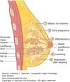
- Tubuloalveolar sweat glands derived from epidermis
- 15-20 lobes connnected by CT (Cooper’s or suspensory ligaments)
- each lobe has lactiferous duct that opens on nipple
- 15-20 lobes connnected by CT (Cooper’s or suspensory ligaments)
After menopause:
-
glandular elements atrophy
- decline in ovarian hormones
- apoptosis