Hemodynamic Monitoring reduced Flashcards
(94 cards)
Minimal Standard Monitors
1 .Electrocardiogram (HR and rhythm)
- Blood pressure
- Precordial stethoscope
- Pulse oximetry
- Oxygen analyzer
- End tidal carbon dioxide
document q5 min minimum
Minimal Standard - On Graphic Display
- Electrocardiogram
- Blood pressure
- Heart rate
- Ventilation status
- Oxygen saturation
continuous display
precordial sthetoscope
a stethoscope that is placed on the chest (just the bell part) that is attached to tubing with an ear piece on the other end
used for continuously listen to breath sounds and heart tones throughout the case.
You can quickly pick up on changes in patient condition (loss of breath sounds, loss of heart tones, etc).
used in peds 100% of the time
esophageal sthetoscope
used for continuous assessment of a pt’s breath sounds and health sounds
placed inside of the ETT - pt has to be intubated
28-30 cm into the esophagus
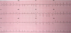
EKG
what is it? what it is not?
Recording of electrical activity of the heart
does NOT tell you about circulation
NOT a pulse rate monitor
EKG - what do you use it for?
- – Detect arrhythmias
- – Monitor heart rate
- – Detect ischemia
- – Detect electrolyte changes
- – Monitor pacemaker function
5 Principle ECG Indicators of Acute Ischemia
- ST segment elevation , ≥1mm
- T wave inversion
- Development of Q waves
- ST segment depression, flat or downslope of ≥1mm
- Peaked T waves
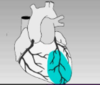
Lead I, AVL, V1-V4

Anterior wall ischemia (left coronary artery)

Lead V1-V4

Anterioseptal ischemia

Lead I, AVL, V5-V6

Lateral wall ischemia
(circumflex branch of left coronary artery)

Lead II, III, AVF
(Posterior)/ Inferior wall ischemia
(right coronary artery)

How far is an esophageal stethoscope inserted into the esophagus?
28-30cm.
This allows us to hear heart sounds and BS internally.
What are precordial and esophageal stethoscopes useful for?
Continuous assessment of heart and breath sounds.
Very sensitive monitor for bronchospasm and changes in pediatric patients
How often should we have a regular stethoscope available?
At all times
What 4 general things are continually evaluated?
Oxygenation, ventilation, circulation, and temperature
Considerations in deciding what type of monitoring to use
1) Indication
2) Risk/benefit
3) Complications
4) Alternatives
5) Cost
6) Skill level
Types of hemodynamic monitoring used
EKG, BP (NIBP and IABP), CVP, PAP, PCWP, TEE, stethoscope
What can the EKG tell you?
- Heart rate
- arrhythmias
- Ischemia
- electrolyte imbalances
- pacemaker function
Aspects of the 3 Lead EKG
Electrodes used: RA, LA, LL
Leads: I, II, III
Number of views of the heart: 3
Aspects of the 5 lead EKG
Electrodes used: RA, LA, RL, LL, chest
Leads: I, II III, AVL, AVR, AVF, V lead
Number of views of the heart: 7
Posterior / inferior wall ischemia is seen in these leads and is due to a blockage in this artery

II, III, AVF
Right coronary

Lateral wall ischemia is seen in these leadsand is due to a blockage in this artery

I, AVL, V5-6
Left circumflex coronary artery

Anterior wall ischemia is seen in these leadsand is due to a blockage in this artery

I, AVL, V1-4
Left coronary artery

Anterioseptal wall ischemia is seen in these leads and is due to a blockage in this artery
V1-4
Left anterior descending coronary artery