Health Assessment Test 3 Flashcards
What are some common CC
- Appetite
- Food intolerance
- Abdominal pain
- Nausea/Vomiting
- Bowel habits/constipations/diarrhea
- Past abdominal history
- S/E of medications
When assessing a pt with abd pain remember….
- History is the most important element in developing and refining your list of diagnostic possibilities
- Localizing the pain to a quadrant helps to refine and narrow your DDX.
- Workup should be targeted based on the history and physical examination.
- Review the list of medicines the patient is taking prior to seeing the patient and validate during the interview. Remember to ask about over-the-counter and herbal medicines.
Abdominal Pain
Diagnostic Approach
DDX abdmoninal pain

- 24-year-old African American male with no significant past medical history presents with abdominal pain 4 days in duration. Pain started as diffused and pressure sensation, most intensely in the mid abdomen
- Vital signs: 120/70, HR:98 RR: 16 T: 99.2 BMI 23.5
- What are your Differential diagnoses with the available data?
he presents today with worsening of symptoms. Pain is more localized to the Right lower quadrant and increased in intensity, he admits mild chills, nausea, vomiting and anorexia and constipation. Denies any other symptoms.
1.Refine your working diagnosis. Give me your top two?
●
2.What is your leading hypothesis at this point?
DD: Appendicitis
Sickle Cell
Gastroenteritis
Gas
Top Two: Gas, Appendicitis
Leading Hypothesis: Appendicitis
Evaluation of Abdominal Pain in Special Populations

Inspection of the abdomen different shapes
Flat, Rounded, Scaphoid, Protuberant
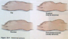
Two ulcers associated with Peptic Ulcer disease
Duodenal Ulcer
Gastric Ulcer
•Nonspecific dyspeptic symptoms: indigestion, nausea, vomiting, loss of appetite, heartburn, and epigastric fullness
Duodenal Ulcer
Midepigastric pain
Gnawing or burning, non-radiating, recurring pain that is often is episodic and relieved by food or antacids because the sphincter closes when food is in stomach stopping the acid from regurgitating
Gastric Ulcer
- Midepigastric pain
- Aggravated by food, relieved by antacids
The pancreas does what and why does pancreatitis happen
The pancreas secretes enzymes to break down protein. This enzyme is not activated until the small bowel. Pancreatitis happens when the enzyme activates prematurely inside the pancreas and it begins to brealk down.
Family history associated with Abdominal Pain
- Colorectal cancer and familial colorectal cancer syndromes
- Gallbladder disease
- Kidney disease
- Malabsorption syndrome
- Hirschsprung disease, aganglionic megacolon
- Familial Mediterranean fever (periodic peritonitis)
Infants at risk for abd pain
- Gestational age and birth weight
- Passage of first meconium stool within 24 hours
- Jaundice
- Vomiting, frequency, projectile
- Diarrhea, colic, failure to gain weight, weight loss, or steatorrhea (malabsorption syndrome)
- Apparent enlargement of abdomen (with or without pain), constipation, or diarrhea
Abdominal disorder causes associated with pregnancy
- Abdominal wall muscles stretch and lose tone
- Organs are displaced and affect functions:
- Heartburn results from alkaline reflux from duodenal contents into stomach
- Gallstones may result from gallbladder changes that produce cholesterol crystals
- Urinary stasis and urgency may occur
- Constipation or flatus is more common
- Hemorrhoids may develop later in pregnancy
- Gastrointestinal concerns common
- Nausea
- Vomiting
- Constipation
- Hemorrhoids
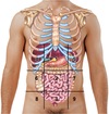
Landmarks of the abdomen
•Nine regions
- Two horizontal lines
- Across the lowest edge of the costal margin
- Across the edge of the iliac crest
Two vertical lines
- Running bilaterally from the midclavicular line to the middle of the Poupart ligament, approximating the lateral borders of the rectus abdominis muscles
Figure. Nine regions of the abdomen. 1, epigastric; 2, umbilical; 3, hypogastric; 4 and 5, right and left hypochondriac; 6 and 7, right and left lumbar; 8 and 9, right and left inguinal.
Order of exam of the abdomen
- Inspection
- Auscultation
- Percussion
- Palpation (light and deep)
Inspection of the Abdomen
•Surface characteristics
- •Skin
- •Venous return
- •Lesions and scars
- •Tautness and striae
Contour
- •Contour (abdominal profile from the rib margin to the pubis, viewed on the horizontal plane)
- •Symmetry
- Surface motion
- Inspect abdominal muscles as patient raises head to detect presence of the following:
- •Masses
- •Hernia
- •Separation of muscles
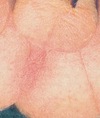
Cullen’s Sign
Ecchymosis around the umbilicus from:
Retroperitoneal hemorrhage
Acute pancreatitis
Pancreatic hemorrhage
Intraperitoneal hemorrhage
Blunt abdominal trauma
Ruptured spleen
Ruptured abdominal aortic aneurysm
Ruptured / hemorrhagic ectopic pregnancy.
Spontaneous bleeding secondary to coagulopathy
Grey Turner Sign
Ecchymosis at the flanks
Retroperitoneal hemorrhage
Acute pancreatitis
Pancreatic hemorrhage
Intraperitoneal hemorrhage
Blunt abdominal trauma
Ruptured spleen
Ruptured abdominal aortic aneurysm
Ruptured / hemorrhagic ectopic pregnancy.
Spontaneous bleeding secondary to coagulopathy
Diastasis recti (abdominal separation)
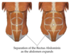
Defined as a separation of the rectus abdominis muscle into right and left halves
Newborns
- increased risk if premature
Postpartum
increased risk if women over 35
after multiple pregnancies
Auscultation of Abdomen

•Auscultate with stethoscope diaphragm for the following:
- Bowel sounds
•Auscultate with bell of stethoscope for the following:
- Bruits over aorta and renal and femoral arteries
Cannot say bowel sounds are absent unless you listen for minutes

Bowel Sounds
- Frequency
- Character
- Usually heard as clicks and gurgles that occur irregularly and range from 5 to 15 per minute
- Generalized so most often they can be assessed adequately by listening in one place
- Loud prolonged gurgles are called borborygmi (stomach growling)
When would you hear increased bowel sounds?
Increased bowel sounds may occur with gastroenteritis, early intestinal obstruction, or hunger
High pitched tinkling sounds suggest:
Intestinal fluid and air under pressure, as in early obstruction
Decreased bowel sounds occur with:
Peritonitis and paralytic ileus
Friction rubs:
- High-pitched sounds that are heard in association with respiration
- Use the diaphragm of the stethoscope
- Rare in the abdomen
- Indicate inflammation of the peritoneal surface of the organ from tumor, infection, or infarct
- Liver and spleen
Bruits:
- Harsh or musical intermittent auscultatory sounds that may reflect blood flow turbulence and indicate vascular disease
- Heard best with the bell of the stethoscope
- Well localized
- Epigastric region and over the aortic, renal, iliac, and femoral arteries
Sites to auscultate for bruits, renal arteries, aorta, and femoral arteries

Venous hum
- Soft, low-pitched, and continuous sound heard best with the bell of the stethoscope
- Occurs with increased collateral circulation between portal and systemic venous systems
- Epigastric region and around the umbilicus
Percuss the abdomen for the following:
- Tone in all four quadrants (or nine regions)
- Liver borders to estimate span
- Splenic dullness in left midaxillary line
- Gastric air bubble
Palpation of the Abdomen
- Keep your palpating hand low and parallel to the abdomen
- For ticklish person: Keep the patient’s fingers under your own with your fingers curled over your patients
Light Palpation: 1 cm
- Start palpating the abdomen using gentle probing with the hands; this reassures and relaxes the patient
- Identify any superficial organs or masses
- Use relaxation techniques to assess voluntary guarding
Deep Palpation: 5 to 8 cm
- Palpate deeply in the periumbilical area and both lower quadrants.
- Observe for Rebound tenderness
- Assess organs
Masses: Identify and note
- Location
- Size
- Shape
- Consistency
- Tenderness
- Pulsation
- Mobility
Movement with respiration
Umbilical ring
- Palpate the umbilical ring and around the umbilicus
- Area should be free of bulges, nodules, and granulation
- Umbilical ring should be round and free of irregularities
- Potential for herniation
•Umbilicus may be either slightly inverted or everted, but it should not protrude
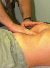
How do you palpate the liver
Palpating the liver.
A, Fingers are extended, with tips on right midclavicular line below the level of liver dullness and pointing toward the head.

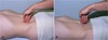
Palpating the Spleen

Palpating the spleen.
A, Press upward with the left hand at the patient’s left costovertebral angle. Feel for the spleen with the right hand below the left costal margin
Palpation of the Aorta
Press firmly deep in the upper abdomen
Identify the aortic pulsations.
Assess the width of the aorta > age 50
Press deeply in the upper abdomen with one hand on each side of the aorta gauge width
Not more than 4 cm wide (average, 2.5 cm

Murphy’s Sign
This is the standard sign of cholecystitis or inflammation of the liver
Pain is felt during inhalation or coughing when the during palpation of the RUQ due to the inflammation of the gallbladder.
Rebound tenderness should be performed….
At the end of the examination
Iliopsoas muscle test
Performed when you suspect appendicitis
Iliopsoas muscle test. A, The patient raises the leg from the hip while the examiner pushes downward against it.

Obturator muscle test
Performed when you suspect an appendicitis, ruptured appendix or a pelvic abscess
Obturator muscle test. With the right leg flexed at the hip and knee, rotate the leg laterally and medially.

McBurney’s sign
Tests for acute appendicitis
To elicit Mcburney’s sign patient should be in supine position with his knees slightly flexed and his abdominal muscles relaxed. Palpate deeply and slowly in the right lower quadrant over McBurney’s point located about 2” from the right anterior superior lliac spine. On a line between the spine and umbilicus. Point pain and tenderness is a positive sign for appendicitis.

In what age group is it more likely to detect the edge of the spleen
Children and Infants
•Assessment of the abdomen of pregnant women includes:
- Uterine size estimation for gestational age
- Fetal growth
- Position of the fetus
- Monitoring of fetal well-being
- Presence of uterine contractions
Abdominal contour is often ________ as a result of loss of muscle tone.
Rounded
Aaron Sign
Appendicitis
Pain or distress occurs in the area of the patient’s heart or stomach on palpation of McBurney’s point
Rovsing’s Sign
Appendicitis
Elicited by palpating the left lower quadrant; this paradoxically causes pain to be felt at the right lower quadrant
Ballance
Peritoneal irritation
fixed dullness to percussion in left flank, and dullness in right flank that disappears on change of position
Blumberg
Peritoneal irritation; appendicitis
Rebound tenderness
Cullen
Hemoperitoneum; pancreatitis; ectopic pregnancy
Ecchymosis around umbilicus
Dance’s Sign
Intussusception (where part of the intestine telescopes into itself)
Absence of bowel sounds in right lower quadrant
Grey Turner
Hemoperitoneum; pancreatitis
Ecchymosis of flanks
Markle
(heel jar)
Peritoneal irritation; appendicitis
Patient stands with straightened knees, then raises up on toes, relaxes, and allows heals to hit floor, thus jarring body. Action will cause abdominal pain if positive
McBurney
Appendicitis
Rebound tenderness and sharp pain when McBurney’s point is palpated
Murphy
Cholecystitis
Abrupt cessation of inspiration on palpation of gallbladder
Murphy
Cholecystitis
Abrupt cessation of inspiration on palpation of gallbladder
Romberg-Howship
Strangulated obturator hernia
Pain down the medial aspect of the hip to the knees
Rovsing Sign
Peritoneal irritation; appendicitis
Right lower quadrant pain intensified by left lower quadrant abdominal palpation
Duodenal ulcer
Peptic ulcer disease
- Midepigastric pain
- Gnawing or burning, nonradiating, recurring pain that is often is episodic and relieved by food or antacids.
- guaiac-positive stool from occult blood loss
Gastric ulcer
Peptic Ulcer Disease
- Midepigastric pain
- Aggravated by food, relieved by antacids
- guaiac-positive stool from occult blood loss
Typical Sx of GERD
•Typical symptoms: acid regurgitation, heartburn, dysphagia (mostly postprandial)
Atypical sx of GERD
•Atypical symptoms: epigastric fullness/pressure/pain, dyspepsia, nausea, bloating, belching, chest pain, lump in throat
Extraesophageal sx of GERD
- Extraesophageal signs and symptoms: chronic cough, bronchospasm, wheezing, hoarseness, sore throat
- Heartburn: retrosternal burning sensation
- Regurgitation; sour or acid taste in mouth (“water brash”)
Gallstones/Choleliathiasis/
Cholecystitis
s/s
- Episodic right upper quadrant or epigastric pain lasting >15 minutes and sometimes radiating to the back (biliary colic—due to transient cystic duct obstruction)
- Pain is usually postprandial.
- Pain sometimes awakens patients from sleep.
- Most patients develop recurrent symptoms after a first episode of biliary colic.
Other symptoms include nausea, vomiting, indigestion or bloating sensation, and fatty food intolerance
Physical exam of patient with Gallstones/Choleliathiasis/Cholecystitis
- Physical exam is usually normal in patients with cholelithiasis in the absence of an acute attack.
- Epigastric and/or right upper quadrant tenderness (Murphy sign)
- Charcot triad: fever, jaundice, right upper quadrant pain
- Reynolds pentad: fever, jaundice, right upper quadrant pain, hemodynamic instability, mental status changes; classically associated with ascending cholangitis
- Flank and periumbilical ecchymoses (Cullen sign and Grey Turner sign) in patients with acute hemorrhagic pancreatitis
- Courvoisier sign: palpable mass in the right upper quadrant in patient with obstructive jaundice most commonly due to malignant tumors within the biliary tree or pancreas
Acute pancreatitis
s/s
- Acute onset of mild to severe constant epigastric pain, which may radiate toward the back
- Chills/Nausea/vomiting
- Alcohol use
- Personal or family history of gallstones
- Medication use (Thiazide, valproic acid)
- Abdominal trauma
- Recent significant rapid weight loss
Acute pancreatitis
Physical Exam
- Abdominal findings: epigastric tenderness, loss of bowel sounds, peritoneal signs
- Other findings, jaundice, rales/percussive dullness
- Rare (with hemorrhagic pancreatitis)
- Flank discoloration (Grey Turner sign) or umbilical discoloration (Cullen sign)
Appendicitis
S/S
- Classic history is vague periumbilical pain with anorexia followed by fever, nausea, and vomiting. Over the next 4 to 48 hours, pain migrates to the RLQ.
- Pain before vomiting, abdominal pain, pain migration
- Anorexia, nausea, vomiting, obstipation (complete constipation)
Appendicitis
Signs
- Fever; temperature >100.4°F (may be absent); tachycardia
- Occasionally hypoactive BS.
- RLQ tenderness; maximal tenderness at McBurney point (1/3 the distance from the anterior superior iliac spine to the umbilicus)
- Voluntary and involuntary guarding
- Rovsing sign: RLQ pain with palpation of left lower quadrant
- Psoas sign: pain with right thigh extension (retrocecal appendix)
- Obturator sign: pain with internal rotation of flexed right thigh (pelvic appendix);
- local and suprapubic pain on rectal exam (pelvic appendix)
MASS
Appendicitis Scoring System
Modified Alvarado Scoring System
- A MASS score >7 suggests appendicitis without the need for further imaging.
- The use of MASS in the diagnosis of acute appendicitis improves diagnostic accuracy and reduces negative appendectomy and complication rates.
- Supplement MASS in female patients with additional investigations (e.g., abdominal ultrasound or laparoscopy).
- Migratory right iliac fossa pain (1 point)
- Nausea/vomiting (1 point)
- Anorexia (1 point)
- Tenderness in right iliac fossa (2 points)
- Rebound tenderness in right iliac fossa (1 point)
- Elevated temperature (1 point)
- Leukocytosis (2 points)
- A MASS score >7 suggests appendicitis without the need for further imaging.
Gastroenteritis
- Sudden onset of diarrhea and/or vomiting
- Symptoms may include:
- •Crampy abdominal pain
- •Fevers
- •Nausea or anorexia
- •Weakness or fatigue
- Abdominal exam; hyperactive BS, mild diffused tenderness, hypertympany, evaluate for peritoneal signs.
- Look for signs of dehydration (minimal, moderate, or severe) which may include:
- Tachycardia, tachypnea, lethargy, mental status changes, dry mucous membranes, mottled or pale skin color and decreased skin turgor, or increased capillary refill time
Colorectal Cancer
Symptoms
- Mostly are asymptomatic
- Symptoms may indicate advanced disease. Common presenting symptoms include:
- •Abdominal pain or cramping
- •Change in bowel habits (constipation, diarrhea, narrowing of stool)
- •Rectal bleeding, dark stools, or blood in stool
- •Weakness or fatigue
- •Unintentional weight loss
- •Other presentations may include symptoms due to the presence of metastatic lesions (lymph nodes, liver, lung, peritoneum), fever of unknown origin.
Colorectal Cancer
Signs
- Signs of anemia
- Weight loss
- Palpable abdominal mass (late presentation)
- Must include digital rectal exam
Diverticulosis/diverticulitis
Symptoms
•Diverticulosis
- •80–85% of patients are asymptomatic. Of the 15–20% with symptoms, 1–2% will require hospitalization, and 0.5% will undergo surgery.
- •The most common symptom is dull, colicky abdominal pain, typically in the LLQ. Pain can be exacerbated by eating and by passing bowel movement or flatus.
- •Diarrhea or constipation is common.
Acute Diverticulitis
Symptoms
- Abdominal pain: acute onset, typically in LLQ
- Fever and/or chills
- Anorexia, nausea (20–62%), or vomiting
- Constipation (50%) or diarrhea (25–35%)
Diverticulosis/diverticulitis
Signs
•Diverticulosis
- •Exam is usually normal.
- •May have intermittent distension or tympany
- •May have heme + stools
Acute Diverticulosis/diverticulitis
Signs
•Acute diverticulitis
- •Abdominal tenderness (usually LLQ) (+ Rovsing signs)
- •Abdominal distension and tympany
- •Rebound tenderness, involuntary guarding, or rigidity suggests perforation and/or peritonitis.
- •Palpable mass in LLQ (20%)
- •Bowel sounds hypoactive (could be high-pitched and intermittent if obstruction is present)
- •Rectal exam may reveal tenderness or a mass.
The Menstrual Cycle
- Estrogen is lowest at the end of the cycle during the start of the follicular phase
- Proliferative phase is influenced by estrogen causing the endometrium to rapidly thicken
- Ovulation happens mid-cycle
- Progesterone is highest right after ovulation during the start of the luteal phase
- Secretory phase is influenced by progesterone. The lining becomes highly vascular and edematous

Cervical eversion is where the endocervical canal extends into the surface of the cervix creates a circular raised erythematous appearance. It is normal nonprurlient cervical mucous

Features of the Vagina of the Infant and Child
- Cervix is 2/3 of the entire length of the organ
- Hymen is a thin diaphragm inside the introitus and has crescent-shaped opening
- Labia major are hairless and nonprominent
- Labia minora are avascular, thin, and pale
Explain the Tanner Stage
Tanner Stage 1: No pubic hair/Infantile
Tanner Stage 2: Initial hair is straight and fine/ 8-11 yo
Tanner Stage 3: Pubic hair coarsens, darkens and spreads/12yo
Tanner Stage 4: Hair looks like adults but limited in area/ 13yo
Tanner Stage 5: Inverted triangular pattern is est./ Age range 12.5-16.5 yo

Vaginal features of Adolescents
- Puberty: functional maturation of reproductive organs
- External genitalia increase in size
- Clitoris becomes erectile
- Pubic hair develops
- Vagina lengthens and secretions become acidic
- Uterus, ovaries, and tubes increase in size
- Uterine musculature and vascular supply increase
- Endometrial lining thickens in preparation for the onset of menstruation
- The average age at menarche in the United States is between 12 and 13 years
- Irregular menstrual cycles are not unusual during adolescence as a result of anovulatory cycles
Female Reproductive Life Span

Any woman that goes into menopause under 40 is premature
Any woman that goes into menopause under 45 is early

Vaginal changes in the Pregnant Woman
Increased estrogen and progesterone cause the enlargement of the uterus to support the growing fetus. After the first trimester, it is the mechanical enlargement due to fetal growth
By 12 week of pregnancy the uterus rises out of the pelvis
- Increased estrogen and progesterone
- Enlarged elastic uterus
- Softened pelvic cartilage
- Strengthened pelvic ligaments
- Pelvic congestion and edema
- Thickened vaginal walls
Increased vaginal secretions
Vaginal changes in the Older Adult
- Ovarian fx begins to diminish in 40’s.
- Median age is 51
- Menopause is defined as 1 year with no menses - amenorrhea
- Menopause
- External and internal genitalia decrease in size
- Tissue loses elasticity and tone
- Pubic hair turns gray
- Libido decreases
- Vagina narrows and loses lubrication
What must be address with the older adult female?
Urinary incontinence and libido
Questions during history in a GYN exam regarding contraceptive history?
Are they happy with current method of BC did they use one previously they liked
What does GTPAL stand for ?

What are the five P’s
Partners
Practices
Protection from STIs
Past history of STIs
Pregnancy prevention
What are Preventative Care Considerations during a GYN exam?
Preventive care considerations
- Imperative to have assessment and discussion of health, wellness, and lifestyle behaviors
- Two specific areas: Personal habits and safety
How to prepare for a GYN exam?
Minimize patient apprehension
Empty bladder
Privacy
Warm room temperature
Warm speculum
What to inspect the Labia Majoria for during an exam
Symmetry
Redness, swelling, or tenderness
Excoriation, rashes, or lesions
Discoloration
Varicosities
Stretching
Trauma or scarring
What to inspect the Labia Minora for during a GYN exam
- Symmetry
- Moisture
- Color
- Soft, homogeneous, and without tenderness
- Inflammation
- Excoriation
- Discharge
- Ulcers
Examination findings of the Vaginal Introitus
- Moisture
- Swelling
- Discoloration
- Discharge
- Lesions, fistulae, or fissures
How to examine the Skene gland
Periurethral location looking for discharge
Insert one finger to the 2nd joint upward pressure. Move finger to both sides of the urethra and directly on the urethra.
Discharge indicates infection, usually gonococcal infection
Bartholin gland
Posterolateral portion of the labia majora
Discharge
Masses
Tenderness or swelling
Can become very swollen in gonorrheaa infection
How to perform an internal inspection of the female genitalia
Examination of the internal genitalia with a speculum. Begin by inserting a finger and applying downward pressure to relax the vaginal muscles. A, Gently insert the closed speculum blades into the vagina. B, Direct the speculum along the path of least resistance. C, Insert the speculum the length of the vaginal canal. D, Speculum is in place, locked, and stabilized. Note cervix in full view.
Evaluation of the vagina
Inspect for color, tissue appearance, moisture, discharge, moles, or lesions during both insertion and removal of the speculum moles
Evaluation of the cervix
Visualize the entire surface of the cervix, note size and shape of the os, color, bleeding or discharge, evaluate the squamocolumnar junction, and assess for lesions or growths
Cervix
- Color
- Position
- Surface characteristics
- Discharge
- Size and shape
A Pap Smear analyzes
Endocervical cells
A DNA probe is used for
Chlamydia and gonorrhea
Wet mount is used to diagnose…
Trichomonas, bacterial vaginosis, or candidiasis
During a Bi Manual exam the cervix is analyzed for:
- Size, length, and shape
- Position
- Consistency
- Movement
- Nodules
- Hardness
- Tenderness
How to access for cervical motion tenderness during a bimanual examination
•Place the index and middle finger on either side of the cervix and gently pull it first to one side and then the other
Features of an Anteverted uterus
Normal positioning
Uterus is tilted forward
Cervix is positioned DOWN
Features of a Retroverted uterus
1/3 of the population
Uterus tips backward
Cervix positioned UPWARD
SE backpain during menses and pregnancy
External genitalia exam of the infant
Expected swelling – swelling
Milky discharge – want to culture b/c not normal
Enlarged clitoris
Ambiguous appearance
Adhesions between labia minora
Labs r/t GYN
•Cervical cytology: Routine screening for cervical cancer
- Conventional Papanicolaou (Pap) smear and liquid-based thin-layer preparation
- Wet mount: Evaluation of a sample of vaginal discharge under a microscope
- Testing for STIs: Nucleic acid amplification tests
- Human papillomavirus testing: Stand-alone test or in conjunction with collection of cervical cytology
Considerations for the Adolescent GYN exam
- Allay anxiety for what may be first examination
- Same examination and positioning as for adult
- Appropriate-size speculum
- Maturational changes of sexual development
- Just before menarche, there is a physiologic increase in vaginal secretions
- Hymen may or may not be stretched across the vaginal opening
- By menarche, the vaginal opening should be at least 1 cm wide
Note Tanner stage
No internal exams
Where is fundal height measured from and to?
Fundal height is from the symphyses pubis to the superior fundus
Week 10-12 uterus is in the uterus measures at symphyses pubis. Fetal heart tones
Week 16 the uterus measures 1/2 way between symphyses and umbilicus
Other pregnancy-associated changes:
- Uterus may become more anteflexed during the first 3 months, which may cause urinary frequency cue to increased progestrone
- Vulvar varicosities occur commonly during pregnancy
- Breast and skin changes that occur with pregnancy
What are some age-related changes with the older adult
Labia appear flatter and smaller
Skin is drier and shinier
Gray and sparse pubic hair
Clitoris is smaller
Urinary meatus may appear as an irregular opening or slit
- Vaginal introitus may be constricted
- Multiparous older women, the introitus may gape
- Vagina is narrower and shorter
- Absence of rugation
- Less-mobile cervix
- Smaller uterus
- Nonpalpable ovaries
- Rectovaginal septum will feel thin, smooth, and pliable.
Diminished rectal tone
PMS
•Collection of physical, psychological, and mood symptoms related to a woman’s menstrual cycle
PMS symptoms occur 5-7 days before menses during the luteal phase and subside with the onset of menses.
Atrophic vaginitis
now known as
Genitourinary Syndrome of Menopause (GSM)
•Inflammation of the vagina due to the thinning and shrinking of the tissues, as well as decreased lubrication - post menopause
Symptoms de to changes in the vulvovaginal area and urethra during the menopause transition
Physiologic Vaginitis
C/O Increase in d/c
Sx: Clear or mucoid discharge
Diag: Wet mount up to 3/5 WBC’s (epithelial cells)
Bacterial Vaginosis
(Gardnerella vaginalis)
CC No itching or edema, Foul smelling d/c “fishy”
Homogenous thin, white or gray d/c, pH >4.5
Diag: +KOH “whiff” test; wet mount +clue cells
Candida vulvovaginitis
(Candida albicans)
CC: Pturitic d/c, itching of labia; itching may extend to thighs
White, curdy d/c, pH 4-5; cervix may be red; erythema of perineum and thighs
Diag: KOH prep: mycelia, budding, branching yeast, pseudohypae
Trichomoniasis
(Trichomonas vaginalis)
CC: Watery d/c; foul odor; dysuria and dyspareunia with severe infection
Profuse, frothy, greenish d/c; pH 5-6.6; red friable cervix with petechiae “strawberry cervix”
Diag: Wet mount: round or pear-shaped protozoa parasite; motile “gyrating” flagella

Gonorrhea
(Neisseria gonorrhea)
CC: Partner with STI; often asymptomatic or may have sx of PID
Purulent d/c from cervix, Skene/Bartholin gland inflammation; cervix and vulva may be inflamed
Diag Gram stain, culture, DNA probe
Chlamydia
(Chlamydia trachomatis)
MOST COMMONLY REPORTED STI
CC: Partner with nongonococcal urethritis; often asymptomatic; may c/o spotting after intercourse or urethritis
Purulent d/c; cervix may or may not be red/friable
Diag: DNA probe
Syphilis
- STI caused by Treponema pallidum
- Spread by direct contact with a painless chancre
First sx a painless chancre at 2 weeks then chancre disappears
Would chart as: Solid firm round lesion, painless, clear base, endulated boarders
Genital Warts
- Most commonly caused by HPV types 6 and 11
- Warty lesions due to STI with HPV
Caused by condyloma acuminatum

Condyloma latum
Lesion of secondary syphilis
6-12 weeks after infection
flat round/oval papules
covered in gray exudate
PID
Discharge, odor, painful intercourse, vaginal bleeding, painful urination, upper abdomen pain, pain during bimanual exam, adnexa thickness and bilateral tenderness
Endometriosis
The cause is due to retrograde reflux of menstrual tissue from the fallopian tube during menstruation
Vulvar Carcinoma
Classified by the type of tissue from which it arises
Squamous
Adenocarcinoma
Melanoma
Basel Cell
Vaginal bleeding, dysuria, pelvic/leg/back pain, painful intercourse
Myomas
(leiomyomas, fibroids)
Common, benign, uterine tumors
Endometrial carcinoma
Cancers of the glandular cells found in the lining of the uterus
Post menopausal women
Red flag is postmenopausal bleeding
Pts taking tamoxifen
Ovarian Carcinoma
Late dx due to vague sx
Increase in girth and weight gain, fatique
Cystitis/urethritis
HISTORY - Frequency, urgency, dysuria, nocturia with low back pain
PHYSICAL FINDINGS - Discharge may be present, may have suprapubic tenderness
DIAGNOSTIC STUDIES - Urine dipstick, microscopic exam, C & S
Pyelonephritis
HISTORY - Fever, chills, back pain, nausea, toxic appearance, frequency, and dysuria
PHYSICAL FINDINGS - Feels and looks ill, temp > 101, CVA tenderness, abdomen may be tender
DIAGNOSTIC STUDIES - Microscopic exam, WBCs, may have casts, urine C & C, blood cultures
Urolithiasis
HISTORY - Pain, hematuria, may have symptoms of secondary infection as above, renal colic; pain that radiates to the inner thigh, nausea, vomiting
PHYSICAL FINDINGS - May have CVA tenderness, looks ill during periods of acute pain, may have abdominal distension
DIAGNOSTIC STUDIES - Urinalysis, gross or microscopic hematuria
KUB xray, ultrasound
Acute prostatitis
HISTORY - Chills, high fever, urinary frequency, perineal pain and low back pain, varying degrees of obstructive symptoms: dysuria, nocturia, arthralgia
PHYSICAL FINDINGS - Fever > 101, tender, swollen, enlarged, warm prostate.
DO NOT massage, can cause bacteremia
DIAGNOSTIC STUDIES - Urinalysis, urine culture, prostate secretion culture
Epididymitis/orhcitis
HISTORY - Abrupt onset over several hours, febrile, pain in scrotum/testicles
PHYSICAL FINDINGS - Tender swollen epididymis and/or testicles, elevation of affected testicle may lessen discomfort, may have fever
DIAGNOSTIC STUDIES - Doppler with flow studies
Testicular torsion
HISTORY - Sudden onset of testicular pain which radiates to groin, may also have lower abdominal pain
PHYSICAL FINDINGS - Exquisitely tender testicle, may be higher because of shortened spermatic cord, cremasteric relflex absent
DIAGNOSTIC STUDIES - Scrotal ultrasound, SURGICAL CONSULT STAT
BPH
HISTORY - Hesitancy, slow urine stream, dribbling, nocturia
PHYSICAL FINDINGS - Prostate protrusion into rectum, median sulcus reduced, prostate boggy
DIAGNOSTIC STUDIES - PSA 1-4
Prostate cancer
HISTORY - Hesitancy, slow urine stream, dribbling, nocturia, low back pain
PHYSICAL FINDINGS - Prostate protrusion into rectum, prostate hard, nontender
DIAGNOSTIC STUDIES - PSA elevated, surgical biopsy
Bladder or kidney tumor
HISTORY - More common in men than women, often history of ETOH or smoking
PHYSICAL FINDINGS - Usually no signs, sometimes hematuria
DIAGNOSTIC STUDIES - U/A, Renal Ultrasound, CT
Condyloma acuminata
HISTORY - Bleeding, itching, discharge
PHYSICAL FINDINGS - Pink or white warty lesions with papilliform surface. May extend into anal canals
DIAGNOSTIC STUDIES - Serology to distinquish from condyloma lata caused by syphilis
Proctitis
HISTORY - Anorectal pain, mucupurulent discharge, tenesmus, diarrhea, constipation, history of anal intercourse, immunocompromise
PHYSICAL FINDINGS - Purulent discharge, inflamed rectal mucosa
DIAGNOSTIC STUDIES - Stool cultures, Stool for O & P, stool exam for fecal leukocytes
Cystitis/urethritis
HISTORY - Frequency, urgency, dysuria, dribbling of urine, nocturia with suprapubic tenderness, hematuria , chills, sometimes n/v
PHYSICAL FINDINGS - Discharge may be present if secondary to STD, prostatitis, may have suprapubic tenderness
DIAGNOSTIC STUDIES - Urine dipstick, microscopic exam, C & S
Pyelonephritis
HISTORY - Fever, chills, back pain, nausea, toxic appearance, frequency, and dysuria
PHYSICAL FINDINGS - Feels and looks ill, temp > 101, CVA tenderness, abdomen may be tender
DIAGNOSTIC STUDIES - Microscopic exam, WBCs, may have casts, urine C & C, blood cultures
Urolithiasis
HISTORY - Pain, hematuria, may have symptoms of secondary infection as above, renal colic; pain that radiates to the inner thigh, nausea, vomiting
PHYSICAL FINDINGS - May have CVA tenderness, looks ill during periods of acute pain, may have abdominal distension
DIAGNOSTIC STUDIES - Urinalysis, gross or microscopic hematuria, KUB xray, ultrasound
Acute prostatitis
HISTORY - Chills, high fever, urinary frequency, perineal pain and low back pain, varying degrees of obstructive symptoms: dysuria, nocturia, penile pain with bowel movement
PHYSICAL FINDINGS - Fever > 101, potential obstructive signs: distended bladder, positive CVA tenderness, tender, swollen, enlarged, warm tendered prostate with or without sulcus. Pain with BM that radiates to tip of penis
DO NOT massage, can cause bacteremia
DIAGNOSTIC STUDIES - Urinalysis, urine culture, prostate secretion culture
Benign prostatic Hyperplasia (BPH)
HISTORY - Frequency, hesitancy, weak urine stream, nocturia, straining, urgency, dribbling post void
PHYSICAL FINDINGS - Enlarged non tendered boggy prostate with no sulcus
DIAGNOSTIC STUDIES - IPSS scoring luts questionnaire, Post void residual, PSA levels
Tanner Stage 1
No signs of puberty
Scrotum testes and penis as in childhood
No pubic hair
Tanner Stage 2
Initial growth of scrotum and testes. The skin on the scrotum has become redder, thinner, and more wrinkled. The penis may have grown a little in length.
Few hairs around the root of the penis. The hairs are straight, without curls and light color.
Tanner Stage 3
The penis has now grown in length. Scrotum and testes have grown. The skin of the scrotum has become darker and more wrinkled.
Hairs are darker and curlier and still sparse, mostly located at the penis root.
Tanner Stage 4
The penis has grown in both length and width. the head of the penis has become larger. The scrotum and testes have grown.
More dense, curly, and dark hair. The hair growth is reaching the inner thighs.
Tanner Stage 5
Penis and scrotum as an adult.
Pubic hair extends upwards to the umbilicus. It is dense and curly.
What part of the prostate can be examined by DRE
Posterior lobes
- Two lateral lobes
- Sulcus between the two lateral lobes
Changes in the older adult (rectum)
•Degeneration of afferent neurons in the rectal wall
- •Interferes with the process of relaxation of the internal sphincter
- •Increased pressure sensation threshold in rectum
- •Stool retention
•Loss of external sphincter tone
- •Fecal incontinence
•Prostate
- •Enlargement of the prostate with aging
- •Pressing on the urethra may lead to urinary dysfunction.
- •Loss of function of the secretory alveoli
- •Decrease androgen level-> decrease secretory function
Anorectal pain associated with moving bowels algorhythm

Anorectal pain NOT directly associated with moving bowels

•Proctalgia fugax
- Non triggered short episodic perianal pain
- Patho:
- • Mostly unknown
- •Normal anorectal pressure but during attack spasm of smooth muscle.
•Presentations:
- •Recurrent attacks of severe anorectal pain unrelated to defecation
- •Few seconds to 30 minutes
•Levator ani syndrome
- Lasting more than 30 minutes usually chronic.
- Patho:
- •Nonrelaxing pelvic floor dysfunction.
•Presentation:
- •Chronic proctalgia with vague, dull pressure pain high in the rectum, worsen with sitting
Examination of the anus and rectum is performed
- As part of a wellness examination for some men and women (no longer performed on all women during papsmear)
- USPSTF new guidelines (2019)
- If not symptomatic no prostate screening for men aged 70 or above
- Men 55- 69: If the patient is a high risk, ƒh + prostate cancer, obesity, AA, or Caribbean men, inherited BRCA1 and 2 genes) After the conversation, and teaching screening on men who expressed a desire to be screened.
- When patient is symptomatic
- When the patient has a specific concern or problem
How to describe the location of anal and rectal findings
•12 o’clock is in the ventral (front) midline and 6 o’clock is in the dorsal (back) midline
Anal Wink
- contraction of the external anal sphincter upon stroking of the skin around the anus
- The absence of this reflex indicates that there is an interruption of the reflex arc, or damage to the spinal cord
Rectal pain is almost always indicative of:
- a local disease
- Irritation, rock-hard constipation, rectal fissures, or thrombosed hemorrhoids
•Lateral and posterior rectal walls
- Nodules, masses, irregularities, polyps, tenderness
- Internal hemorrhoids not ordinarily felt unless they are thrombosed
•Anterior rectal wall
- Contact with the peritoneum
- Peritoneal inflammation
- Nodularity of peritoneal metastases
- Shelf lesions
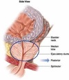
- Posterior surface of prostate
Levator ani muscle

Prostate
Via anterior rectal wall
Size
Contour
- Median sulcus
- Lateral lobes
- Remember you can not palpate the median lobe
Consistency
Mobility
Tenderness

•Anal fistula
- Inflammatory tract that runs from the anus or rectum and opens onto the surface of the perianal skin or other tissue
- Caused by drainage of a perianal or perirectal abscess
•Pruritus ani
•Commonly caused by fungal infection in adults and by parasites in children
•Enterobiasis (roundworm, pinworm)
•Adult nematode (parasite) lives in the rectum or colon and emerges onto perianal skin to lay eggs while the child sleeps
•Imperforate anus
•Rectum may end blindly, be stenosed, or have a fistulous connection to the perineum, urinary tract, or, in females, the vagina
Pinworms
•Intestinal infection with Enterobius vermicularis
- Characterized by perineal and perianal itching
- Usually worse at night
- Vulvovaginitis
- Dysuria
- Abdominal pain (rare)
- Insomnia (typically due to pruritus)
- PE:Perineal and perianal exam; particularly in early morning to look for evidence of migrating worms
Anatomy of the penis understand male genitalia esp prostate, testes, and vas deferens
Anatomy of the testes

What do the testes do:
- spermatogenesis (production of sperm)
- secretion of the male sex hormone testosterone, which induces and maintains male sex characteristics.
- Localization: Lower temperature to promote spermatogenesis.
- Consist of numerous seminiferous tubules divided into segments within the testicle itself.
- Collecting tubules transmit spermatozoa into the epididymis, that lead into the vas deferens.
- Vas deferens and seminal vesicles part of the spermatic cord enter abdomen, and end in elongated structure, the seminal vesicle

Where is most prostate cancer found?
Most CA is found on the posterior aspect of the prostate
The male penis•
The structures in the male reproductive system include the testes, the vas deferens (ductus deferens) and seminal vesicles, the penis, and accessory glands such as the prostate gland and Cowper’s gland (bulbourethral gland; see Figure 9.1, right).

•THE PENIS 1st note is whether it is circumcised or not
The body of the penis: Composed of erectile tissues containing numerous blood vessels that become distended, leading to an erection during sexual excitement
The rigidity of the erection results from blood distending the paired penile corpora, chambers that are covered with very tough connective tissue (the tunica albuginea).
The urethra extends from the bladder through the prostate to the distal end of the penis, ending at the meatal opening.

•CONSIDERATIONS FOR SPECIAL POPULATIONS
Male Adolescents
oConsider patients’ developmental stage and needs
Pay attention to issues affecting this age group and assess as needed, including the following:
Mental health§Substance abuse
Sexuality and family planning
Relationship issues
Suicide risk - 50% of ppl who commit suicide have been to see their doctors in the prev 4 weeks
Unintentional injury and violence
Use of social media platforms to obtain health information
- CONSIDERATIONS FOR SPECIAL POPULATIONS (CONT’D)
- Transgender Population
Create an inclusive, welcoming environment.
Transgender patients may have specialized health needs, especially related to genital complaints and reproductive health. Be open to all questions and concerns.
Be aware of sexual health concerns:
- Trans individuals may have partners of both sexes.
- Patients may need access to a reliable provider for hormone management and birth control.
- Assessment of high-risk sexual practices is vitally important in this population.
Transgender patients may desire to medically transition and may or may not proceed with full sexual reassignment surgery.
Common health issues and needs of trans men (biological females that identify as male):
Egg or embryo cryopreservation
Patient’s desire to medically transition: May seek hysterectomy, testosterone management, breast reduction, and/or aesthetic procedures such as liposuction
Common health issues and needs of trans women (biological males that identify as female):
Testicular pain or prostate infection
Cryopreservation of sperm
Patient’s desire to medically transition: May seek breast implants, bilateral orchiectomy, thyroid cartilage reduction, and/or electrolysis
Anatomic response in the older adult
- Penis decrease in size and thickness
- Pendulous scrotum
- Pubic hair finer/balding
- Prostate gland enlarges
- Testes smaller and less firm
- Decrease in sperm viability
- Decreased ejaculate volume
- Decrease testosterone level
•CHANGES IN THE MALE SEXUAL RESPONSE CYCLE WITH AGE
- Lengthening of the excitement (plateau)
- Decreased penile rigidity.
- Longer interval to ejaculation (plateau) phase
- Fewer and less forceful contractions of the urethra
- Lower ejaculatory volume
- Less well-defined sense of impending orgasm
- Shortening of the ejaculatory event and orgasmic phase
- Increased occurrence of resolution without ejaculation
- More rapid detumescence (erection to relaxation)
- Lengthening of the refractory period
- Functional and obstructive incontinence
Elite athletes are at risk for
Paradoxical hypogonadism; their extreme exercise regimen can result in such significant physiologic stress that the hypothalamic-pituitary-gonadal axis becomes suppressed.
The health history should include the following
The patient’s sexual history: Number and gender of partners; any history of unprotected sex
Family history: Any testicular or other GU malignancies; a general history of any cancers and prostate or bladder problems in other family members (including female relatives with bladder conditions)
Surgical history: Any previous procedures that may affect physical examination findings or have compromised the structure/function of the patient’s reproductive or GU system
Young patients: Early surgeries affecting the male GU system include orchidopexy, hernia repair (ask age at repair), and hypospadias or epispadias repair.
Adult men: Ask about hernia repair and presence/absence of mesh used for the repair, previous prostate procedures, history of vasectomy
Flank Pain Algorhythm
Dysuria in men

Erectile Dysfunction Algorithm

Scrotal pain algorithm

Peyronie Disease
Contracture of penis

- progressive connective tissue disorder affecting the tunica albuginea.
- aberrant fibrosis and inelastic scar (plaque) formation due to abnormal wound healing
- Formation of plaque results in penile deformities: curvature, indentation,
- painful erections, inability to penetrate
- PD is often accompanied with erectile dysfunction (ED)
- history including history of trauma, diabetes, hypertension, hypercholesterolemia, tobacco use
- Duration and onset of symptoms
- Degree of curvature
- Ability to penetrate
- Erectile capacity
Case Study overactive bladder, benign prostate hypertrophy (BPH), medications, cancer

Differential Diag
Older male with dysuria
OBSTRUCTIVE
•Obstructive
- •Prostate cancer
- •Urethral stricture or valves
- •Bladder neck contracture (usually secondary to prostate surgery)
- •Inability of bladder neck or external sphincter to relax appropriately during voiding
Differential Diag
Older male with dysuria
NEUROGENIC
•Neurogenic
- •Spinal cord injury or stroke
- •Parkinsonism
- •Multiple sclerosis
DIFFERENTIAL DIAGNOSIS:
Older male with dysuria
PHARMACOLOGIC
•Pharmacologic
- •Diuretics
- •Decongestants
- •Anticholinergics
- •Opioids
- •Tricyclic antidepressants
DIFFERENTIAL DIAGNOSIS:
Older male with dysuria
OTHER
•Other:
- •Bladder carcinoma
- •Overactive bladder
- •Nocturnal polyuria (>33% of the 24-hour urine volume occurs at night.)
- •Bladder calculi
- •UTI
- •Prostatitis
- •Urethritis/sexually transmitted infections
- •Obstructive sleep apnea (OSA) (nocturia)
- •Caffeine
- •Polyuria (either isolated nocturnal polyuria or 24-hour polyuria)
•American Urological Society Symptom Index (AUA-SI) or the International Prostate Symptom Score (IPSS).

American Urological Society Symptom Index (AUA-SI) or the International Prostate Symptom Score (IPSS).

Tanner Staging

Teach testicular self-examination
T = Timing
S = Shower
E = Examination points

Areas of fibrous plaque along shaft
Peyronie’s disease, previous injury
Phimosis
Difficulty with foreskin retraction
Paraphimosis
(Emergency)
Difficulty moving foreskin forward
CDC recommendations for HPV vaccine for ages 11 - 12
•The Centers for Disease Control and Prevention (CDC) recommends routine use of quadrivalent HPV vaccine in males ages 11 or 12 years
CDC recommendations for HPV vaccine for ages 13 - 21
•The CDC also recommends vaccination with HPV4 for males ages 13 through 21 years who have not been vaccinated previously or who have not completed the 3-dose series
What is the recommended top age for men receiving the HPV vaccine?
26
Testicular Torsion

HISTORY: Sudden onset of testicular pain which radiates to groin, may also have lower abdominal pain
PHYSICAL EXAM; Exquisitely tender testicle, may be higher because of shortened spermatic cord, cremasteric reflex absent
DIAGNOSTIC TESTS: Scrotal ultrasound SURGICAL CONSULT STAT
DUDONAL ULCER
Improves with eating bc stomach sphincter closes URQ pain
Epididymitis
- Spread of infection: Bladder or urethra
- Sudden onset with potential urethral discharge and constitutional signs.
- Increased risk in uncircumcised male, male with a catheter, and with BPH
- Causative pathogens:
- •Heterosexual men younger than 35: causative organisms Neisseria gonorrhoeae and Chlamydia trachomatis;
- •Homosexual men: the causative organism is usually Escherichia coli

Orchitis
- Abrupt onset over several hours, febrile, pain in scrotum/testicles
- Tender swollen epididymis and/or testicles, elevation of affected testicle may lessen discomfort, may have fever
- Doppler with flow studies
Hydrocele
A collection of fluid between the parietal and visceral layers of the tunica vaginalis within the scrotum
Most often unilaterally
Origin:
Idiopathic
Infection
Tumor
Trauma
Nephrotic syndromes
Symptoms:
- Usually painless unless acute onset
- Sensation of heaviness or pressure in the scrotum
- Pain radiating to the flank/back
Signs
Swelling in the scrotum or inguinal canal
Scrotal mass,
Scrotal mass that transilluminates
Spermatocele
Definition - Usually asymptomatic, small mass of the epididymis (equivalent of a berry aneurysm of the epididymis)
Benign
Diagnosis - confirmed with u/s although the only definitive diagnosis is via bx or aspiration returning spermatozoa - not necessary
Trmt - surgical excision reserved for chronic pain or extensive
Varicocele
Bag of worms

- A varicocele is an abnormal tortuosity and dilation of the testicular veins
- Often asymptomatic
- If symptomatic:
- Heaviness, dull ache
- PE:
- Supine and standing:
- Decompress in supine position
- Palpate at rest and with Valsalva
- Grade 1: Palpable only with Valsalva
•Grade 2: Easily palpable but not visible
•Grade 3: Visible (attached picture)
Increased risk of infertility
Balanitis
- Inflammation of the glans penis
- Causative factors:
- •Allergic reaction (condom latex, contraceptive jelly)
- •Infections (Candida albicans, Borrelia vincentii, streptococci, Trichomonas, HPV)
- •Fixed-drug eruption (sulfa, tetracycline)
- •Increased risk in uncircumcised or with high BMI male
Symptoms: Pain, tenderness, drainage, dysuria,
Signs: odor, redness, ulceration, edema, discharge
plaque
Hypospadias
Hypospadias is one of the most common congenital anomalies of the male external genitalia. It is characterized by a urethral meatus that opens proximally on the ventral surface of the penis,
AT THE BOTTOM (VENTRAL)

Epispadias
Maldevelopment results in the meatus opening dorsally on the glans, shaft, or at the penoscrotal junction. It is often associated with exstrophy of the bladder. (abnormal shape, and sometimes exposed to the abdomen)
AT THE BOTTOM (DORSAL)

Gonococcal Urethritis
HISTORY - Unprotected sexual activity, abrupt onset of symptoms 3-5 days after exposure. Yellow-green discharge. Frequency, urgency, dysuria may be worse at beginning of urine flow
PHYSICAL FINDINGS - Yellow-green discharge, spontaneous or copious with stripping of penis
DIAGNOSTIC STUDIES - Collect specimen 1-4 hours after last voiding. Gram stain. Culture
Nongonococcal urethritis
HISTORY - Unprotected sexual activity, longer incubation period 8-21 days, meatal itching, scant, mucoid discharge. May also have urinary symptoms as above
PHYSICAL FINDINGS - Thin mucoid discharge, may be minimal or absent with stripping penis
DIAGNOSTIC STUDIES - Gram stain. Culture for chlamydia
Male Genital Lesions
- Chronic, recurrent herpes simplex virus (HSV) type 1 or 2 infection of any area innervated by the sacral ganglia
- HSV-1 causes anogenital and orolabial lesions.
- HSV-2 causes anogenital lesions.
- History:
- Many patients are asymptomatic
- Common presenting symptoms: multiple genital vesicles dysuria, pruritus, fever, tender inguinal lymphadenopathy, headache, malaise, myalgias, cervicitis/dyspareunia, urethritis (watery discharge)
- More severe symptoms in primary episode
- PE: Vesicular single or cluster rash to groin, anus, and/or penis
- Syphilitic chancre
- Primary syphilis
Firm, round, Painless chancre
Heal whether treat or not in 4 to 6 weeks, if not treated syphilis progresses to secondary stag
Lymphogranuloma Venereum (LGV)
Initially presents as a painless anogenital, papular, vesicular, ulcer, or ulcerative lesion(s)
at the site of inoculation, followed tender inguinal/femoral lymphadenopathy,
Progress into rectal pain, pruritis, rectal discharge
STD caused by C Trachomatis
“Groove Sign”

Inguinal Hernia

Examination of the breast includes:
Examination of the axillae
Relevant lymph node chains
Parenchyma
The functional or glandular breast tissue, known as the parenchyma, is divided into 15 to 20 lobes that come together at the nipple in a radial configuration
Life-Span Differences and Considerations
Adolescence/Tanner staging
- Synergistic effects of estrogen and progesterone for ductular–lobular–alveolar maturation
- Menstruation by third or fourth Tanner stage
- Thelarche (breast development) early sign of puberty in adolescent girls
Life-Span Differences and Considerations
Pregnancy/lactation
- Significant ductular, lobular, and alveolar growth in breast due to estrogen, progesterone, and placental hormonal secretion
- Lactation induced by prolactin and oxytocin
Life-Span Differences and Considerations
Menopausal changes
- Decline in production of estrogen and progesterone and reduction in breast density
- Decrease in glandular tissue is replaced by fat
- Inframammary ridge thickens
- Breasts hang loosely
- Result of the tissue changes and relaxation of the suspensory ligaments
- Nipples are smaller and flatter
- Skin may take on a relatively dry, thin texture
- Hair decrease in axilla
Tanner stage Breast development

Phase 1 <10 Preadolescent elevation of the nipple with no palpable glandular tissue or areolar pigmentation
Phase 2 10-12 Presence of glandular tissue in the subareolar region; nipple and breast project as a single mound from the chest wall
Phase 3 11 - 13 Increase in the amount of readily palpable glandular tissue with enlargement of the breast and increased diameter and pigmentation of the areola; the contour of the breast and nipple remains in a single plane
Phase 4 12 - 14 Enlargement of the areola and increased areolar pigmentation; nipple-areola form a secondary mound above the level of the breast
Phase 5 13 - 17 Final adolescent development of a smooth contour with no projection of the areola and nipple
Gynescomastia
- Breast enlargement in men: Gynescomastia
- May cause discomfort, and embarrassment.
- Common during puberty and middle age or older man
- Common Etiologies:
- •Hormonal changes (teenagers)
- •Underlying illness (hyperthyroidism, testical tumor, klinefelter syndrome) middle age, older adults
- •Drugs (spironolactone, antifungal, omeprazole, cimetidine, some anti prostate cancer treatment, Hiv/Aids tx,
- •Use of products containing tea oil or lavender oil, anabolic steroid, marijuana: Teenagers and adults.
•Do not confuse with fat deposit in men with high BMI.
Nonmodifiable Risk Factors: Breast Cancer
Nonmodifiable
- Age: increase with age specially after 40
- Gender: Higher in female
- Race: Caucasian non hispanic
- Genetic: Inherited BRAC1 or 2
- Personal hx of breast cancer
- Family hx: One first degree relative
- Previous breast biopsy
- Radiation
- Menstruation before 12 yo
- Menopause after 55
- Breast density: women with dense breast tissue
Modifiable Risk Factors: Breast Cancer
Modifiable
- Childbirth: Nulliparity or first child after 30
- Hormone therapy post menopause
- ETOH
- High BMI
- Sedentary lifestyle
When is the best time to perform a self-breast exam?
3 - 5 days post menstruation
Should a woman taking abx feed breast milk to her chld?
No, pump and dump
Five segments for breast exams
Five segments and a tail
- Upper outer quadrant: greatest amount of glandular tissue
- Upper inner quadrant
- Lower inner quadrant
- Lower outer quadrant
- Tail of Spence
Inspection of the breast includes
- Size, symmetry, and contour
- Retractions or dimpling
- Skin color and texture
- Venous patterns
- Lesions
- Supernumerary nipples
Breasts, with patient seated and arms hanging loosely at the sides―inspect both breasts and compare the following:
Palpation of the breast
Sitting up:
Chest wall sweep. With the palm of your hand, sweep from the clavicle to the nipple, covering the area from the sternum to the midaxillary line.
Supine:
Nipple facing the ceiling
Palpate each breast sep for lump and nodules.
Breast tissue extends from the 2-3 ribs to the 6-7 rib and from the sternal margin to the mid axillary line
Must include the tail of spence in palpation

•Breast cyst
•Benign fluid-filled cyst formation caused by ductal enlargement
most common cause of discrete breast lumps of women in their 40s
•Fibroadenoma
- Benign tumors composed of stromal and epithelial elements that represent a hyperplastic or proliferative process in a single terminal ductal unit
- typically presenting in women under 25
- Most common benign breast masses: Fibroadenomas and breast cysts
Age Scale for Breasts
- 20-49 breast mass r/t fibrocystic changes
- 15-55 fibroadenomas
- 30-80 higher chance of breast cancer
Breast Cancer
Ba Ca irregular hard fixed poorly delineated non tender indicative breast cancer painless
•Malignant breast tumors
- Ductal carcinoma arises from the epithelial lining of ducts
- Lobular carcinoma originates in the glandular tissue of the lobules
•Fat necrosis
•Benign breast lump that occurs as an inflammatory response to local injury
•Intraductal papillomas and papillomatosis
•Benign tumors of the subareolar ducts produce nipple discharge
•Duct ectasia
•Benign condition of the subareolar ducts that produces nipple discharge
Galactorrhea
•Galactorrhea produced by a prolactin-secreting pituitary tumor
