GI Tract Absorption and Secretion Flashcards
Draw a diagram detailing the mechanism of fluid absorption from the small intestine

Draw a diagram detailing the mechanism of fluid secretion from the small intestine

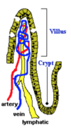
Draw a diagram of the small intestine lining, outlining the cells that are responsible for absorption and those responsible for secretion

What are the four cells that line the epithelium of the villi?
- Enterocytes: Responsible for fluid and nutrient absorption
- Goblet Cells: Secrete mucus to protect intestinal epithelium from pathogenic bacteria
- Crypt Cells (of Lieberkuhn): Invaginations of the epithelium around the villi, involved mainly in secretion
- Importantly, toward the base of the crypts are Stem Cells, which continually divide and provide the source of all the epithelial cells in the crypts and on the villi.
Describe the pathophysiology of Secretory Diahorrea
- Pathogenic bacteria attaches to the host enterocytes.
- Once adhered, the organism releases enterotoxin, which induces the intestinal epithelial cell to secrete a fluid rich in chloride ions.
- H20, Na+, K+, and HCO3- follow Cl-, creating a massive efflux of electrolyte-rich fluid into the intestinal lumen.
- Although some of this fluid is reabsorbed in the colon, the efflux of secreted fluid exceeds the colonic capacity for fluid absorption, and watery diarrhea results.
Describe the pathophysiology of Osmotic Diahorrea
Occurs when a osmotically active substance -e.g. lactose - is ingested yet poorly digested.
If the substance is taken as an isotonic solution, the water and solute will simply pass through the gut unabsorbed, causing diarrhoea.
If taken as a hypertonic solution, water (and some electrolytes) will move from the ECF into the gut lumen until equilibrium, This causes dehydration owing to the loss of body water.
Because the loss of body water is greater than the loss of sodium chloride, hypernatraemia also develops (see below).


