GI Part I (Pre-Midterm) Flashcards

1
Q

Your favorite snack while hiking is trail mix. This mix contains salty peanuts, sweet dried fruit and chocolate bits. You can enjoy the various flavors in this snack because the neuroepithelial cells in the taste bud:

Choices:

  1. have five receptor genes and each one encodes receptors for sweet, sour, bitter, salt and umami.
  2. extend their axons directly to the ventral posteromedial nucleus in the thalamus.
  3. synapse with pseudounipolar neurons in the facial nerve, glossopharyngeal or vagus nerves.
  4. phagocytose bits of ingested food for processing in secondary lysosomes.
  5. exchange ions between cells via gap junctions and therefore respond as a syncytium.
A
  1. synapse with pseudounipolar neurons in the facial nerve, glossopharyngeal or vagus

Explanation:

  • Have five receptor genes and each one encodes receptors for sweet, sour, bitter, salt and umami. is incorrect because salt and sour tastes are activated by ion channels, not receptors.
  • Extend their axons directly to the ventral posteromedial nucleus in the thalamus. is incorrect because the neuroepithelial cells do not extend into the CNS like olfactory cells do.
  • Exchange ions between cells via gap junctions and therefore respond as a syncytium. is incorrect because the cells respond individually, not as a syncytium.
2
Q

Which ionic component of saliva is most likely to be higher in saliva than in its concentration in plasma when saliva is secreted at low rates (resting state)?

  1. Sodium (Na)
  2. Potassium (K)
  3. Chloride (Cl)
A
  1. Potassium (K)

Explanation:
Na and Cl are absorbed, but K is secreted into saliva.

3
Q

When you smell freshly baked “Mom’s apple pie” which ions increase in concentration in your saliva?

  • *Choices:**
    1. Na and Cl
    2. Na and K
    3. K and Cl
A
  1. Na and Cl

Explanation:
At high flow rates the duct cells can’t reabsorb the Na and Cl fast enough to recover all of the ions from the
flow.

4
Q

In an unfortunate surgical mishap, the vagal innervation of a patient’s lower esophageal sphincter is severed. The effect on esophageal function will be:

  • *Choices:**
    1. minimal because the vagus plays no role in esophageal function.
    2. to disrupt control of the initiation of swallowing because the resting tone of the esophagus will be lost.
    3. to induce gastric reflux since the resting tone of the lower esophageal sphincter will be lost.
    4. to disrupt passage of food into the stomach because LES relaxation will be compromised.
    5. rapid atrophy of the muscles of the lower one-third of the esophagus, resulting in decreased ability to protect the esophagus from stomach acid.
A
  1. to disrupt passage of food into the stomach because LES relaxation will be compromised.

Explanation:

LES relaxation is induced by activation of the vagus nerve, therefore loss of vagus innervation disrupts the ability to pass food into the stomach.

5
Q

Where does the main pancreatic duct enter into the duodenum?

  • *Choices:**
    1. Ampulla of Vater
    2. Ileocecal valve
    3. Ligament of Trietz
    4. Pyloric sphincter
A
  1. Ampulla of Vater
6
Q

Thrombotic occlusion of the superior mesenteric artery would lead to infarction of which of the following GI organs?

Choices:

  1. Esophagus
  2. Large Intestine
  3. Liver
  4. Pancreas
  5. Small intestine
  6. Stomach
A
  1. Small Intestine

NOTE: After discussing it w/ Dr. Grahame, the colon could/would also be affected. The small intestine would be completely destroyed, though, so you can argue that it’s the “more correct” answer. That being said, Dr. Grahame said the question was unfair, so it will be removed/changed. (Our percentage breakdown on QStream also shows that most of us picked either small or large intestine.)

7
Q

A 72-year-old male with a 5-decade history of smoking and chronic alcoholism has lost 15 kg over the past 3 months. Following a biopsy, an upper esophageal mass is resected and sent to pathology for examination.

Which one of the following clinical findings is most likely to be associated with the lesion shown above?

Choices:

  1. 30% 5-year survival
  2. Gastroesophageal reflux
  3. Progressive dysphagia
  4. Celiac lymph node metastases
  5. Past Epstein Barr virus infection
A
  1. Progressive dysphagia.

Explanation:

The pictures show a moderately to well differentiated squamous cell carcinoma. Esophageal carcinomas typically manifest with progressive dysphagia, initially with solids, then liquids; the correct choice is

  • Progressive dysphagia. Additional clinical manifestations include odynophagia,, weight loss, hemorrhage following rupture, and pneumonias from aspiration or bronchoesophageal fistulas. The extensive infiltration of the wall is an adverse prognostic feature; the overall 5-year survival is 9%.
  • An upper esophagus squamous cell carcinoma would most probably metastasize to cervical lymph nodes, mid-esophagus neoplasms to mediastinal, paratracheal or peribronchial lymph nodes, and those involving lower esophagus tend to involve gastric and celiac nodes. Plummer-Vinson syndrome, human papillomavirus infections, smoking and alcoholism are some risk factors or associations described in the literature.
8
Q

Which one of the following is the most likely cause of adenocarcinoma of the esophagus?

Choices:

  1. Acid reflux
  2. Alcohol
  3. Human papilloma virus
  4. Lye ingestion
  5. Smoking
A
  1. Acid Reflux

Explanation:
Acid reflux is associated with gastric metaplasia which is considered precancerous and may lead to
adenocarcinoma of the esophagus. The other risk factors listed are associated with squamous carcinoma.

9
Q

The condition that occurs lateral to the umbilicus, in which there is a failure of the abdominal wall to completely form, leaving portions of the viscera fully exposed to the amniotic fluid, is called

Choices:

  1. Gastroschisis
  2. Hirschprung disease
  3. Intussusception
  4. Meckel diverticulum
  5. Omphalocele
  6. Volvulus
A
  1. Gastroschisis

Explanation:

  • Hirschprung’s disease (also known as congenital aganglionic megacolon) is caused by the incomplete migration of neurons derived from the primitive neural crest down the GI tract. The result is the absence ofganglion cells in distal regions such as the anal sphincter, compromising the ability to pass stool.
  • Intussusception is the “telescope-like” invagination of a segment of intestine into the adjoining intestinal lumen.
  • Meckel diverticulum is the most frequent congenital malformation of the GI tract. Essentially, it is a pouch-like structure located on the wall of the distal illium (~40-60 cm from the ileocecal valve) formed as a vestigial remnant of the omphalomesenteric duct (also called the vitelline duct or yolk stalk).
  • Volvulus is a complete twisting of a loop of intestine around its mesenteric site of attachment.
  • Omphalocele is the condition where there is failure of the intestinal loops to return to the abdomen during development. The viscera remain covered by the thin peritoneum-amnion membranes.
10
Q

With respect to esophageal atresia (EA) and tracheoesophageal fistula (TEF), which one of the following conditions is least likely to require immediate surgical intervention?

Choices:

  1. TEF without EA
  2. EA without TEF
  3. EA with TEF to proximal esophagus
  4. EA with TEF to distal esophagus
  5. EA with TEF to both proximal and distal esophagus
A
  1. TEF without EA

Explanation:
Anything with EA needs to be corrected ASAP because there is no patent esophagus. If there is a EA with TEF, food and water (i.e., formula/milk) will have to enter the respiratory tract if it will ever make it to the stomach. IF there is no TEF but there is EA, then there’s no way of even getting into the stomach.
In the case of TEF without EA there are the expected separate paths to the stomach and to the lungs, but there’s also an additional connection. It will probably still need surgical intervention, but at least the child can breathe and eat.

http://2.bp.blogspot.com/_kE4lQ4oqHVc/S9BbqM53c1I/AAAAAAAAB4g/fwX2gTAQc7s/s1600/tea.gif
Explanation and link provided by 2LT Rayad Barakat USU Class of 2018.

11
Q

The development of the gastrointestinal tract requires an epithelial-mesenchymal interaction for tube patterning into the different components. Which one of the following transcription factors is expressed in the endoderm and up-regulates expression of factors in the mesoderm (such as the HOX genes)?

Choices:

  1. NKX2.5 (NK2 homeobox 5)
  2. PAX2 (paired box gene 2)
  3. PITX2 (paired-like homeodomain 2)
  4. SHH (Sonic hedgehog)
  5. WT1 (Wilms tumor 1)
A
  1. SHH (Sonic hedgehog)

Explanation:

  • NKX2.5 is responsible for the induction of the heart-forming region, is expressed in mesoderm and induced by endoderm (p169 Langman’s text). Mutations of NKX2.5 may produce atrial septal defects (ASDs) and other cardiac abnormalities.
  • PAX2 is expressed in the metanephric mesenchyme and causes the mesenchyme to epithelialize during the development of the collecting ducts of the kidney. Mutations in the PAX2 gene may cause renal coloboma syndrome.
  • PITX2 is involved in cardiac looping and is expressed in the lateral plate mesoderm of the left side. Mutations in the PITX2 homeobox gene are known to cause Axenfeld-Rieger syndrome (ARS).
  • WT1 is expressed in the metanephric blastema and regulates the production of factors that stimulate growth and branching of the uteric buds. Mutations in WT1 may cause Wilms’ tumor.
12
Q

Which of the following substances are the cells surrounding the asterisk most likely secreting?

Choices:

  1. Enzymes
  2. Ghrelin
  3. Mucus
  4. Serotonin
  5. Sodium
A
  1. Mucus

Explanation:

  • Cells that compose the bulk of the acini are pyramidal or flask shaped (like an Erlenmeyer flask). The lumen of an acinus is quite small and usually difficult to see in the light microscope. Mucus-secreting cells are lightly stained with flattened nuclei pushed to the bottom of the cells. Their secretory products are Mucins, proteins that are heavily glycosylated and poor staining.
  • Serous secreting acinar cells contain secretory granules that are often visible in the light microscope. Examples of serous secreting cells are visible above. They are frequently secreting Enzymes, such as in the pancreas. Cells actively involved in ion pumping (e.g. Sodium) often have regularly arranged mitochondria, e.g, striated ducts.
  • Cells secreting Ghrelin and Serotonin would also have secretory granules that will stain with eosin but these cells tend to be found as singles or small groups in the GI tract.
13
Q

The asterisk is in the lumen of a structure. This structure is most likely a/an
Choices:

  1. Excretory duct
  2. Intercalated duct
  3. Mucus-secreting acinus
  4. Serous acinus
  5. Striated duct
A
  1. Striated duct

Explanation:

  • Examination of the indicated structure shows that the cells that make up the wall of the structure are regular cuboidal in shape. This suggests that we may be looking at a duct of some kind. In comparison, the cells that compose the bulk of the acini are pyramidal or flask shaped (like an Erlenmeyer flask). The lumen of an acinus is quite small and usually difficult to see in the light microscope.
  • Mucus-secreting cells would be lightly stained with flattened nuclei pushed to the bottom of the cells. Serous secreting acinar cells contain visible secretory granules and some signs of this can be seen in the cells located at either sides of the indicated structure.
  • If the structure is not part of the acinar component of the gland, then the choices are of various ducts. Excretory ducts are very large and generally have stratified layers of cells. This structure has a simple epithelium (one layer of nuclei). The lumen is about equivalent to the height of a cell, too large for a typical intercalated duct and there is evidence of lines radiating from the apical to basal aspect of the cells. These are the staining patterns of mitochondria that give the striated appearance of “striated ducts”.
14
Q

Dental infections have been linked to an increased risk for all of the following conditions/diseases EXCEPT:

Choices:

  1. Asthma
  2. Cardiovascular disease
  3. Diabetes
  4. Endocarditis
  5. Pre-term low birth weight infants
A
  1. Asthma

Explanation:

  • Dental infections are associated with an increased risk of each of the above except asthma.
  • The correlation with cardiovascular disease is significant, however, a direct cause and effect relation has not yet been proven.
15
Q

Which of the following recommendations applies to pregnant patients who seek dental care during pregnancy?
Choices:

  1. No dental treatment during pregnancy because it increases the risk for miscarriage.
  2. Treat acute periodontal problems during pregnancy to reduce risk for pre-term delivery.
  3. Do not debride (scale and root plane) until the 2nd trimester.
  4. Provide emergency dental care only.
A
  1. Treat acute periodontal problems during pregnancy to reduce risk for pre-term delivery.

Explanation:
Acute periodontal problems should be treated during pregnancy to reduce the risk for pre-term delivery.

16
Q

The surface monolayer of chylomicrons consists mainly of phosphodiglycerides such as a lecithin and which one of the following?
Choices:

  1. Bile salts
  2. Cholesterol
  3. Cholesterol esters
  4. Monoglycerides (e.g., lysolecithin)
  5. Ttriglycerides
A
  1. Cholesterol

Explanation:
To be in the surface monolayer, a molecule must be amphipathic. Cholesterol is not only amphipathic, but the steroid nucleus also packs tightly between the fatty acyl chains of the phospholipids. Cholesterol esters and triglycerides are too nonpolar to be part of the surface layer and therefore migrate to the core of the lipoprotein particle. Bile salts and monoglycerides are detergents. In high concentrations they would simply emulsify any lipid structure. The physiological concentration of monoglycerides is therefore kept very low. Bile salts are not found in plasma or lymph, where lipoproteins are present, and therefore are not incorporated into the structures.

17
Q

All of the following are significant products of pancreatic amylase action on amylopectin EXCEPT which one?
Choices:

  1. Glucose
  2. Maltose
  3. Maltotriose
  4. Isomaltose
  5. Alpha-dextrins
A
  1. Glucose

Explanation:
In the small intestine, pancreatic amylase continues the process of starch digestion started in the mouth by salivary amylase (but, that was largely interupted by denaturing effects of acid in the stomach). Products of pancreatic amylase are the disaccharide maltose, the trisaccharide maltotriose, the alpha 1,6-linked disaccharide isomaltose, and oligosaccharides known as limit dextrins that are usually 4-9 glucosyl residues long. Alpha dextrins are the shortened oligosaccharide products of alpha amylase action. Amylase does not release free glucose.

18
Q

Reduced activity of which one of the following enzymes would have the most deleterious effect on protein digestion?
Choices:

  1. Chymotrypsin
  2. Elastase
  3. Pepsin
  4. Carboxypeptidase
  5. Enteropeptidase
A
  1. Enteropeptidase

Explanation:
The pancreas secretes digestive enzymes mainly as inactive zymogens or proenzymes that require proteolytic cleavage to become active. Enteropeptidase (often termed enterokinase, which is actually a misnomer because no ATP is involved) is secreted by the brush border cells in the intestine, and its job is to get the ball rolling. It cleaves trypsinogen to form trypsin, which in turn “autoactivates” more molecules of trypsin. Once activated, trypsin becomes a very important player in the whole process; it not only acts by itself directly on dietary proteins in the gut, but also plays a central role in converting various other pancreatic zymogens into their active forms.

19
Q

Amino acids and glucose in the lumen of the small intestine are taken up by intestinal absorptive cells mainly by which kind of transport mechanism?

Choices:

  1. Primary-active, using ATP driven pumps
  2. Secondary-active, using Na+ driven symporters
  3. Secondary-active, using K+ driven antiporters
  4. Passive-facilitated, using substrate selective transport proteins
  5. Passive-facilitated, using non selective, ungated channels
  6. Passive-facilitated, using substrate selective voltage gated channels
A
  1. Secondary-active, using Na+ driven symporters

Explanation:
Recall that the kidney uses a similar symport system that recovers glucose by co-transport with sodium.

20
Q

Which one of the following organisms is most often the causative agent of “hand, foot, and mouth disease?”
Choices:

  1. Candida albicans
  2. Coagulase-negative Staphylococci
  3. Coxsackievirus
  4. HSV-1
  5. HSV-2
A
  1. Coxsackievirus
21
Q

The enzyme urease is encoded by Helicobacter pylori and is required for colonization and survival in the stomach. The reaction that is catalyzed by this enzyme results in the production of which one of the following?

Choices:

  1. Ammonia
  2. Carbon monoxide
  3. Hydrogen peroxide
  4. Oxygen
  5. Urea
A
  1. Ammonia

Explanation:
Urease catalyzes the reaction:
urea + H2O -> CO2 + 2NH3
The ammonia produced acts as a base to neutralize stomach acid in the microenvironment of the bacterium, which depends on urease for survival.

22
Q

A 42-year-old female is admitted to the intensive care unit following a motor vehicle accident. On physical examination she exhibits decerebrate posture, papilledema, absent response to pain and unilateral, dilated and unreactive right pupil. On the second day she becomes hypotensive; endoscopic evaluation discloses gastric and duodenal ulcers. Which one of the following alternatives is correct, regarding the lesions developed on the second day?
Choices:

  1. Probably caused by vagal nuclei stimulation.
  2. Rare with intracranial diseases.
  3. Perforation is unexpected.
  4. Gastric obstruction probable if patient survives.
  5. Associated with intractable vomiting.
A
  1. Probably caused by vagal nuclei stimulation

Explanation:
The clinical scenario describes acute gastric and duodenal ulceration arising in patients with intracranial disease (Cushing ulcers). Cushing ulcers are considered to arise from gastric acid hypersecretion caused by direct stimulation of the vagal nuclei, The correct answer is: Probably caused by vagal nuclei stimulation. Cushing ulcers are common in patients with severe intracranial diseases and carry a high incidence of perforation. Obstruction, associated with intractable vomiting if total, is associated typically with chronic ulcers.

23
Q

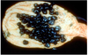
A 57-year-old male has worsening midepigastric pain and 8 kg weight loss over the last 6 months. Stool samples reveal occult blood, there is conjunctival pallor on physical examination. Upper gastrointestinal radiographic series show an ulcerated lesion involving the gastric antrum. The mass is resected; the pathology is attached.
Which one of the following alternatives regarding the epidemiology or pathogenesis of this entity is correct, based on the clinicopathologic features of this case?

Choices:

  1. Strongly associated with peptic ulcer disease.
  2. Association to familial adenomatous polyposis (FAP) proves the preeminent role of genetic changes in its pathogenesis.
  3. Associated with Barret esophagus.
  4. Increased second-generation Japanese inmigrant incidence.
  5. Decreasing USA overall incidence.
A
  1. Decreasing USA overall incidence.

Explanation:
Gastric cancer, more specifically intestinal-type adenocarcinoma, is the diagnosis of this case. The overall incidence is decreasing in the United States, although those neoplasms arising in the gastric cardia are becoming more frequent due to their association with Barrett esophagus.
The correct answer is: Decreasing USA overall incidence. Gastric cancer is more common in patients with multifocal mucosal atrophy and intestinal metaplasia, not in individuals with peptic ulcer disease (PUD). Partial gastrectomy for PUD imparts a slightly increased risk. Japanese patients with FAP have an increased incidence of intestinal-type adenocarcinomas; this incidence is not as elevated for individuals with FAP residing in low-risk areas, thus supporting the importance of environmental factors. By their second generation, migrant populations from high to low incidence countries have the incidences of the countries of destination.

24
Q

A 62-year-old male with Addison disease (primary chronic adrenocortical insufficiency) has progressive memory loss and moderate personality changes over the past 11 months. Physical examination reveals a smooth and beefy-red tongue, numbness, loss of vibration sense and conjunctival pallor. Endoscopy shows diffuse loss of rugal folds over the body and fundus, the antrum is spared. Which of the following pathology findings is most likely to be associated with this disease?
Choices:

  1. Antibodies to Helicobacter pylori
  2. Hyperplastic gastric polyps
  3. Decreased gastrin
  4. Gastric neuroendocrine cell hyperplasia
  5. Neutrophils within gastric lamina propria
A
  1. Gastric neuroendocrine cell hyperplasia

Explanation:
The clinical findings of Addison disease, neurologic/behavioral changes, glossitis, conjunctival pallor (probable anemia), in conjunction with the endoscopic changes are consistent with autoimmune gastritis. Well-developed autoimmune gastritis presents as diffuse atrophy of the body and fundus, with sparing or only mild involvement of the antrum. The antral neuroendocrine cell hyperplasia is a physiologic response to decreased acid production (damage of acid-producing oxyntic body and fundus mucosa). Neuroendocrine cell hyperplasia is responsible for increased gastrin secretion. Antibodies to parietal cells and intrinsic factor are also associated with pernicious anemia, which in turn is associated with megaloblastic anemia and neurologic/behavioral changes. Antibodies to Helicobacter pylori, hyperplastic gastric polyps, normal to decreased gastrin and antral location (occasionally involving body and fundus) are characteristics of
Helicobacter pylori chronic gastritis.

25
Q

A 49-year-old male with a 3-year history of intermittent nausea and abdominal discomfort is evaluated by his primary physician. Serologic tests for antibodies to H. pylori and urea breath test are positive. Endoscopic evaluation discloses erythematous antral and proximal gastric body mucosa. Spiral-shaped Warthin-Starry positive organisms within the surface mucus are detected on the gastric biopsy specimen. Which one of the following statements regarding the complications of this disease is correct?
Choices:

  1. Adenocarcinoma is most commonly associated with this disease.
  2. Peptic ulcer is most commonly associated with this disease.
  3. Submucosal involvement indicates dysplasia.
  4. Chronic ulcers carry high risk of malignant transformation.
  5. Zollinger-Ellison syndrome is a rare complication.
A
  1. Peptic ulcer is most commonly associated with this disease.

Explanation:
The clinical features of this case are typical of chronic gastritis caused by Helicobacter pylori. Peptic ulcer disease is most commonly associated with H. pylori chronic gastritis; developing a lymphoma would be less common, and adenocarcinomas arising from peptic ulcers are distinctly unusual.

The correct answer is: Peptic ulcer is most commonly associated with this disease. Adenocarcinomas frequently may ulcerate; ulcers do not tend to become malignant. Dysplasia is a preneoplastic change that by definition is confined to the mucosa. The risk for adenocarcinoma is greatest in autoimmune chronic gastritis. Zollinger-Ellison syndrome is caused by gastrin-producing tumors, gastrinomas, typically of small intestine or pancreatic origin. Gastric adenocarcinoma is associated with the presence of H. pylori in the stomach, however, much less commonly, when compared to the number of colonized individuals who suffer from peptic ulcers.

26
Q

In a patient who has a mutation that produces a defective SGLT molecule, the transport of glucose and which one of the following molecules will be adversely affected?
Choices:

  1. Chloride
  2. Water
  3. Short chain fatty acids
  4. Monoglycerides
  5. Bile salts
A
  1. Water

Explanation:
Sodium is co-transported with glucose in this transporter. This supports part of the water reabsorption process that takes place in the gut, by contributing to the sodium gradient that pulls water into and around the cells.

27
Q

In which section of the GI tract would one encounter skeletal muscle?

Choices:

  1. Upper esophageal sphincter
  2. Lower esophageal sphincter
  3. Upper and lower esophageal sphincters
  4. Pyloric sphincter
  5. Ileocecal sphincter
  6. Pyloric and ileocecal sphincters
  7. Internal anal sphincter
  8. External anal sphincter
  9. Internal and external anal sphincters
  10. Pyloric and external anal sphincters
  11. Upper esophageal and external anal sphincters
A

Upper esophageal and external anal sphincters

Explanation:

Smooth muscle under involuntary control is used for all of the sphincters in the gastrointestinal tract except for the two located at ends of the “tube”. Those two, the UES and external anal sphincters, contain striated muscle and are under voluntary control (anal sphincter) or can be trained to be under voluntary control (UES).

28
Q

Which of the following components of the abdominal examination is associated with the correct areas to
assess? (Choose all that apply.)

Choices:

Inspection: contour, skin, pulsations, movement

Auscultation: bowel sounds, bruits, hums and rubs

Percussion: sounds, four quadrants, liver, spleen

Palpation: superficial, deep, rebound tenderness, liver, spleen, kidney, aorta

A

Inspection: contour, skin, pulsations, movement

Auscultation: bowel sounds, bruits, hums and rubs

Percussion: sounds, four quadrants, liver, spleen

Palpation: superficial, deep, rebound tenderness, liver, spleen, kidney, aorta

Explanation:

All of the parts of the examination sequence are associated with the correct areas to assess.

In the patient with the undifferentiated complaint, a complete exam may be most helpful – findings from the physical may narrow the list of differential diagnoses. When the history has already narrowed your considerations, a focused examination is appropriate, and may include any combination of the component parts of the examination sequence.

29
Q

The basal electrical rhythm (BER) of smooth muscle in the gut is set by

Choices:

the sympathetic nervous system.

the parasympathetic nervous system.

the enteric nervous system.

reflexes induced by stretch receptors.

interstitial cells of Cajal.

A

interstitial cells of Cajal.

Explanation:

Named after the Spanish neuroanatomist Santiago Ramón y Cajal (who you remember from the NES module) the interstitial cells of Cajal (ICC) are the “pacemakers” of the system (and also probable progenitor cells of gastrointestinal stromal tumors, GIST). All other inputs can influence whether slow waves reach threshold and achieve an action potential, but only the ICC sets the BER. Not surprisingly, malfunction in the ICC network can result in motility disorders associated with obstruction. And, the complete or partial loss of ICC cells may be involved in a number of conditions including, pseudoobstruction, achalasia, ulcerative colitis, infantile pyloric stenosis, Chagas’ disease, diabetes, slow-transit constipation, and idiopathic gastric perforation.

30
Q

Which one of the following acts to increase the frequency of spike potentials in the gut?

Choices:

Nitric oxide (NO)

Vasoactive intestinal peptide (VIP)

Acetylcholine

Sympathetic stimulation

Cholecystokinin (CCK)

A

Acetylcholine acts on the ICC to depolarize them, making them more likely to reach threshold. NO and VIP are inhibitory and act on the ICC to hyperpolarize. Sympathetics are also inhibitory, releasing norepinephrine to hyperpolarize. CCK, in addition to its various other actions, reduces gastric emptying by inhibiting muscle activity in the stomach.

31
Q

Which portion of the GI tract demonstrates the following characteristics: Reverse BER gradient, absence of a migrating motor complex (MMC), and frequent retrograde movement?

Choices:

Stomach
Duodenum
Jejunum
Ileum
Colon

A

Colon

Explanation:

The stomach and small intestine both demonstrate an MMC. The small intestine shows anterograde movement with a BER gradient that is aboral.

32
Q

Antibodies are able to enter the lumen of the GI system because

Choices:

  1. lymphocytes migrate between the enterocytes, differentiate into plasma cells and secrete the immunoglobulins into the lumenal space.
  2. lymphocytes develop into plasma cells in the environment of M cells and the M cells present the antibodies on MHC complexes.
  3. plasma cells secrete antibodies that diffuse into the lumen of the gut through lesions in the tight junctions of the enterocytes.
  4. plasma cells secrete antibodies that complex with carrier proteins that permit the uptake and transcytosis of the antibodies.
  5. activated M cells secrete antibodies directly into the GI tract.
A
  1. plasma cells secrete antibodies that complex with carrier proteins that permit the uptake and transcytosis of the antibodies.

Explanation:

Receptor-mediated transcytosis is a specialized transport process that permits molecules to travel through the epithelium to the space on the other side of the cells.

33
Q

The cells indicated by the arrows below most likely secrete

Choices:

gastrin.
motilin.
acid.
pepsinogen.
lysozyme.
lipase.

A

lysozyme.

Explanation:

These are Paneth cells, located at the base of the intestinal glands/crypts, throughout the small intestine and appendix. Paneth cells secrete lysozyme and antimicrobial peptides (defensins) that are able to insert into bacterial cell membranes and create pores, disrupting the bacterium. It is thought that Paneth cells function to protect nearby stem cells, defending them from bacterial attack, thereby preserving the regenerative ability of the epithelium.

34
Q

Several days following abdominal surgery, a patient still has not had a bowel movement and now complains of nausea. What is taking place in the patient’s GI tract that most likely explains these signs and symptoms?

Choices:

  1. The BER is increased, leading to sustained smooth muscle contractions.
  2. Inflammation has caused an increase in nitric oxide, leading to reduced muscle contractions.
  3. Inflammation has caused a decrease in NO, leading to muscle spasm.
  4. The sympathetic nervous system is activated, leading to increased release of acetylcholine.
  5. The enteric nervous system remains “paralyzed” as a result of incomplete clearance of the anesthetics used during the operation.
A

Inflammation has caused an increase in nitric oxide, leading to reduced muscle contractions.

Explanation:

The patient is experiencing ileus, which is a result of inflammation following open abdominal surgery. Inflammation induces the increase in NO, which is inhibitory to the smooth muscle of the intestines, reducing contractions. The BER cannot be changed and the sympathetic nervous system uses norepinephrine, not acetylcholine as a neurotransmitter. Much of the small intestinal motility is regained as anesthesia lightens and the patient regains consciousness, and normalizes within a few hours post-operation. So, longer-term paralysis of the ENS by anesthetics is unlikely.

35
Q

Which one of the following pathways is not significant in an individual who has fasted for a week?

Choices:

Ketone body production by liver
Gluconeogenesis in liver
Glycogenolysis in the liver
Fatty acid oxidation in muscle
Anaerobic glycolysis in red blood cells

A

Glycogenolysis in the liver

Explanation:

The first four options are pathways that are active in a fasted state to provide energy for tissues and maintain blood glucose. However, liver glycogen stores are exhausted after 1-2 days so this pathway is not relevant in the third day of a fast. Anaerobic glycolysis must be active in red blood cells no matter what the nutritional state is because, lacking mitochondria, it is their only mechanism of ATP production.

36
Q

Insulin leads to an increase in the activity of all the following pathways in liver EXCEPT:

Choices:

Glucose transport across the plasma membrane

Glycogen synthesis

Glycolysis

Fatty acid synthesis

Citrate-pyruvate cycle

A

Glucose transport across the plasma membrane

Explanation:

Insulin signals the well fed state, so binding to its receptor leads to the activation of pathways that store fuel as glycogen or fat. Hence, the pathways for glycogen synthesis and fatty acid synthesis are activated. Glucose provides the carbon atoms for fatty acid synthesis, so glycolysis must be activated to produce the precursor, ultimately acetyl-CoA. The citrate-pyruvate cycle increases in activity to shuttle acetyl-CoA out of the mitochondrial matrix into the cytoplasm, and to produce NADPH for reduction reactions.

The glucose transporter in liver is not insulin sensitive, probably because the transporter must be active in both the fed and starved state; in the fed state, the liver must be able to take up glucose from the blood at a rapid rate, and in the starved state it must be able to release glucose into the blood at a rapid rate.

37
Q

The major precursor for gluconeogenesis after a 36 hour fast is

Choices:

Glutamate
Alanine
Glycerol
Lactate
Acetoacetate

A

Alanine

Explanation:

Any of the other options (except acetoacetate) are theoretically possible, but alanine is the major precursor in the early fasted state. In deep starvation, when muscle protein breakdown is greatly reduced and fat mobilization is accelerated, glycerol becomes a major precursor. Acetoacetate cannot be converted to glucose because it is broken down to acetyl-CoA, and there is no route from acetyl-CoA to net synthesis of a gluconeogenic precursor; both carbons in the acetyl moiety are lost as CO2 in the TCA cycle.

38
Q

What is the key activator of pyruvate carboxylase, the committed enzyme in gluconeogenesis?

Choices:

Pyruvate
NADH
Alanine
Acetyl-CoA
ATP

A

Acetyl-CoA is the answer; the logic to this is as follows. Gluconeogenesis is an endergonic pathway that requires fatty acid oxidation to supply ATP. Since the beta oxidation pathway for fatty acids produces acetyl-CoA, a rise in acetyl-CoA levels is the signal that beta oxidation is taking place.

39
Q

A 23-year-old male returns from a camping trip to Mexico. He presents to the ER suffering from abdominal pain, nausea, fever, and bloody diarrhea. Stool specimens are collected and sent to the laboratory for bacterial cultures and ova and parasite (O&P) examination. Bacterial cultures are negative for enteric pathogens but fecal leukocytes are 4+. The O&P examination reveals the organism shown in the image.
Which host is the primary reservoir for this parasite?

Choices:

Cockroach
Dog
Fly
Human
Mosquito

A

Human

Explanation:

There are no reservoir hosts for Entamoeba histolytica; man is the only host.

40
Q

Which one of the following sequellae is associated with infections due to Shiga toxin-producing Escherichia coli?

Choices:

Gram negative sepsis

Guillain-Barre syndrome

Hemolytic uremic syndrome (HUS)

Mesenteric lymphadenitis

Reiter’s syndrome

A

Hemolytic uremic syndrome (HUS)

Explanation:

Shiga toxin-producing E. coli (STEC) do not disseminate into the blood stream to cause sepsis, and are not associated with the other choices. Guillain-Barre syndrome is associated with Campylobacter jejuni infections, mesenteric lymphadenitis is caused by the gastrointestinal Yersina species, and Reiter’s syndrome is associated with inflammatory diarrheas (shigellosis, salmonellosis, yersiniosis and campylobacter infection) but not hemorrhagic colitis caused by STEC.

41
Q

Several attendees of a church picnic developed diarrhea 2-3 days after attending the picnic that was characterized as being of low volume with trace amounts of blood and pus. One of the victims, an 82 year old woman, was hospitalized with fever, chills, and hypotension. Gram-negative rods that do not ferment lactose were isolated from her blood. Which one of the following is most likely the agent responsible for her condition?

Choices:

Enterotoxigenic Escherichia coli

Escherichia coli O157:H7

Salmonella enterica, var. Enteriditis

Shigella sonnei

Vibrio parahaemolyticus

A

Salmonella enterica, var. Enteriditis

Explanation: Due to the inflammatory nature of the diarrhea and the culture characteristics of the bacteria, the most likely cause of her condition is Salmonella enterica, var. Enteriditis. Of the other organisms listed, only Shigella sonnei causes a similar gastrointestinal illness and is a nonlactose fermenter. However, the shigellae, unlike the nontyphoidal salmonellae, do not disseminate into the bloodstream. While Salmonella enterica, var. Enteriditis usually stays localized to the small intestine, it can cause bacteremia with septicemia in the elderly. (Most strains of E. coli are happy to ferment lactose, entero-invasive EIEC is an exception.)

42
Q

Large volume stools that do not contain blood or white blood cells are suggestive of

Choices:

dysentery.
enteric fever.
inflammatory diarrhea.
peptic ulcer disease.
secretory diarrhea.

A

secretory diarrhea.

Explanation:

  • Stools from individuals with dysentary and inflammatory diarrhea contain neutrophlis and sometimes blood.
  • Enteric fever does not result in diarrhea until possibly late during the disease and it is not large volume.
  • Peptic ulcer disease does not cause diarrhea.
43
Q

Several people who share dinner at a Chinese restaurant develop sudden onset of vomiting within 3 hours of eating. The only item shared by all the diners was Chinese fried rice. Which one of the following is the most likely cause of this illness?

Choices:

Bacillus cereus toxin in the rice
Contamination of the rice with raw chicken juice containing Campylobacter jejuni
Contamination of the rice with raw egg containing Salmonella enteriditis
Listeria monocytogenes in the cold chicken used to flavor the rice
Shigella sonnei on the server’s hands contaminating the rice

A

Bacillus cereus toxin in the rice

Explanation:

The major clue here is that the onset of illness was only 3 hours after ingestion of the rice. That indicates preformed toxin in the rice. Therefore, the correct answer is the choice indicating toxin already in the rice.

44
Q

The wife of an American diplomat in Morocco presents to an ER with production of copious volumes of diarrhea and muscle cramping. She showed signs of severe dehydration and was becoming nonresponsive. A stool sample, which was watery and contained flecks of mucus, was sent to the laboratory. She was given oral rehydration therapy and recovered within two days. The laboratory reported that the stool was negative for lactoferrin and that the culture yielded a cytochrome oxidase-positive Gram-negative organism. The organism that was most likely responsible for his condition produces a toxin that causes

Choices:

  1. constitutive production of cyclic AMP via ADP-ribosylation of cellular G proteins.
  2. elongation and effacement of the small intestinal microvilli.
  3. hemolysis of human erythrocytes.
  4. inhibition of protein synthesis due to deamidation of a ribosomal subunit.
  5. massive cytokine induction due to nonspecific binding of T-cell receptors and major histocompatibility (MHC) II molecules.
A

constitutive production of cyclic AMP via ADP-ribosylation of cellular G proteins.

Explanation:

Based on the clinical manifestations and case history the scenario suggests infection by the facultative anaerobe Vibrio cholerae. The question also requires knowledge of the mechanism of cholera toxin, the principal virulence factor of V. cholerae, which acts as an enzyme to ADP-ribosylate the regulatory G protein (G-alpha-s) that controls the activity of adenylate cyclase, resulting in constitutive production of cAMP.

Elongation and effacement of the small intestinal microvilli, is descriptive of the pathogenesis of enteropathogenic E. coli.

Hemolysis of human erythrocytes, is descriptive of a hemolysin (i.e. V. parahaemolyticus Kanagawa toxin).

Inhibition of protein synthesis due to deamidation of a ribosomal subunit., describes the mechanism of Shiga toxin;

Massive cytokine induction due to nonspecific binding of T-cell receptors and major histocompatibility (MHC) II molecules. describes the mechanism of action of superantigens.

45
Q

A 6-year-old girl is admitted to a hospital in suburban Boston following several days of fever and vomiting. Ulcerative, vesicular lesions in the oral mucosa and vesicles on the hands and soles of the feet are observed upon physical examination. The girl develops aseptic meningitis and encephalomyelitis 3 days after entering the hospital. With which one of the following viruses is she most likely infected?

Choices:

Measles virus

Herpes simplex virus type 2 (HSV-2)

A strain of Coxsackie A virus

Rubella virus

Varicella zoster virus (VZV)

A

A strain of Coxsackie A virus

Explanation:

The patient is a child with fever and gastrointestinal symptoms for several days along with vesicular lesions:

  • the child’s illness is hand-foot-and-mouth disease, and Coxsackie A viruses are often a cause of this disease manifestation. Further progression to signs of CNS involvement are also quite common with enteroviruses like a Coxsackie A virus.
  • The child would likely be vaccinated against measles (a paramyxovirus) and rubella virus (a togavirus) and both measles and rubella cause a maculopapular rash not vesicular lesions.
  • HSV-2 is normally associated as a sexually-transmitted disease with fewer vesicular lesions associated with primary infection and reactivation in the genital area.
  • VZV causes chickenpox, and would result in more wide-spread vesicular lesions on the body, not in the oral cavity, encephalitis caused by VZV is rare, and the child would also likely be vaccinated against VZV.
46
Q

Humans and various other animals often come into close contact with one another on farms. In one example a farmer and a pig both appear to have flu-like symptoms. A duck then becomes co-infected with different Influenza A virus strains, one from the farmer and another from the pig, and a new virus strain resulted from this infection. Formation of the new strain is an example of what genetic process:

Choices:
Complementation

Reassortment

Recombination

Transcapsidation

Conjugation

A

Reassortment

Explanation:

  • Reassortment is the mixing of gene segments within a single infected cell to produce new virus strains with components of each of the parental strains. Like Influenza, Rotavirus has segmented genomes and generates new strains by reassortment.
  • Complementation occurs when one virus or cell expressing viral proteins provides an enzyme or protein required by another virus.
  • Recombination is the process in which co-infection and exchange of genes occur within a single strand to create two hybrid viruses. Recombination is common in DNA viruses or viruses that have a DNA phase such as HIV.
  • Transcapsidation or camouflage occurs when the genome of one virus is wrapped in the shell or envelope of a different virus. For example, Hepatitis D virus (RNA virus) does not code for its own envelope but uses the envelope of Hepatitis B virus (DNA virus).
  • Conjugation usually refers to exchange of genetic material between compatible bacterial strains (not done by viruses) with a physical interaction of the bacterial cells, usually involving a bridging structure of some kind, such as the F pilus.
47
Q

A 72-year-old female presents with severe abdominal pain and reported lower GI bleeding to a local emergency room. Her acute abdominal series is shown below for review, demonstrating dilated colon and small bowel. Based on this image, which one of the following is the most likely cause for this patient’s symptoms?

Choices:

Diverticulosis

Malignancy

Intussusception

Adhesions

A

Malignancy

Explanation:

Based on the image and description you can tell that both colon and small bowel are dilated. This suggests a distal obstruction affecting the colon. The most common cause of colonic obstruction is malignancy such as a colorectal carcinoma. If only the small bowel were dilated as may be seen in a small bowel obstruction, then adhesions would be the most likely cause.

48
Q

The colon segment below was removed from a 28-year-old male with a 12-year history of recurrent diarrhea and cramps after he developed profuse bleeding through the anus. Previous microbiologic, immunologic and vascular studies failed to disclose a specific etiology. Which of the following would be most likely?

Choices:

Osteomalacia and bleeding due to vitamin D and K deficiency from malabsorption.

Abdominal rigidity and sepsis secondary to acute peritonitis.

This disease having a high incidence of developing colonic lymphoma.

This disease having increased incidence of developing sclerosing cholangitis.

Microscopic sections will show crypt abscesses and granulomas.

A

This disease having increased incidence of developing sclerosing cholangitis.

Explanation:

The gross picture shows continuous involvement of the colon consistent with ulcerative colitis; sclerosing cholangitis, migratory polyarthritis and colon adenocarcinoma (not lymphoma) are some of the most common associations or complications. Acute peritonitis is uncommon since this process is mainly limited to the mucosa; peritonitis would develop only within the very rare instance of toxic megacolon. Since the disease does not involve small intestine, malabsorption is not a complication of ulcerative colitis. The microscopic sections may show crypt abscesses; but granulomas, skip lesions and transmural inflammation would be more typical of Crohn disease.

49
Q

A 14-year-old male with rectal bleeding undergoes a rectosigmoidoscopy that shows approximately 200 elevated lesions. A sample from his partial colectomy is shown below. Which one of the following clinical conditions or complications is most likely to develop?

Choices:

Colonic adenocarcinoma
Sclerosing cholangitis
Gastric adenomas
Toxic megacolon
Turcot syndrome

A

Colonic adenocarcinoma

Explanation:

The multiple elevated lesions correspond to multiple adenomas that in this case would be diagnostic of familial adenomatous polyposis. Notice on the high magnification view the contrast between normal (left side-abundant mucus) and dysplastic/neoplastic (right side-decreased mucus and hyperchromatic nuclei) glands typical of adenomas. Gastric adenomas and Turcot syndrome can both be associated with familial adenomatous polyposis, but in this case the development of adenocarcinoma is almost certain since the treatment was partial colectomy, not total colectomy. Sclerosing cholangitis and toxic megacolon are diseases associated with inflammatory bowel disease and ulcerative colitis respectively.

50
Q

A 48-year-old man is being treated for 2 weeks with a combination of antibiotics (nafcillin, clindamycin) for aspiration pneumonia. He has recently developed abdominal pain, fever, and diarrhea over the past 3 days.
The gross appearance of his resected colon is attached. What is your diagnosis?

Choices:

Amebiasis
Pseudomembranous colitis
Ulcerative colitis
Ischemic bowel disease
Angiodysplasia

A

Pseudomembranous colitis

Explanation:

The gross specimen shows an erythematous mucosa with an overlying tan pseudomembrane. These findings, in the context of the clinical history, are consistent with pseudomembranous colitis.

51
Q

A 28-year-old female with an intermittent history of bloody diarrhea and abdominal pain is admitted to the emergency room after an episode of severe bleeding per rectum, hypotension and clammy skin. After initial stabilization imaging studies disclose multiple colonic ulcers with intervening normal mucosa, changes consistent with active colonic bleeding, colon to colon fistulas and marked narrowing of the distal 26 cm of terminal ileum. A partial colectomy is performed. Images of the resected colon segment with corresponding histology are shown below. On the basis of this information, which one of the following pathologic findings is most likely?

Choices:

Multiple pseudopolyps

Most lesions on sigmoid colon

Backwash ileitis

Transmural inflammation

Acute necrotizing lymphadenitis

A

Transmural inflammation
Explanation:

The clinical case described above is consistent with Chron disease, an inflammatory bowel disease characterized by transmural inflammation, granulomas, skip lesions and involvement of any portion of the gastrointestinal tract. The correct answer is transmural inflammation. Although any segment of the gastrointestinal tract may be involved, terminal ileum, ileocecal valve and cecum are the most commonly affected sites. Non-caseating granulomas are identified in about 35% of instances. Granulomas, not acute necrotizing lymphadenitis, may also be present in lymph nodes and skin. Pseudopolyps may be seen in ulcerative colitis when islands of regenerating mucosa bulge into the colonic lumen.

Backwash ileitis refers to the mild ileal inflammation that may be seen in ulcerative colitis cases involving the whole colon.

52
Q

A 32-year-old female comes to your consult complaining of diarrhea, bloating and fatigue gradually worsening over the last 7 months. Physical examination reveals conjunctival pallor, joint pains and clustered itchy skin blisters. You submit samples for serologic testing. Serologic testing was followed by a biopsy. The proximal jejunum biopsy was later reported as showing crypt hyperplasia, villous atrophy and increased intraepithelial CD8+ T-lymphocytes. Which one of the following tests would be most helpful to confirm your clinical impression?
Choices:
IgA antibodies to tissue transglutaminase
Presence of HLA-DQ2 alleles
Monoclonal mucosal B-lymphocytes
Anti-endomysial antibodies
Serum iron levels

A

Anti-endomysial antibodies.

Explanation:

The clinicopathologic features are consistent with celiac disease. IgA antibodies to tissue transglutaminase is the most sensitive test for celiac disease of those listed above. IgG antibodies to deamidated gliadin are most sensitive in patients with IgA deficiencies. Anti-endomysial antibodies are the most specific, although less sensitive, of the tests shown above. The correct answer is d. Anti-endomysial antibodies. Monoclonal lymphocytes are consistent with lymphoma. In contrast to most gastrointestinal tract lymphomas that are of B-lymphocyte origin, lymphomas arising from celiac disease are typically of T-lymphocyte origin. The absence of HLA-DQ2 or HLA-DQ8 alleles is useful for their negative predictive value, not to confirm the diagnosis. Serum iron levels may be requested to evaluate iron deficiency anemia, but is not confirmatory of celiac disease. Serum iron studies are a first step; further steps should aim at discovering an underlying cause (nutritional, iron loss, decreased absorption,etc).

53
Q

A 12-year-old female is admitted to the emergency room due to nausea, vomiting and severe right lower quadrant abdominal pain. Laboratory studies show leukocytosis with shift to the left. Exploratory laparotomy shows a slightly edematous and hyperemic vermiform appendix. Microscopic sections disclose acute transmural inflammation. Which one of the following alternatives regarding the pathogenesis or epidemiology of this disease is correct?

Choices:
Initiated by arteriolar vasodilation from acute inflammation.
Typically caused by lymphoid hyperplasia.
Initiated by progressive intraluminal pressure increase.
Gangrenous necrosis is an early event.
Hepatic abscesses are predisposing conditions.

A

Initiated by progressive intraluminal pressure increase

Explanation:
The clinicopathologic features are consistent with acute appendicitis. This disease is probably initiated by progressive intraluminal pressure increases that compromise venous outflow, not from arteriolar vasodilation. There is overt luminal obstruction in 50-80% of cases, usually from a fecalith. Gangrenous necrosis and hepatic abscesses are late events that may be associated with severe complications such as peritonitis and sepsis.

54
Q

The organ that is “secondarily” retroperitoneal is the

Choices:
right lobe of the liver.
part of the gallbladder that touches the liver.
tail of the pancreas.
spleen.
pyloric region of the stomach.

A

Tail of the pancreas

Explanation:
Organs that are closely attached to the abdominal wall and only partially covered with peritoneum are said to be retroperitoneal. Originally, the pancreas is covered with peritoneum but later fuses to the posterior body wall and so becomes retroperitoneal. It is ‘secondarily’ retroperitoneal because originally the pancreas was not retroperitoneal but later acquired this location. Please see page 216 of your embryology text for more details.

The other organs listed are referred to as intraperitoneal viscera.

55
Q

The epithelium lining of the bile duct is derived from
Choices:
ectoderm.
endoderm.
mesoderm.
neural crest.
neuroectoderm.

A

Endoderm

Explanation:
The liver bud is an outgrowth of the endodermal lining of the gut tube. The bile duct is formed when the connection between the liver diverticulum and the gut narrows. The connective tissue and smooth muscle of the bile duct are derived from mesoderm, septum transversum. For more information see page 219 of your embryology text

56
Q

A blockage of the common bile duct can cause bile to back up into the liver. Microscopic examination in the early stages of this problem will reveal

Choices:
dilation of the small passages at the apical surface of hepatocytes as the bile secretion is blocked.
dilation of the space of Disse as the bile secretion is blocked.
dilation of the right hepatic vein.
increased lymphatic flow out of the hilum of the liver.
disruption of the continuity of the endothelium of liver sinusoids.

A

dilation of the small passages at the apical surface of hepatocytes as the bile secretion is blocked.

Explanation:
Bile is secreted apically into the bile canaliculi. Increased pressure in the drainage system (the ducts into the duodenum) will cause backup into canaliculi. If it goes on long enough, major pathological changes will result.

57
Q

What substance would have been present in vivo in the space indicated by the arrow?

Choices:

Gas
Lymph
Bile
CSF
Blood

A

Blood.

Explanation:

This is a liver sinusoid, which carries blood.

58
Q

Which one of the lumens in the slide below likely carried oxygenated blood?

A

3

Explanation:
The largest vessel (1) is a branch of the portal vein. The irregular-shaped empty space (2) is a lymphatic duct. (3) is a branch from the hepatic artery. Lumen (4), surrounded by cuboidal epithelium, is a bile duct.

59
Q

Which gastrointestinal hormone is responsible for causing relaxation of the hepatopancreatic sphincter (Sphincter of Oddi) when a fatty meal is ingested?
Choices:
Secretin
Gastrin
Cholecystokinin (CCK)
Motilin
Vasoactive intestinal peptide (VIP)

A

CCK

Explanation:
CCK acts at the level of the pancreas and the hepatopancreatic sphincter. Recall that CCK also stimulates contraction of the gallbladder – the name cholecystokinin signifies that function – in a coordinated response that releases bile and pancreatic enzymes together to promote fat emulsification and lipolysis.

60
Q

Centroacinar cells perform all of the following functions EXCEPT which one?
Choices:

  • They secrete mucin.
  • They have receptors that bind secretin.
  • They act as a part of the pancreatic duct system despite their location in the center of the acinus.
  • They contain a chloride-bicarbonate antiporter protein located on the apical face.
  • They use primary active transport of bicarbonate ions to increase the pH of pancreatic secretions.
  • They take up bicarbonate and sodium ions from the plasma using a Na/HCO3- symporter.
  • They contain a Na+,K+-ATPase located in the plasma membrane on the basolateral face.
A

They use primary active transport of bicarbonate ions to increase the pH of pancreatic secretions.

Explanation:
Centroacinar cells are spindle-shaped cells that form an extension of the intercalated duct cells extending into the pancreatic acinus. Their transport functions are similar to duct cells, they produce mucin (MUC6) and secrete bicarbonate when stimulated by secretin. The likely mechanism for secretion involves bicarbonate being first taken into the cell from the blood by a sodium-dependent symporter – this is secondary active transport because it is coupled to Na+ efflux driven by the Na+,K+-ATPase located on the basolateral face. Transport of HCO3- into the lumen of the duct/acinus on the apical side of the cell then takes place in exchange for chloride using the Cl-/HCO3- exchange antiporter. Hence, bicarbonate is transported up its concentration gradient into the lumen NOT by primary active transport, but by exchange for chloride which moves down its concentration gradient. The ion transport processes are initiated by secretin, which binds to a basolateral receptor that links via a G-protein to adenylate cyclase. The primary target of the cAMP thereby produced is protein kinase A, which phosphorylates the CFTR chloride channel located in the apical membrane of the cell. This triggers opening of the CFTR channel, allowing outflow of chloride ions that now become available to exchange for bicarbonate and provide for net movement of bicarbonate ions into the duct lumen. Water and sodium ions are thought to follow paracellularly.

61
Q

A patient with chronic obstructive lung disease develops E. coli sepsis after initiation of oral steroids. The patient was most likely infected with which helminth?
Choices:
Echinococcus multilocularis
Necator americanus
Strongyloides stercoralis
Taenia solium
Trichuris trichiura

A

Strongyloides stercoralis

Explanation:

  • Strongyloides stercoralis is able to complete its lifecycle inside a human host. There is probably some component of immunological control of Strongyloides worm numbers, since administration of oral steroids or other immunosuppressives can lead to the Strongyloides hyperinfection syndrome, in which worm numbers increase tremendously.
  • The high worm burdens lead to increased larval penetration of the intestinal walls during autoinfection, compromising the integrity of the intestinal tract and increasing the likelihood that bacteria from the intestinal tract enter the bloodstream.
  • Remember that eosinophilia is often absent during Strongyloides hyperinfection when bacterial sepsis is concurrently present.
62
Q

When fatty acids are being synthesized de novo in the cytoplasm of liver, oxidation of the newly formed fatty acids to acetyl-CoA is prevented by inhibition of which step in fatty acid catabolism?
Choices:
Conversion of fatty acids to their CoA derivatives
Conversion of fatty acyl-CoA to fatty acyl carnitine
Import of fatty acyl carnitine into the mitochondrial matrix
Dehydrogenation of fatty acyl-CoA in the matrix to its enoyl-CoA intermediate
Thiolytic cleavage of the beta-keto fatty acyl-CoA intermediate

A

Conversion of fatty acyl-CoA to fatty acyl carnitine

Explanation:
The “game plan” is to block fatty acid import into the mitochondrial matrix, where the pathway for betaoxidation resides. The most logical place to inhibit a pathway is the first step that is unique to that pathway, which in this case is conversion of fatty acyl-CoA to fatty acyl carnitine. A block in the conversion of fatty acids to their CoA derivatives is a bad idea because this would block all other pathways of fatty acid metabolism that rely on the activated derivative. It is also a bad idea to block beta oxidation at a downstream step because this would result in accumulation of fatty acyl intermediates in the mitochondrial matrix. This would do harm for many reasons, one of which is that most of the coenzyme A in the cell would be tied up in the matrix as fatty acyl-CoA derivatives, and unavailable for other reactions.

63
Q

All of the following fatty acids can be synthesized from myristic acid (C14:0) in humans EXCEPT which one?
Choices:
C-16 (delta 9)
C-18 (delta 8,11)
C-18 (delta 8,11,14)
C-20 (delta 7,10,13)
C-20 (delta 5,8,11)

A

C-18 (delta 8,11,14)

Explanation:
The fatty acid C-18 (delta 8,11,14) is an omega-4 fatty acid, which humans cannot make. We cannot put the double bond so close to the end of the chain. C-16 (delta 9) can be made from C-14 (0) by elongation (adds 2 carbons) and desaturation at 9-10. The other fatty acids can be made by elongation and desaturation of C-16 (delta 9).

64
Q

In the post prandial state, the main precursor(s) for the synthesis of the glycerol backbone of triglycerides in adipose tissue is/are
Choices:
amino acids.
glycerol (from chylomicron lipolysis).
glucose.
lactate.

A

Glucose

Explanation:
All answer choices are plausible, but glucose is the source of the glycerol backbone when insulin levels are high, as it normally is in the post prandial state (after a meal). It is not clear why the system is “designed” that way, but maybe this mechanism offers the body another way to lower blood glucose after a meal. Lactate and amino acids do provide the precursors for glycerol backbone synthesis in the fasting state. See the last few slides of the “Synthesis of Fatty Acids, Triacylglycerols and VLDL” (Fat Metabolism II) lecture.

65
Q

Which of the following are characteristic of the process of bile acid conjugation? (Choose all that apply.)
Choices:

  • Conjugation increases the solubility of bile acids.
  • Conjugation promotes the reuptake of bile acids, whereas deconjugation promotes excretion.
  • Conjugation decreases the ionization of bile acids in the intestinal lumen.
  • Conjugation involves addition of glycine and taurine residues to a coenzyme A derivative of cholic acid.
  • Conjugated bile acids can be de-conjugated by bacteria in the small intestine.
  • Approximately 95% of the conjugated bile acids is excreted after undergoing deconjugation in the colon.
A
  • Conjugation increases the solubility of bile acids.
  • Conjugation promotes the reuptake of bile acids, whereas deconjugation promotes excretion.
  • Conjugation involves addition of glycine and taurine residues to a coenzyme A derivative of cholic acid.
  • Conjugated bile acids can be de-conjugated by bacteria in the small intestine.

Explanation:
Bile acids are made from cholesterol. (They are also called “bile salts” when they dissociate a proton to produce a negative charge.) In the liver, cholesterol is modified by reactions that include hydroxylation and cleavage of the side chain, ultimately to produce chenocholic and cholic acid. The COOH group is then activated by (thio)esterification with coenzyme A to form a bile acid CoA-thioester.
Next, bile acids conjugates are formed by adding glycine or taurine. The amino group of glycine/taurine reacts with the thioester group of the bile acid CoA derivative to yield the bile acid conjugated to glycine or taurine. An amide linkage now connects the COOH group from the bile acid to the amine of glycine or taurine.
The carboxylic acid group of the attached glycine, and the sulfonic acid part of taurine, remain free and uncombined. These groups are much more acidic than the original bile acid COOH, so they undergo ionization which is essentially complete at the pH of the intestinal lumen. Ionizaton increases the solubility of bile acids, as well as their detergent-like properties, as needed to act as good emulsifiers of fat from the diet.
Over 95% of the bile acids is normally reabsorbed in the ileum and returned to the liver via the enterohepatic circulation. A small amount of deconjugation takes place by bacterial action in the small intestine. The unconjugated forms are less well reabsorbed, and the small amount of bile acids that do end up being excreted are essentially all in the unconjugated form.

66
Q

An upper gastrointestinal endoscopic examination of a 40-year-old woman with a history of gastrointestinal esophageal reflux disease (GERD) reveals areas of erosion in the lower esophagus. Her physician prescribes the proton pump inhibitor esomeprazole to treat her condition, with instructions to take it twice a day, 30 minutes before breakfast and 30 minutes before dinner. Which one of the following best explains the rationale for this approach?

Choices:
Esomeprazole has a low affinity for the H+,K+-ATPase and therefore needs to be administered immediately prior to eating.

  • Esomeprazole must be absorbed into the circulation, taken up by the parietal cells of the stomach, transported into the acidic canaliculi, activated, and irreversibly bind to the H+,K+-ATPase.
  • Esomeprazole dissolves slowly in the acidic environment of the stomach before irreversibly binding to the H+,K+-ATPase.
  • Esomeprazole must be activated by hepatic cytochrome P450 (CYP2C19) before being taken up by the parietal cells of the stomach, a process that takes approximately 30 minutes.
  • Esomeprazole has a very long elimination half-life and therefore needs to be administered only twice a day
A

Esomeprazole must be absorbed into the circulation, taken up by the parietal cells of the stomach, transported into the acidic canaliculi, activated, and irreversibly bind to the H+,K+-ATPase.

Explanation:
Esomeprazole is encapsulated to prevent its breakdown in the acidic environment of the stomach. The capsule is dissolved in the duodenum where the drug is absorbed. It enters the parietal cells via the general circulation. Once inside the acidic environment of the parietal cell canaliculus, it is activated and trapped by its protonation. The reactive form of the drug forms a covalent disulfide bond with the H+/K+-ATPase (the proton pump) and inactivates it. The activated drug inhibits both basal and stimulated secretion of protons into the stomach. The maximal effect of the PPI on stimulated secretion occurs if the PPI is taken 30-60 min before eating.
Explanations for incorrect answers:

  • The drug needs time to be activated (30-60 min) so it should not be taken immediately before eating.
  • The drug is encapsulated to prevent its breakdown in the acidic environment of the stomach, so it cannot bind to the H+/K+-ATPase until after it is absorbed in the duodenum and transported to the stomach via the circulation.
  • The drug is activated in the parietal cell canaliculus, not in the liver.
  • The t1/2 of esomeprazole is 1.5 h, which is not a very long half-life.
67
Q

A 54-year-old woman with ovarian cancer who is receiving chemotherapy that includes cisplatin experiences significant chemotherapy-induced nausea and vomiting. Which one of the following is a NK1 receptor antagonist that could be used to control these side effects?
Choices:
Aprepitant
Dimenhydrinate
Loperamide
Metoclopramide
Ondansetron

A

Aprepitant

Explanation:

  • Aprepitant is the correct answer. Aprepitant acts as an antagonist at NK1 receptors in the Chemoreceptor Trigger Zone of the area postrema. Aprepitant is often used to counter chemotherapy-induced nausea and vomiting.
  • Dimenhydrinate is an antihistamine that blocks H1 receptors, not NK1 receptors. Antihistamines are often used to prevent nausea associated with motion sickness.
  • Loperamide is an opiate agonist that is used to treat diarrhea. Loperamide acts on mu opioid receptors to inhibit acetylcholine releasein the enteric nervous system.
  • Metoclopramide is a D2 dopamine receptor antagonist. By blocking D2 receptors in the area postrema and the solitary tract nucleus, it prevents emesis.
  • Ondansetron is a 5HT3 receptor antagonist. It is particularly effective in suppressing emesis in patients receiving radiation therapy or cisplatin cancer chemotherapy, but it does not antagonize NK1 receptors.
68
Q

A major difference between the synthesis of N-linked versus O-linked glycoproteins is that formation of N-linked oligosaccharide chains requires
Choices:

  • a polyisoprene lipid derivative that contains phosphate as its head group.
  • enzymes located mainly in the Golgi.
  • activated monosaccharide donor substrates.
  • incorporation of N-acetyl neuraminic acid (sialic acid) residues.
  • an amino acid sequence of Arg-X-Tyr in the protein.
A

a polyisoprene lipid derivative that contains phosphate as its head group.

Explanation:

  • Dolichol phosphate is the lipid carrier upon which polysaccharide chains are assembled for synthesis of N-linked glycoprotein carbohydrate chains. Once formed, the entire oligosaccharide group is transferred “en bloc” from dolichol to protein asparagine (Asn) residues at sites with a Asn-X-Ser/Thr peptide motif (glycosylation sites). By contrast, O-linked glycoprotein chains are assembled directly on the protein, and extended by sequential addition of sugar residues to the growing chain already attached to the protein.
  • The structure of dolichol is dominated by its polyisoprene chain, which contains nineteen isoprene groups (5-carbons each), making it very hydrophobic and anchoring it firmly in the membrane of the rough endoplasmic reticulum.
  • Both processes of O-linked (e.g., mucins) and N-linked glycoprotein synthesis rely on glycosyltransferases to form glycosidic linkages. Both use enzymes located in the ER and Golgi, but the high mannose, N-linked carbohydrate chains are pre-assembled attached to the polar head group of dolichol, with its polyisoprene tail embedded in the membrane of the RER. N-Acetyl-neuraminic acid (sialic acid) groups add negative charge to the polysaccharide chains; they are present in both classes of glycoproteins, and are added in the Golgi.
69
Q

The different blood types A, B, AB, and O result from allelic variation in a gene encoding which type of
enzyme?
Choices:
Glycosidase
Glycosyltransferase
Phosphatase
Phosphorylase
Pyrophosphorylase

A

Glycosyltransferase

Explanation:

  • The presence or absence of specific terminal sugar residues on the carbohydrate chains of cell-surface glycoproteins and glycolipids determines structural properties that define the ABO blood group system of antigens.
  • With rare exceptions, most humans are able to produce a complex carbohydrate structure known as the H antigen that ends with a terminal fucose(1->2)galactose disaccharide grouping. Addition of a single N-acetyl galactosamine to the 3-OH of the galactose portion produces the A antigen, whereas, addition of a galactose at that position produces the B antigen (see Fig. 30.17 in Marks textbook). The sugars are transferred from activated monosaccharide donor substrates (either UDP-galactose or UDP-N-acetyl galactosamine) to the H substance by a glycosyltransferase enzyme. Slight variation in the gene sequence encoding the enzyme accounts for the different substrate specificity.
  • The bottom line: glycosyltransferases are involved in forming glycoside linkages by transfer of carbohydrate groups from the activated building blocks (in anabolic synthetic processes), whereas, glycosidases hydrolyze the glycoside linkages (for catabolic breakdown of carbohydrate chains).
70
Q

In I-cell disease, the deficiency of a single transferase enzyme results in the extracellular mislocalization of many different lysosomal enzymes. What sugar residues, normally found only on the oligosaccharide chains of these glycoproteins, are missing?
Choices:
N-Acetyl-glucosamine
N-Acetyl-galactosamine
N-Acetyl-neuraminic acid
Glucose
Galactose
Mannose
Glucose 6-phosphate
Galactose 6-phosphate
Mannose 6-phosphate
Fucose
Glucuronic acid
Iduronic acid

A

Mannose 6-phosphate

Explanation:

  • I-cell (inclusion cell) disease gets it name from the massive amount of undegraded materials that accumulate in lysosomes, forming Inclusion bodies in the lysosomes of cells from affected individuals.
  • Lysosomal enzymes are themselves glycoproteins. We can remember the various enzymes found in the lysosome by recalling the macromolecules that are broken down there. Thus, lysosomal enzymes include: proteases, nucleases, phosphatases, lipases and glycosidases, and other hydrolases, all of which are glycoproteins.
  • The carbohydrate chains help to protect these glycoprotein-enzymes from denaturation and degradation in the harsh environment inside the lysosome. For example, the chains act to physically/sterically shield the peptide backbone from attack by proteases.
  • Glycoproteins are synthesized in the RER and Golgi and are targeted to the lysosome by modification of one or more terminal mannose residues by adding a phosphate group to the 6 position of the sugar. Two steps are used in the Golgi to add the phosphate to these glycoproteins, the first of which involves action of a specific transferase enzyme (see 30.15 in Marks text). Once added, the mannose 6-phosphate group is the marker, recognized by a specific receptor, that targets the enzyme glycoprotein into vesicles that ultimately fuse with the lysosome – delivering the cargo of lysosomal enzymes.
  • A deficiency of the first enzyme (the transferase) of the two used to attach the phosphate causes I-cell disorder. The result is mislocalization of the entire group of lysosomal enzymes – lacking the mannose 6-phosphate marker groups, they instead end up being secreted from the cell, and not targeted to the lysosome. Dysfunctional lysosomes, lacking their enzymes, then accumulate macromolecules that would normally by broken down, among which, undegraded proteoglycan polysaccharides and glycolipids are especially notable. The overall effects on cell and tissue functions are severe.
71
Q

What is the most likely clinical presentation of the patient with the upper GI fluoroscopic study shown below?

Choices:

Two-week-old with bilious vomiting
Two-year-old with currant jelly stool
Four-week-old with nonbilious vomiting
Four-week-old with right lower quadrant pain

A

Two week old with bilious vomiting

Explanation:

  • The image demonstrates a corkscrew appearance of the proximal small bowel. Notice how the small bowel does not rise back to the level of the duodenal bulb and does to cross beyond the left paraspinal line.
  • The findings are consistent with a midgut volvulus in a patient with malrotation and would be expected to present in a young child with bilious vomiting.
  • Other options describe clinical presentations of intussusception (two-year-old with currant jelly stool), pyloric stenosis (four-week-old with nonbilious vomiting) and appendicitis (four-week-old with right lower quadrantpain). These options would not have this imaging appearance.
72
Q

A pregnant 20-year-old female presents with right lower quadrant pain. She reports that she thinks she is still in her first trimester based on her last menstrual period. Following your physical exam you suspect appendicitis. General surgery agrees with your assessment but would like to obtain imaging to support their suspicions. Given this information, what is your preferred first step in imaging to evaluate her pain?
Choices:
CT
MRI
Ultrasound
Acute abdominal series

A

Ultrasound

Explanation:

  • Appendicitis is a clinical diagnosis but will often be supported by imaging preoperatively in order to increase accuracy and manage therapy. In pregnant patients, it is good practice typically to avoid unnecessary radiation exposure to the mother and fetus, particularly in the first trimester.
  • Therefore CT and acute abdominal series would not be advisable. Similarly, MRI is avoided in the firsttrimester as the risk of exposing the developing fetus to magnetic fields is not well understood and may pose some harm. As a result, the best first step in imaging evaluation in a pregnant first trimester patient is ultrasound. Not only is there no radiation but the adnexa can also be evaluated during the same exam.
73
Q

A 50-year-old man gives a history of chronic alcohol abuse, several bouts of abdominal pain in the past year, and worsening abdominal pain for the past month. Physical examination reveals right upper and left upper quadrant pain with guarding. An abdominal plain film radiograph reveals no free air. An abdominal CT scan reveals a 7 to 8 cm cyst in the tail of the pancreas. Which one of the following is the most likely diagnosis?

Choices:

Pancreatic endocrine tumor
Peptic ulcer disease
Pancreatic pseudocyst
Metastatic carcinoma
Pancreatic adenocarcinoma

A

Pancreatic pseudocyst

Explanation:

  • The clinical scenario suggests a patient with pancreatitis secondary to alcohol use. A recognized complication of pancreatitis is pseudocyst formation which accounts for the cystic lesion in this case.
  • Pancreatic endocrine tumors present with hypoglycemia (insulin), hyperglycemia (glucagon)or Zollinger Ellison syndrome (gastrin) and have a discrete mass on imaging.
  • Peptic ulcer disease is not associated with cyst in tail of pancreas.
  • Metastatic cancer presents with multiple lesions that are small and non-cystic and are usually present in the liver and less common in pancreas.
  • Pancreatic adenocarcinoma occurs usually in older individuals and most often presents with painless jaundice. It may occur in the tail, but imaging would show a mass. Most carcinomas in the tail would also have signs of metastatic disease that would be present on imaging.
74
Q

A 15-year-old African-American male has acute-onset right upper quadrant abdominal pain. Ultrasound reveals a dilated calculous gallbladder with thickened wall; the opened gallbladder following a laparoscopic cholecystectomy is shown. Which one of the following conditions does this male likely have?

Choices:

Obesity
Sickle cell anemia
Hyperparathyroidism
Familial hypercholesterolemia

A

Sickle cell anemia

Explanation:

The image shows highly pigmented gallstones in the gall bladder. Pigmented stones are associated withhemolytic anemias. Among the choices, sickle cell anemia is the only disorder with red cell hemolysis. The other choices are more consistent with cholesterol based stones.

75
Q

A 24-year-old man with microcytic hypochromic anemia and family history of colon cancer with onset at a young age undergoes colonoscopy and there are over 200 tubular adenomas ranging in size from 0.2 to 1 cm on gross inspection and microscopic examination of biopsies. Which of the following genetic diseases is he most likely to have?

Choices:

Turcot syndrome

Gardner syndrome

Adenomatous polyposis coli

Peutz-Jeghers syndrome

Multiple endocrine neoplasia

A

Adenomatous polyposis coli

Explanation:

The very large number of adenomatous polyps is consistent with familial adenomatous polyposis (FAP). Turcot syndrome designates the autosomal recessive association of adenomatous polyps and brain tumors (astrocytomas and medulloblastomas). Gardner syndrome designates the autosomal dominant association of adenomatous polyps, osteomas and desmoid tumors. Peutz-Jeghers syndrome includes hamartomatous (not adenomatous) polyps and melanin pigmentation of lips and oral mucosa.

76
Q

A 52-year-old male with a 18-year history of chronic alcoholism suffered episodic abdominal pain associated with jaundice and indigestion for 10 months. The pain recently became more severe and constant. This was followed by development of aspiration pneumonia that progressed to adult respiratory distress syndrome and demise. During the last admission the patient was anemic, had bilateral radiographic pulmonary infiltrates and elevated alkaline phosphatase. Gross and microscopic images of pancreas are shown below.

On the basis of this information, which one of the following statements regarding the anatomic or clinical pathology of this case is correct?

Choices:
Serum lipases were significantly elevated.
The head of the pancreas was involved.
A pseudocyst compressed ductal structures.
The tail of the pancreas showed marked acinar tissue loss.
The disease was at an early stage of its natural history.

A

The head of the pancreas was involved.

Explanation:
The case describes pancreatic adenocarcinoma arising from a background of chronic pancreatitis secondary to alcoholism. The images show an infiltrating pancreatic ductal adenocarcinoma with perineural invasion.
Obstructive jaundice is typical of those neoplasms involving the head of the pancreas. Neoplasms of the body and tail remain silent for some time as they do not compress the biliary tree. Significant serum lipase elevations are consistent with acute pancreatitis. Acinar tissue loss and pseudocysts are features or complications of chronic pancreatitis. By the time pain manifests, the cancer is at an advanced stage of its natural history.

77
Q

A 42-year-old obese and slightly febrile female is admitted to the emergency room complaining of severe abdominal pain that radiates to the back. Laboratory tests show elevated serum lipase and amylase, elevated blood urea nitrogen, and there is mild to moderate leukocytosis with shift to the left. During the previous 14 months she had 3 episodes of right upper quadrant abdominal pain associated with mild scleral icterus and elevated alkaline phosphatase. These episodes typically resolved within 6-10 hours.

Given the clinical history, which one of the following mechanisms affecting acinar cells is most likely?

Choices:

Alcohol causes release of intracellular proenzymes.

Duct obstruction causes primary acinar injury.

Inactivating mutations of SPINK1 gene.

Defective intracellular transport of proenzymes.

Interstitial edema leads to ischemia.

A

Interstitial edema leads to ischemia.

Explanation:

The clinical history and laboratory tests are consistent with acute pancreatitis secondary to gallstones or biliary sludge. No significant history of alcohol abuse is provided as to consider it as an etiology in this case.
Inactivating mutations of the SPINK1 gene are associated with a rare autosomal recessive form of pancreatitis, usually beginning during childhood. Defective intracellular transport of proenzymes is a pathogenetic mechanism studied in animals, where proenzymes are inappropriately delivered to lysosomal hydrolases and thus activated. The role of this mechainsms in humans is unclear. In this case the gallstones or sludge are thought to cause an interstitial edema that impairs blood flow, resulting in secondary ischemic acinar injury.

78
Q

Both glucagon and epinephrine are counter-regulatory hormones to insulin but their targets are sometimes different. Which one of the following is activated by epinephrine but not by glucagon?

Choices:
Glycogenolysis in liver
Glycogenolysis in muscle
Gluconeogenesis in liver
Triglyceride hydrolysis in adipose tissue

A

Glycogenolysis in muscle

Explanation:
Muscle does not have a glucagon receptor. The rationale appears to be that muscle must maintain its glycogen stores for a “fight or flight” response even in a fasted state. To mobilize fuels for fight or flight, the muscle needs an epinephrine receptor. Liver and adipose must mobilize stores in both a fasted state and an exercise state, so they have receptors for both hormones.

79
Q

Hyperglycemia in Type I diabetics is caused by all of the following EXCEPT which one?
Choices:
Induction of a (normally) minor pathway in liver that converts fatty acids to glucose
Reduced GLUT4 activity in adipose and muscle tissue
Hyperactive gluconeogenesis in liver
Reduced glycogen synthesis in muscle and liver
Hyperactive glycogenolysis in liver

A

Induction of a (normally) minor pathway in liver that converts fatty acids to glucose

Explanation:
In the absence of insulin, the insulin-sensitive GLUT4 transporter is not brought to the surface of adipose and muscle cells, so less glucose can be taken out of the blood by muscle and adipose.
One of the effects of insulin is to modulate the effect of glucagon, in part by inhibiting the release of glucagon from the alpha cells of the pancreas. Without this modulating effect, glucagon sensitive pathways that put glucose into the blood, namely, glycogenolysis and gluconeogenesis, are too active.
Glycogen synthesis, which takes glucose out of the blood, is underactive under these conditions. The result is too much glucose output and not enough glucose uptake.
There is no pathway in humans to convert fatty acids to glucose.

80
Q

A 42-year-old male with a 14-year history of chronic alcoholism suffered episodic abdominal pain associated with jaundice and indigestion for 10 months. This was followed by development of aspiration pneumonia that progressed to adult respiratory distress syndrome and demise. Gross, microscopic and radiologic images are shown below. Which of the following lab test results would have suggested gallstone-induced obstruction as the cause of episodic abdominal pain and jaundice?

Choices:
Elevated serum amylase
Elevated serum alkaline phosphatase
Elevated indirect (unconjugated) bilirubin
Elevated serum lipase
Elevated aspartate and alanine aminotransferases

A

Elevated serum alkaline phosphatase

Explanation:
The clinical history is consistent with chronic pancreatitis secondary to chronic alcoholism. These patients may also develop gallstone-induced obstructions at different levels of the hepatobiliary tree, malabsorption and pancreatic cancer. The images further confirm the diagnosis as the gross specimen and radiologic image show the ductal concretions. The histology shows fibrosis, loss of acinar tissue and dilated ducts with luminal concretions typical of chronic pancreatitis. Elevations of serum amylase or lipase are consistent with pancreatitis, but by themselves do not suggest gallstone-induced obstruction. Due to marked destruction of acinar tissue, lipases and amylases may not be elevated in some cases of chronic pancreatitis. The jaundice associated with elevations of unconjugated bilirubin most commonly results from excess production of bilirubin (ie hemolysis), reduced hepatic uptake, or impaired conjugation. Ductal obstruction would be associated with conjugated hyperbilirubinemia, but this test would not be as suggestive of gallstone obstruction as the presence of an elevated alkaline phosphatase.

81
Q

A 9-month-old male infant has been evaluated for jaundice that started at about 5 weeks and gradually worsened over the last 5 months with the addition of increased abdominal girth and pruritus. A fluid wave consistent with ascites is detected on abdominal palpation. Imaging studies show splenomegaly, and a liver of slightly decreased size. Laboratory studies disclose decreased serum albumin, slightly elevated AST and ALT, elevated alkaline phosphatase, conjugated hyperbilirubinemia and prolonged PT. The biopsy shown below is taken as part of his evaluation.

Which one of the following additional studies would most likely support the correct diagnosis?
Choices:
Serum iron and biopsy iron quantification
Hepatic glucoronyl transferase assay
Ultrasonography and cholangiography
Renal imaging studies and biopsy
Hepatic glucose-6-phosphatase assay

A

Ultrasonography and cholangiography
Explanation:
The clinical findings and biopsy changes showing marked fibrosis, bile plugs and bile lakes are consistent with extrahepatic biliary obstruction. Ultrasonography and cholangiography would further help delineate the nature of the obstruction (i.e. extrahepatic biliary atresia or choledochal cyst), patency of the portal vein, or presence of collateral circulation. The biopsy does not show iron deposits as to suggest hereditary or perinatal hemochromatosis. Decreased glucoronyl transferase activity is observed in Crigler-Najjar syndrome, autosomal recessive or dominant disorders characterized by unconjugated hyperbilirubinemia. In a similar fashion, decreased glucoronyl transferase activity is typical of von-Gierke disease, the hepatorenal glycogen storage disorder (GSD). A liver biopsy of GSD involving liver would show enlarged and optically clear hepatocytes due to glycogen accumulation. Liver and kidney lesions may be a feature of either autosomal dominant or autosomal recessive polycystic kidney and liver disease. The liver biopsy above does not show cyst formation or irregular bile duct configurations typical of these diseases. Portal hypertension (ascites, splenomegaly) would not be observed this early when the liver is involved with these autosomal diseases.

82
Q

A 38-year-old male comes to your office complaining of worsening malaise and fatigue, and occasional bouts of mild jaundice over the last 18 months. The patient has a history of heroin abuse, but refers no IV drug use over the last 4 months. He also refers unprotected sex with prostitutes. On physical exam he is a thin male with clear lung fields. No visceromegaly, fluid accumulations, skin lesions or jaundice are detected. The eyes are unremarkable. The testes are normal-sized. The liver span is normal per percusion, the palpable lower liver border under the ribs is smooth. Imaging studies disclose a normal-sized liver with no parenchymal masses; biliary structures are patent throughout. Images from a liver biopsy are shown below (panoramic and high magnification of portal triad).

Which one of the following serum abnormalities is most likely?
Choices:
Elevated anti-smooth muscle antibodies (anti-SMA) and antinuclear antibodies
Elevated alkaline phosphatase
Elevated anti-HVA antibody
Elevated HCV-RNA
Elevated copper levels

A

Elevated HCV-RNA

Explanation:
The clinical findings, history and biopsy in conjunction suggest chronic hepatitis. No clinical signs related to well-established cirrhosis with portal hypertension are described and the liver is smooth to palpation. At small magnification the biopsy shows bridging inflammation; high power view depicts piecemeal necrosis with inflammation beyond the limiting plate. Autoimmune hepatitis, hepatitis C and Wilson disease may all evolve to chronic liver disease. HCV virus is the most common cause of chronic hepatitis and should be suspected given his parenteral drug use, unprotected sex and evolution of symptoms over the last 18 months.
Autoimmune hepatitis is more common in females (78%), with certain drugs, and associated with other autoimmune disorders. Elevated serum copper levels may be detected in the much less frequent Wilson disease; these levels could also be normal or decreased. No Kayser-Fleisher ocular rings were seen on physical examination. Hepatitis A does not develop chronic liver disease. Elevated serum alkaline phosphatase is more consistent with biliary tract obstruction.

83
Q

A 22-year-old female medical student is cut while performing an emergency room procedure to treat a well-known patient with hepatitis B virus chronic liver disease and actively bleeding esophageal varices. The cut is deep; she is exposed to a significant amount of blood. The student undergoes post exposure prophylaxis with Hepatitis B immune globulin within 1 week of the episode as she had no prior vaccination. She also reviews the patient’s clinical record. Presence at the time of the cut of which one of the following laboratory findings on the patient’s serum would pose the highest risk of seroconversion to her?

Choices:

HBsAg

Anti-HBc

Anti-HBsAg

HBeAg

HBcAg

A

HBeAg

Explanation:

The risk of seroconversion is greatest if the patient is HBeAg positive at the time of the incident since this is associated with active viral replication and infectivity. HBcAg does not circulate in serum. Anti-HBc does not clear the virus or protect against infection and although a marker of previous HBV infection, it does not give information concerning the risk of seroconversion. HBsAg is the first marker to appear in serum, but does not help quantify the risk of infectivity. Anti-HBsAg is associated with complete recovery or immunity.

84
Q

You evaluate a 22-year-old female has developed behavioral changes that adversely affect interaction with her co-workers over the past 5 months. Physical examination reveals moderate discomfort upon hepatic palpation, tremor and abnormal gait. Brown-green deposits are observed in the limbus of the cornea, bilaterally. Past medical/social history discloses moderate alcohol consumption, 1 pack/day smoking, and 8 prior episodes of jaundice and elevated liver enzymes diagnosed as hepatitis. During the last episode, 5 months ago, a biopsy was reported as having changes consistent with chronic hepatitis, as well as Mallory bodies and focal fatty change. Which one of the following results or findings would help confirm the diagnosis most?

Choices:

Elevated serum alcohol

Liver function tests

Blood copper levels

Liver biopsy copper quantification

Liver iron quantification

A

Liver biopsy copper quantification

Explanation:

The combination of liver damage, neurologic symptoms and ocular changes (Kayser-Fleischer rings) is for practical purposes diagnostic of Wilson disease. Copper quantification would be the next logical step to corroborate the diagnosis. Other cholestatic diseases may show stainable copper on liver biopsy, thus quantification is required. Depending on the stage of disease serum copper levels can be low, elevated or normal, thus they are of no diagnostic value. Moderate alcohol consumption by itself is not likely to cause chronic liver changes this early, the ocular accumulations are not part of alcohol consumption either. As you may surmise from this case, Mallory bodies and fatty changes are not necessarily diagnostic of alcoholic liver damage. Liver iron content may be elevated in hemochromatosis or hemosiderosis, either of which would be characterized by the previously described ocular or behavioral changes.

85
Q

A 38-year-old male with a 12-year history of intermittent, painful and bloody diarrhea goes to your office because he has developed jaundice and pruritus over the last 8 days. Laboratory studies disclose microcytic hypochromic anemia, low serum iron, mild AST elevation, negative antimitochondrial antibodies, positive ANA, elevated alkaline phosphatase and elevated total bilirubin, with a high conjugated fraction. A colonoscopy reveals continuous involvement from rectum to mid-transverse colon by a hyperemic mucosa. Endoscopic retrograde cholangiopancreatography (ERCP) and liver biopsy are shown below.

Which one of the following is the most likely diagnosis?

Choices:

Extrahepatic biliary atresia

Primary biliary cirrhosis

Primary sclerosing cholangitis

Autoimmune hepatitis

Autosomal dominant polycystic kidney and liver disease

A

Primary sclerosing cholangitis

Explanation:

The intermittent and painful bloody diarrhea with continuous involvement from distal to proximal colon is consistent with ulcerative colitis. The ERCP shows multiple extrahepatic biliary tree strictures; circumferential periductal fibrosis is the evident histologic lesion. These previously described features are consistent with primary sclerosing cholangitis (PSC). Extrahepatic biliary atresia is a childhood disease that manifests signs and symptoms within the first 6 months of life. Primary biliary cirrhosis is characterized by positive antimitochondrial antibodies and does not involve extrahepatic bile ducts. Autoimmune hepatitis may also be associated with ulcerative colitis, but lacks the extrahepatic biliary lesions of PSC; in addition it is characterized by the presence of autoantibodies and biopsy changes within the spectrum of chronic hepatitis to include clusters of plasma cells near the limiting plate. The salient morphologic features of autosomal dominant polycystic kidney and liver disease are the cyst formation, more prominent of kidneys.

86
Q

You examine in your office a 54-year-old African-American male that complains of moderate upper abdominal pain and paroxysmal nocturnal dyspnea for the last 5 days. He also refers multiple past episodes of palpitations during the last 2 years, as well as decreased libido. He relates moderate alcohol consumption over the past 12 years. His 73-year-old mother is in relatively good health. He is a single child and lost contact with his father after his parents divorced 26 years ago. On physical examination his heart is moderately enlarged, crackles are heard on both lung bases, there is moderate edema on both lower extremities, and the pulse is irregular. Sun-exposed skin is hyperpigmented; a biopsy shows increased melanocytes, but no iron deposits. Laboratory studies reveal elevated fasting blood sugar and abnormal glucose tolerance test as well as elevated serum ferritin. A liver biopsy shows moderate portal fibrosis. Which one of the following findings would confirm a diagnosis of primary hemochromatosis (PH)?

Choices:
Increased iron on hepatocytes
Increased iron on Kupffer cells
Absent iron on the skin biopsy
Sickle cell disease mutations
HFE gene mutations (both alleles)

A

HFE gene mutations (both alleles)

Explanation:
The clinical combination of cirrhosis, skin pigmentation, heart failure and diabetes mellitus are hallmarks of advanced hereditary (primary) hemochromatosis. Hereditary hemochromatosis is an autosomal recessive disease characterized by excessive iron accumulation on parenchymal organs. Cardiac accumulations may produce a cardiomyopathy with ensuing heart failure or arrhythmias. In the liver the deposits on the hepatocytes produce fibrosis, or cirrhosis in more advanced cases. In cases of secondary hemochromatosis (best designated as hemosiderosis), the iron deposits predominantly affect the liver macrophages (Kupffer cells). Such deposits are nonspecific as to etiology as they may be seen with other etiologies such as excessive alcohol ingestion. The skin pigmentation in hemochromatosis is caused largely by melanin and not by iron, thus absence of iron would not confirm or rule out the diagnosis. Cutaneous iron deposits, more commonly of legs, may also be detected on areas of venous stasis, or as a consequence of previous inflammation no longer evident. Secondary hemochromatosis may be associated with hemolytic anemias, such as sickle cell disease and thalassemias. The HFE gene is located on chromosome 6. The mutated gene codes for a protein that regulates intestinal absorption of dietary iron. A current hypothesis holds that the mutated protein does not promote iron uptake at the duodenum. As a result, the duodenal crypt cells sense a deficiency and in turn upregulate expression of molecules such as ferroportin, that markedly increase iron absorption. Alternatively, HFE mutations may decrease hepcidin levels. Under normal circumstances hepcidin binds to the ferroportin channel of intestinal cells and prevents iron release into the circulation.

87
Q

A 17-year-old male comes to your office complaining of general malaise and anorexia. On physical examination there is conjunctival icterus and a slightly enlarged liver that is painful to palpation. Review of the medical history reveals neonatal hepatitis, as well as 7 prior episodes of jaundice associated to elevated bilirubin and elevated liver enzymes (AST, ALT). During this last episode AST and ALT are significantly elevated. A biopsy discloses mild portal fibrosis, hepatocellular disarray and mild canalicular cholestasis. Special stains on the biopsy confirm the portal fibrosis; there are no significant iron or copper deposits, but several periodic acid Schiff (PAS)-diastase resistant pink intracytoplasmic globules are visualized within the hepatocytes. Which one of the following diseases or complications is most likely to be associated with his disease?
Choices:
Acute pancreatitis
Panacinar emphysema
Renal cysts
Intracranial bleeding from ruptured aneurysm
Fulminant liver failure

A

Panacinar emphysema

Explanation:

The clinical description and biopsy results are consistent with alpha-1-antitrypsin deficiency. The presence of several PAS positive and diastase resistant globules is consistent with the more severe PiZZ genotype (PiMM genotype is the normal). All PiZZ individuals accumulate the globules in the liver, but only about 10% develop overt liver damage. PiZZ is an important cause of early-onset panacinar emphysema in these patients; this complication usually develops during the 3rd or 4th decades. Although chronic pancreatitis may occur in A-1-AT deficient persons on the basis of protease/antiprotease imbalances, acute pancreatitis is not a typical complication of this disease. Renal cysts and Berry aneurysms are features of polycystic kidney and liver disease, not of A-1-AT deficiency. Fulminant liver failure is more typical of drug toxicities, shock or some rare cases of viral hepatitis, but is not characteristic of this condition.