GI-CM Flashcards
what is included in the upper GI tract?
brief function?
mouth to stomach
“food intake”
what is included in the middle GI system?
brief function?
small intestine
- duodenum
- jejunum
- ileum
“digestion and absorption”
what is considered part of the lower GI?
function?
cecum to rectum
“storage channel for elimination of waste”
what are considered accessory organs to the GI tract? 3
salivary glands
liver
pancreas
what are the 5 functions of the mouth?
2 enzymes released here and their functions?
- directs food
- mastication
- moistens/lubricates
- initial digestion
- receptacle for saliva
amylase break down starches
function of the esophagus?
what does it do?
conduit
- smooth muscle
- mucosal and submucossal glands that protect and lubricated
explain the two spincters that are found in the esophagus and what their function is?
pharyngoesophageal
keeps air from entering the esopahgus while breathing
concious and unconcious
gastroesophageal
prevents gastric reflux
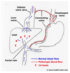
what is the function of the stomache?
name the 6 parts?
mostly storage untl ready to head into the duodenum since very little digestions actually occurs here
- cardiac region
- fundus
- pyloric region
- antrum
- pyloric canal
- pyloric spincter

what are the 3 segements of the SI? and major function?
whats one important thing that happens in teh first?
DIGESTION AND ABSORPTION
-
duodenum
a. contains opening for bile duct and main pancreatic duct - jejunum
- ilieum
what does bile break down?
what does pancreatic juices break down?
bile: lipids!!!
pancreatic juices: lipids, carbs, proteins
what are the two main functions of the LI?
what are the 8 parts?
STORAGE and WATER ABSORPTION
- cecum
- colon
- ascending
- transverse
- descending
- sigmoid - rectum
- anal canal
what are the four layers of the wall of the intestin and what are their functions?
4
1
2
1
- mucosal layer

a. changes shape to increase SA
b. produce mucous to protect and lubricate the GI tract
c. digestion and absorption
d. protection barrier against pathogens
- submucosal layer
vascular, lymphatics, nerves to supply tissue
- muscularis externa
a. contains both circular and longitudal laters
b. contracts to move food along GI tract
- serosa
single layer of cells that makes the mesothelium
what are the 2 different types of movement found in the GI system? characteristics?
2 each
- rythmic
a. moves food forward and keeps GI contents mixed up “oscillations”
b. present from esophagus to SI
- tonic
a. constant levels of contaction or tone without regular periods of relaxation
b. think always contracted like spincters and upper region of the stomact
what are the 2 types of cells of the GI tract?
characterstics?
- unitary

cells are electrically coupled so that signals can move quickly initiating SM contractions
“many cells that function as 1”
this is how you get things moving in 1 direction and make a smooth rythmic motion
- pacemaker
“slow waves” of interstitial cells
the resting waves are the pacemaker cells that keep the oscillations at slow waves and keep it primed
timulated by stretch, acetylcholine or parasympathetic then the AP is prompted and you get depolarization and contraction
don’t cause any contractions but just keep the tissue primed for when stimulated
enteric nervous system of the GI system

what is this and why is it unique?
2 divisions?
location of each?
functios of each?
intrinsic nervous system
means it functions on its own without influence from the brain or higher systems
- submucosal plexsus
a. controls the function of each section of the GI tract
b. takes in the signals from the mucosa in that specific region and adjust the motility, secretions, and absorption appropriately
between the mucosal and submucosal layers
- myenteric plexsus
a. linear chair along the muscular externa all the way down the GI tract, causing motility along the entire aspect

parasympathetic stimulation
what overall effect does this have?
what 2 nerves control this and what areas do they cover?
INCREASES FUNCTION!!
stomach-transverse colon= vagus nerve
transverse-rectum=pelvic nerve
sympathetic stimulation
overall effect?
inhibitory, slows it down
swallowing
voluntary/involuntary?
4 nerves that control the first
1 nerve control the second
whta are they?
VOLUNTARY MOST OF THE TIME BUT CAN BE INVOLVUNARY
ORAL AND PHARYNGEAL
TRIGEMINIAL 5
GLOSSPHARYNGEAL 9
VAGUS 10
HYPOGLOSSAL 12
ESOPHAGEAL PHASE
VAGUS 10

ORAL PHASE OF SWALLOWING
VOLUNTARY OR NOT?
WHAT HAPPENS IN THIS?
STARTS VOLUNTARY
bolus of food is in the mouth until the posterior tongue lifts it to the posterior wall
pharyngeal phase of swallowing
voluntary/involuntary?
when does this occur?
4 things that happen?
INVOLUNTARY
**point when food is at the posterior roof of mouth to esophagus**
- respiration halts so you don’t breath in food
- pharyngeal spincter relaxes
- larynx closed
- soft palate closes of the nasopharyngeal folds so food doesn’t go up into the nose
esophageal phase of swallowing
what are the two types?
where and when do they occur?
- primary peristalsis
upper 1/3
begins when food enters the esophagus
- secondary peristalsis
lower 2/3
where peristalsis realy occurs, occurs if primary peristalsis can’t handle the load
as it comes down it triggers stretch receptors so the spincters relax and let the food enter the stomach
dysphagia
5 things that can contribute
- lubrication loss-xerostomia
- size of bolus, poor mastication
- paralysis-stroke (aspiration)
- strictures (scar tissue in esophagus)
- cancer (obstruction)
gastric motility

function of peristalsis?
explain emptying process?when?
- peristalsis
a. used the make chyme
b. starts in the body and moves out to the antrum
c. as the chyme moves towards the antrum, the antrum contracts blocking the pyloric spincter so it doens’t exit the stomach and continues to be churned
2. emptying
a. chyme is empyting into duodenum between antrum contractions because the spincter is relaxed during this time, lets SMALL amounts out at a time!
rate of gastric emptying
explain the neural and hormonal controls of this?
3
2
- neural control
a. hypertonic solutions in duodenum: (indicates lots of particles already present, so digestion needs to occur first)
b. pH below 3.5 (indicates recent release of acidic stomach contents
c. presence of fatty acids, amino acids, and peptides (food)
2. hormonal control
a. cholecystokinin
b. glucose-dependent insulinotrophic peptides
**these are released in response to fats being released into the duodenum, so if these are elevated it means food as already been released from stomach so don’t want to release more**
explain two conditions that are cause the gastric emptying to be too slow?
4
3
- hypertrophic pyloric stenosis
a. very young in life, babies
b. thick pylorus so don’t get anything through
c. causes vomiting in the baby and can’t keep anything down
d. “grape” in pylorus
2. gastric atony
a. no tone so no contraction
b. stomach just sits there and is a problem with the nerves
c. causes: stroke, diabetes, surgical complications
what is a condition that can cause the gastric emptying to occur too fast?
dumping syndrome
-no control over pyloric spincter
often complication from gastric surgery
leads to:
diarrhea
higher chance of getting ulcers in the duodenum since higher amount of acid!
what the two types of motility in the SI?
- segmentation waves
- peristaltic movements

SI:
segmentation waves
’
what is this?
what happens in this?
what is the goal of this?
local mixing!
exposes more chyme to more surface area
circular msucle occludes the lumen and drives some of the contents forward and some backwards
**imagine squeezin a long balloon**
**mixing it up in one spot allowing for digestion**

SI:
peristaltic movements
what is this?
accomplished by?
goal?
stimulus?
contraction in proximal portion and subsequent relaxation in distal
accomplished by the myenteric plexus
goal: smooth muscle** **propel the chyme along the gut, moving it foward
stimulated by MORE CHYME! NEED TO MOVE IT ALONG!!!

what are two probles that can occur with SI motility? what might you ifnd with each othese?
2
3
- inflammation
a. increased motility
b. hyperacitve bowel sounds because the body wants to get it out
2. ileus
a. no motion or bowel movement
b. chyme just sitting there
c. see air fluid levels on xray (cmes from eating or bacteria)
colonic motility
what are the 2 types?
when does this occur?
what allows this to occur?
goals of each?
contracts longer here than in SI since more mass and less chyme ad this poinr and occurs 24-48 hours after ingestion
- segemental
a. HAUSTRATIONS: produce digging like movements to insure fecal material is exposed to mucosa
b. e-segment allows this to occur - propulsive
propels contents forward using LARGE segments!! close to 20 cm contract at the same time as one segment since it is mass instead of chyme that needs to be pushed along
lasts 10-30 minutes a couple times a day
explain the differences between the internal and external spincters?
what type of muscle?
voluntary/involuntary?
control?
internal
SMOOTH MUSCLE
INVOLUNTARY
external
striated muscle
VOLUNTARY
pudenal nerve control
what are the 2 reflexes that influence defecation?
- intrinsic myenteric reflex
stimulated by the local enteric nervous system from distention of the rectal wall with inititiartion of the preistaltic waves that push the fecal material into area
- parasympathetic reflex
sacral cord level
when nerve endings in the rectum are stimulated its feedback increases the peristaltic movements and relaxes internal spincter
explain the difference of what happens when it is an appropriate time to defecate vs inappropriate time to defecate?
appropriate time
the external sphincter is under conscious control by the cortex
when the rectum is stimulated from distention it sends messages to the cortex to relax the sphincter
if appropriate, cortex sends message to relax this sphincter
inappropriate time
same process as above BUT
if inappropriate cortex sends impulses to constrict external sphincter and inhibit efferent parasympathetic activity
keeps butt hole clenched
eventually this feedback loop fatigues and the urge to defecate stops
gastric hormones:
gastrin
what is this released by?
what does it cause?
what stimulates it?
produced by G-cellsin antrum and prompts gastric acid secretion, HCL
stimulus: vagus nerve from presence of food
gastric hormones:
ghrelin
what is this produced by?
what does it stimulate to be released?
what does it cause?
what are two things that stimulate its release?
produced by endocrine cells in the fundus and stimulates the release of growth hormone and causes stimulatory effect on food intake and digestive function with decreased energy expenditure
stimulus:
- nuitritional like fasting
- hormonal, decreased GH
what are 3 hormones that are secreted by the intestines?
- secretin
- cholecystokinin
- incretin hormones (GIP, GLP-1)
intestinal hormones:
secretin
what cells is this release by and why?
what causes its release?
what does it inbit and stimulate?
overall effect?
secreted by S CELLS** in the mucosa of the **duodenum and jejunum in response to acidic PH of the chyme entering the duodenum from the stomach
inhibits gastric secretion since this increases the HCL relase form the stomach
response:
prompts** **pancreas** **to release large quantities of fluid with high bicarbonate and low chloride to equalize the chyme (acid) that was released from the stomach

gastric hormones:
cholecystokinin (CCK)
what is this secreted by?
this is a?
3 things that it stimulates?
2 things that stimulate its release?
secreted by I cells and is used as a neurotransmitted
stimulates:
- pancreatic enzyme release (lipase-fats)
- contraction of gall bladder
- potentiates secretion of secretin to increase bicarbonate in repsonse to acidity of chyme in SI
Stimulated by:
chyme release into SI and products of protein digestion and long chain fatty acid

intestinal secretions:
incretin hormones
what do these 2 hormoens do?
what are the two hormones?
what are they secreted by and where?
4
2
increase insulin release after an oral glucose load /carb load decreasing blod sugar levels
- glucagon-like peptide (GLP-1)
secreted by L cells** in the **distal small bowel
supresses glucagon release
decreases gastric emptying
get cells to release insulin
- glucose dependent insulinotrophic polypeptide (GIP)
k cells in jejunum
decrease blood glucose by increasing insulin
what are the 5 regulations of GI secretions?
- pH
- osmolarity
- chyme
- hormones
- neuroregulation
salivary secretions
3 functions?
1 breakdown?
stimulated and inhibited by?
- lubrication
- antimicrobial-lysozyme
- digestion-amylase break down starches
stimulated by parasympathetic
inhibited by sympathetic
parietal cells
2 things it secretes?
in stomach
secrete:
- gastric acid HCL
- intrinsic factor needed for Ca++ absorption
chief cells
2 things it secretes?
- pepsin(ogen)
- gastric lipase
D-cells
1 secretion?
somatostatin (inhibits acid)
G-cells
1 secretion
gastrin (stimulates acid)
gastric lining
what is this? junctions? balance? 3
what is one major component that creates this? 3 functions
mucosal barrier
impermeable cell surface because of tight junctions
coupled secretions of H and HCO3, remains balanced
PROTECTIVE LAYER!!
prostaglandins
improve blood flow
decrease acid secretion
increase mucous protection
provides protection
what are 3 things that disrupt the gastric mucosal lining? how?
- ASA
penetrates the lipid layer and can damage the endothelium
- ETOH
lipid soluble so may disrupt mucosal barrier
- bile acids
job is to break down lipis so this breaks down the wall
**ALL OF THESE INHIBIT PROSTAGLANDINS WHICH HAVE A GASTRIC PROTECTIVE EFFECT SO THIS IS WHY YOU GET STOMACHE UPSET becuase H+ ions move into the cells an cause
- ischemia
- vscular stasis
- hypoxia
- necrosis**
what are the 3 things that stimulate the release of HCL and intrinsic factor from the parietal cells?
- gastrin from G-cells
- acteylcholine
- histamine

intestinal secretions:
Brunner glands
location?
funciton?
location: where the contents of the stomach and secretions from the liver ender the duodenum
function: secrete large amounts of alkaline mucous to neutralize the acid content from the stomach
intestinal secretions:
serious fluids
where do they come from? what is it? 3 functions?
secreted from crypts of lieberkuhn
secrere isotonic alkaline solution that acts as a vechicle for absorption
function:
- protection
- replaced erpithelial cells that have sloughed off
- antibacterial
what is the function of the surface enzymes in the SI?
aid in absorption,
secrete pesidases that split sugars
what are the 3 functions of the intestinal flora?
- metabolic-absorb nuitrients and help with energy
- trophic-growth and regeneration
- protection against foreign bugs
what are the 4 functions of the large intestine?
2 for the last
- anaerobes
- metabolic
- vitamin synthesis/absorption K, Mg
- protetion
a. mucous secretion
- protects linin and facilitates compaction of feces
b. bicarbonate secretion
-attaches to mucous and coats stool so the acid by products of bacteria in feces don’t damage the intestine
what are the 3 ways that digestions occurs in the SI? what are each of these?
1. hydrolysis
breakdown of one compound that involves a chemical reaciton with water
2. enzyme cleavage
requires enzymes to cut down substances into smaller pieces and is accomplished by
brush border enzymes
3. fat emulsification
breakdown of large globules of fat into smaller pieces
what are 3 things that contribute to the power of the SI to be good at absorption?
1. large surface area with vili
- brush border enzymes
secrete enzymes that digest proteins and carbohydrates
3. goblet cells
secrete mucous
intestinal absorption:
carbohydrates
what must they be broken down to before they can be absorbed?
expain this process? 5
must be monosacchrides before they can be absorbed in the small intestine
process:
- mouth: starch starts being broken down in the mouth by amylase
- pancreatic secretions also contain amylase
- amylase breaks down starches (carbs) to disaccarides
- dissacchrides are broken down into monosacchrides by brush border enzymes
- absorption in SI
what are the 3 common dissarcharides that must be broken down by brush border enzymes before being absorbed in the SI? what are they broken down into?
1. sucrose: glucose and fructose
2. lactose: glucose and galactose
3. maltose: glucose and glucose
intestinal absorption:
fats
explain three main steps of this and what is included in each step?
- emulsification

breaks down large globules triglycerides into smaller particles so that digestive enzymes can break down
stomach to duodenum
- pancreatic lipase
released in duodenum cleaves triglyceriddes into fatty acids and monglycerides
- micelles
made from bile salts and monoglycerides and transport products to brush borden enzymes to be absorbed, they are then transformed into chylomicrons that are triglyercerides and sent to the lymphatic system
protein digestion/absorption
explain where things occur
2
- pepsinogen
released y chief cells in the stomache in response to food, which ins then activated by the low pH to PEPSIN, then becomes inactivated in the duodenum from alkaline pH
- pancreatic enzymes trypsin
chymotrypsin
carboxypeptidase
elastase
malabsorption syndrome
get osmotic diarreah** with **fatty stool “steatorrhea”
sxs
muscle wasting
weight loss
failure to thirve
what are 6 things that can influence annorexia?
influences on appetite
hunger
hypothalamus
smell
emotional factors
drugs and disease
vomiting
5 ways this system can be triggered?
3 things these triggers stimulate?
limits the possibility of damage from ingested noixious agents by emptying contents of the stomach and portion of SI
triggers:
1. GI tract/organs
2. CNS system respinding to sights, sounds, or emotions
3. vestibular apparatus-mostion sickness
4. chemoreceptors-drugs or toxins
5. hypoxia
stimulates:
1. salivary centers
2. respiratory center
-stops breathing
3. abdominal muscles
- contraction/increased pressure
- LES relazation
gastroesophageal reflux disease
what is this?
population common in?
percent in US?
5 complications it can lead to
most common dxs of esophagous 15-20% US, common in pregnancy

transient relaxation of lower esophageal spincter LES leading to gastric acid reflux that causees damange to esophagus and spincter and can lead to:
1. esphagitits- 50% will get this!!!
2. esophageal stricutre
3. barrettes esophagous
4. esophageal adenocarcinoma
- hiatial hernia
-
GERD
what are 6 sxs?
1 thing to keep in mind about sxs?
- heartburn, restrosternal and postprandial
substernal pain/discomfort most commong 30-60 minutes after a meal
worsens when laying down or recumbant
- regurgitation (vomit burp)
spontaneous reflux of sour bitter gastric contents in mouth
- dysphagia (discomfort)
cough at night from acid asipiraiton
- reccurent pneumonia
- sxs temp relieved with antacids
- can radiate to arm/jaw
***keep in mind sxs don’t correlate with dxs progression so can’t tell how much damage has been done**
GERD
3 dx options
6 reasons of when it is not appopropriate to use the first line dx
- empirically first line unless (below)
- esphagogastroduodenoscopy(EGD) if high risk or tx has failed
a. over 50
b. weight loss
c. melena
d. odynophagia pain with eating
e. heavy alcohol or tabacco
f. non repsonsive to tx - modified/full barium swallow
what is the pathway for txing GERD?
6
- lifestyle adjustments
- OTC antacids-2 weeks
- H2 receptor antagonists
- Proton pump inhibitors
**if these fail EDG**
- prokinetics
- surgical

what are the lifestyle changes a patient should make to relieve sxs of GERD?
5
- avoid eating 2-3 hours before bed
2. elevate head of bed
3. loose weight
- avoid acidic food, chocolate, peppermint, ETOH, coffee
- stop smoking
why is it important to treat GERD?
prevent cancer aka barrette esophagus because the damange from acid makes this more likely to occur
what is the emergency cocktail you give someone in the ED for heart burn?
- benadryl
- lidocaine
- maalox
in child with asthma, what is one thing you want to look for? why?
look for GERD! asthma and GERD coexist in 50% of children with asthma dx
what is the most common cause of esophagitits?
GERD, 50% of patients with GERD have esophagitits
esophagitits
what is this?
5 general causes?
4 sxs
inflammation of the esophagus, esp in immunocomprimised
- viral
- bacterial
- paraistic
- abx induced
- radiation or chest cancers
sxs;
- odyniaphagia
- dysphagia
- substernal chest pain
- oral thursh
esophagitis
herpes liabilis (HSV)
3 SXS?
2 DX?
1 TX
N/V/ chills
herpetic vesicles on nose/lips
dx:
1. endoscopy showing small vesicles or superficial lesions
2. culture esophageal lesions
TX:
ACYCLOVIR 7-21 DAYS!!!
ESOPHAGITIS
VARICELLA-ZOSTER
1 dx?
tx?
N/V fever chils
DX:
endoscopy: vesicles or confluent ulcers
Tx:
- usually resolve spontaneously but can cause necrotizing esophagitits
- ACYCLOVIR!!!!
esophagitis
CMV
who does this occur in?
characteristics of ulcer? 2
3 sxs?
1 tx?
only occurs in immunocomprimised patients
CREEPING ULCER or can be GIANT ULCER
sxs:
odyniaphagia
persistent CP
hematememis
Tx:
IV GANCICLOVIR
esophagitis
candidia
who does it occur in?
3 complications?
1 dx?
1 tx?
occurs in immunocomprimised host
can cause complications:
- bleeding
- perforation
- stricture
dx:
endoscopy: small yellowwhite raised plaques
tx:
oral or IV fluconazole!!
barrett’s esophagus

how does this occur? what hcanges?
what are they at increased risk for? how much?
metaplastic changes in which the stratified squamous** is replaced by the **columnar epithelium that is typically found in the duodenum….extens poximally from LES from repeated exposure to acid esp with GERD (thats why the cells change to this type because its the same exposure the duodenum gets)
increases risk for adenocarcinoma 5-10%
this change increases risk for neoplastic changes/cancer 40-100 times greater than general public
barettes esophagus

2 dx rules?
3 tx?
DX:
EDG every 2 years with bx to check for neoplastic changes
if there is high risk dysplasia, consider surgrical resection
TX:
**more txing symtpoms unless surgical intervention**
- antacids
- H2 blockers OTCs
- PPIs
achalasia
what is this?
what is it caused by?
3 things you are at greater risk for?
4 sxs?
loss of peristalsis in distal esophagus and failure of the LES to relax** (LES tone increased) caused by **dennervation to LES
obstruction of esophagus from LES relaxtion failure from failed stretch receptors or nerves
increased risk for:
- aspiration
- irritation
- cancers
sxs:
dysphagia is most common
substernal cp
regurgitation
difficulty belching
achalasia

4 dx? 1 key word
4 tx options?
DX:
- CXR
- Barrium swallow showing BIRD BEAK APPEARANCE!!!
- manometry
- endoscopy
TX:
- nitrates/CCBs
2. botulinum toxin
3. pneumonic dilation with balloon procedure
4. myotomy

mallory-weiss tear
what is this?
what does it occur from?
who commonly seen in?
MC location?
3 RF?
2 sxs?
linear mucosal tear in the distal esophagus or gastric cardia from forceful vomiting or retching, causing hematemesis commonly seen in alcoholics

most common location: gastroesophageal junction
RF:
alcholic
hiatial hernia
eating disorder BULEMIA
SXS:
- multiple bouts of vomiting and retching followed by PAINLESS HEMATEMESIS
- abdominal pain
***keep in mind the bleeding usually stops spointaneously as teh condition is usually benign**
mallory weiss tear
1 dx?
2 tx?
DX:
ENDOSCOPY
TX:
-
stabliazation
- transfusion/gastric lavage if needed - control bleeding via endoscopy
**keep in mind most bleeding stops spontaneously and condition is usually benign**
in mallory weise syndrome what is somethign you want to do before releasing someone from hospital?
obtain occult negative stool to insure not bleeding still or hemmoraging
esophageal cancer
what are the two types?
frequency?
prognisis?
who is more common?
types:
- adenocarcinoma
- squamous cell carcinoma
prognosis typicaly poor, 5 year survival 10-13%
both appeare with equal frequency
males more common than females
esophageal cancer:
adenocarcinoma
where do you find this?
4 most important RF?
2 protective

distal esophagus
RF:
- reflux over 20 years
- Barrettes esophagus-almost all cases
- obesisty
- caucasion males
- smoking increases risk (not main)
Protective effects:
- fiber
- NSAIDS (seems counterintuitive)
esophageal cancer:
squamous cell carcinoma
prevalence?
location?
4 RF?
key point to remember?
prevalence is decreasing
middle esophagus
RF:
- smoking
- alchohol
- diet low in fruits and veggies
- achalasia increases risk 16 x
*** accounts for 90% of all squamous carcinoma in US***
esophageal cancers
sxs
1 early
3 late
SXS:
early:
1. transient “sticking” of food that turns to PROGRESSIVE DYSPHAGIA
later:
- retrosternal pain/burning
2.iron deficient anemia -loss from chronic cancer, but not enough to notice hememensis or occult
3. tracheobronchial fistula
late complication where the esophageal wall infilates the stem bronchus causeing intractable coughing with frequent pneumonia
_***if this occurs person has less than four weeks to live***_
in esophageal cancer, when would you expect to see the weight loss and dysphagia?
when the lumen is less than 13 mm
esophageal cancer
2 DX
2 STAGE
dx:
- barium studies
- endoscopy
Staging:
- CT OF CHEST AND UPPER ABDOMENT
2. PET SCAN
of the esophageal cancers, which is dxed more?
adenocarcinoma and squamous cell carcinoma are both dxed with equal amouth these days
what is the 5 year survival rate if dxed with esophageal cancer?
10-13% :-/
is smoking a RF for developing esophageal cancer?
yep! just like everything else!!!
where are adenocarcinomas and squamous cell esophageal cancers found?
adenocarcinoma: lower 1/3
squamous: middle esophagus
esophageal varices

what is this?
what causes this?
mortaltiy rate?
MOST COMMON CAUSE?
emergency!!!
dilation of the submucosal veins that develop in pts with portal HTN
patho: blood flow through the liver is diminished causing blood flow increase through the microscopic bood vessesl within the esophageal wall and the vessels dilate profoundly, and then continue to dilate until they are large enough to rupture
patient acutely ill, mortality rate 40-70%
MOST COMMON CAUSE OR PORTAL HTN IS CIRROSIS that causes this

where is the most common stie for esophageal varices? why?
distal esophagus at gastroesophageal junction because veins are most superficial here!!
what is a random syndrome that can cause portal HTN?
budd chiari syndrome
esophageal varices
6 sxs
3 dx
sxs:
1. hematemesis!!! over 50% stop bleeding spontaneously
- melena
- tachycardia
- hypotension
- syncope
- jaundice
DX:
1. emergent endoscopy
CBC
BUN/creatine
type and cross
esophageal varices
4 immediate tx options
- immediate tx-stop bleeding because mortaltiy approaches 75%
- visceral ligation- rubber band
- sclerotherapy
- balllon tamponade
esophageal varices
long term tx considerations
5
- abx
- decrease portal HTN BB/nirates
- shunts
- liver transplant
- STOP ETOH
what is boerrhave syndrome
esophageal rupture
oropharyngeal dysphagia
what is this?
what does it ususally come from?
diffiiculty inititating the swalling reflex
usually comes from neuromuscla disorders that cause weakness or lack of coorindatio of the muscles involved in swallowing
Ex: CVA, parkinsons
esophageal dysphagia
what is this?
where?
arises within the body of the esophagus, LED, or cardia most commonly from mechanical or motility disturnances
ex: stricuture, radiation
odynophagia
what dioes this present as?
reflect what?
substernal sharp pain on swalloing
usually reflects SEVERE erosive disease
esophageal spasm
what is this?
what is a specific subset of this? qualification?
2 tx options
motiltiy disorder usually associated with CP or dysphagia
NUTCRACKER ESOPHAGUS: increased pressure over 180 during peristalsis which creates the dysphagia
DX:
mamometry
TX:
- nitrates (relaxes)
- CCB diltiazem (relaxes)
esophageal manometry
what is this test and what are 2 things it is helpful in dxing?
measures the pressure in the esophagus, signifying the effectiveness of peristalsis
used frequently for achalsaia (NO PERISTALSIS) and NUTCRACKER ESOPHAGUS (hyper peristalsis/contractions)
esophageal ring
what is this?
sxs?
dx?
1 tx?
ring of tissue located at the gastroesophageal junction called
schatzkis ring
sxs:
- dysphagia with foods, but not typically liquds
DX:
barium esophagram
TX:
mechanical dilation with balloon

esophageal web
what is this?
where?
1 simple example of this?
1 dx?
1 tx?
mid to upper esophagus
membranes of squmous mucosa that causes intermittent dysphagia with solid food
plummer vinson syndrome-WEB WITH IRON DEFICIENT ANEMIA AND GLOTTITIS
DX:
barium esophagram
Tx:
mechanical dilation with balloon
pearl: in eldery patient esp after a stroke who is getting reccurent pnuemonia, what should you always check?
modified barium swallow
nissen fundoplication
what is this procedure used to tx?
how does it work?
laproscopic procedure used to treat GERD or hiatial hernia
- gastric fundus is wrapped around the lower end of the esophagus
- stablaizes this area and spincter and prevents hernia and GERD

hiatial hernia
what is this?
what are the sxs?
tx?
occurs at the LES where the upper part of the stomach moves into the upper chest and through the small opening in the diaphram
the diaphragmatic hiatus acts as an additional spincter around the lower end of the esophagus
SXS:
produces symptoms of GERD or dysphagia
TX:
surgical

systemic scleroderma
what is this? what are the 5 common presentations? what is the #1 think you worry about in this? what test do you do in the lab? what are the treatments?
thickening and harderning of the skin via collagen deposition
- raynauds (75%)
- vascular changes in nail bed
- GI dysmotility “watermelon stomache”
- puffy hands
- fixed face
*****WORRY ABOUT PULMONARY FIBROSIS AND ACUTE RENAL FAILURE*******
DX: ANA-SPECKLED
Tx: treat system effected
renal-ACE inhibitors
raynauds-calcium channel blockers
Gi: promotility
lungs: cyclophosphamide

CREST Syndrome
what is the pneumonic to remember the symptoms and what do you need to monitor annually in these patients?
LIMITED SCLERODERMA
C- calcinosis of joints leading to puffy hands
R- raynauds
E-Esophageal dysmotility
S-sclerodactyly of MCPs
T: telangiectasis
**complication=pulmonary hypertension so need to get annual PFT/DLCO to make sure no lung fibrosis**
Tx: symptoms

what do you need to avoid in schleroderma pts because it can cause a RENAL CRISIS?
high dose corticosteroids.
don’t do it!
gastritis

what is this?
5 causes?
2 sxs
dx
inflammation or irritation of the gastric lining
causes:
ETOH
H. pylori
NSAIDS
STRESS
autoimmune
SXS:
often asymptomatic
initial presentation may be GI blleeding
“coffee ground, melena, emesis
DX:
endoscopy
differentiates from PUD
gastritis-ETOH
how do you tx this?
nausea, dyspepsia, hematemesis
TX:
H2, PPI, or sucralfate
4 week tx
gastritis-H. pylori
how presents?
2 things associated with?
usually symptomatic
cofactor for PUD
two things associated with:
1. gastric adenocarcinoma
2. B-cell gastritis-mucosal associated lymphoid tissue (MALT)
NSAID gastritis
common?
recognized early?
2 tx? (how)
VERY COMMON 25-50%
MOST GO UNREGOGNIZED BECAUSE OF NO SXS
TX:
1. D/C NSAIDS
2. TX EMPIRICALLY IF ON NSAIDS
3. PPI 2-4 WEEKS
stress gastritis
who do you find this in?
because?
3 causes?
1 tx?
prophylaxsis
common in ICU patients
develop quickly in this population
caused by decreased mucosal flow
Seen in:
- trauma
- respiration failure/mechanical ventilation
- coagulation problems
prophylaxisis in high risk: PPI oral or NG tube to decrease bleeding
tx: PPI
stomach neoplasms
where do these occur?
appearance? 4
sxs?
3 dx?
1 tx
occur in antrum, MC in lesser curvature
bulky, irregularly shaped
firm, jagged edges
usually asymptomatic till late disease
early detection is therefore difficult
DX:
- barium swallow xray
- endoscopic studies with bx
- cytologic (screening in atrophic gastritis/polyps)
TX:
surgery-radical subtotal gastrectomy TOC
what are 6 RF for stomach neoplasms?
- genetic predisposition
- carcinogenic diet
smoked food/perserved food
- autoimmune gastritis
increased inflammation
- gastric adenomas
- polyps
- h. pylori, cofactor for some
Peptic ulcer disease

what this this?
2 causes?
1
3
size?
break in the gastric or duodenal mucosa that extends through the muscularis mucosa that comes from
1. impaired normal mucosal defense factors
NSAIDS
2. defense factors overwhelmed by aggressive luminal factors
acid
pepsin
infection
greater than 5 mm in diameter
what are the 5 common causes of PUD?
- NSAIDS
- H. pylori
- idiopathic
- hypersecretory states
- smoking
3 locations of PUD?
which one is most common?
how long do they take to heel?
what do you need to keep in mind that is very important depending on location?
- duodenal
a. MOST COMMON LOCATION!!
b. 90% heal in 4 weeks
2. pylorus
3. gastric
**CAN BE MALIGNANT**
**must get bx at endoscopy time**
a. take longer to heal 8 weeks!
b. increased length of tx
peptic ulcer disease
NSAIDS
cause?
why?
4 RF?
2 tx? length of time?
caused by long term NSAID use esp nonselective COX1 and COX2 blockers
COX1 decreases prostaglandins which have a protective effect on gastric mucosa, it the lack of this impairs gastric mucous and HCO3 secretion
**this is why COX2 selective are better option since decrease risk of bleeding**
RF:
- ASA
- corticosteroids
- over 60
TX:
1. PPI or H2
***4 weeks duodenal***
***8 weeks gastric***
2. D/C NSAID
peptic ulcer disease:
h. pylori
KEY?
characteristic?
acute/chronic characteristics?
what is key about this?!
nesscary cofactor for 75-90% of duodenal/gastric ulcers
characteristics:
PRODUCE UREASE
ACUTE:
a. infectious “gastroenteritis”
CHRONIC:
a. ASYMPTOMATIC
b. DIFFUSE SUPERFICIAL MUCOSAL INFLMMATION WITH POLYPS
**ERRADIACATION IS ESSENTIAL OTHERWISE 85% WILL RECURR!!!!!**
what is a RF for all ulcers?
smoking!
explain the patho for duodenal ulcer with h. pylori?
6 steps
- H. pylori infection increases acid production
- causes gastric metaplasia in duodenal bulb
- H. pylori infection
- causes duogenitis
- causes mucosal breakdown
- causes duodenal ulcer from breakdown
explain te patho of stomach ulcers caused by h. pylori?
- infection in body
- causes gastritis and chronic inflamation that overwhelms immune system
- causes mucosal breakdown
- creates gastric ulcer
PEPTIC ULCER DISEASE
2 sxs
3 dx
when do you use each?
1 think must do?
SXS
- epigastric like pain “hunger like” in 80-90%
- 50% have relief by eating antacids within 2-4 hours
**physical exam is often unremarkable so need to watch for changes in pattern, indicating perforation or penetration**
DX:
- endoscopy EDG TOC!
allow so for visulaization and also bx!!
- fecal antigen test (noninvasive)
- c-urear breath test (noninvasive)
****OPTIONS 2 AND THREE AT THE TOC FOR ACTIVE PUD AND PROOF OF ERADICATION*** MUST D/C PPI 7-14 DAYS PRIOR OR MAY GET FALSE POSITIVIVE!
peptic ulcer disease
tx without h. pylori
2
3 for first
1 second
- antisecretory agents
A.PPIs 1st DOC-inhibit 90% acid secretion
ompreazole
lansoprazole
B. H2-inhibit histamine mediated secertion
C. OTC anti-secretory
- mucosal defense agents
2nd line can be used as adjunct for symptom relief
sacralfate, busmuth
peptic ulcer disease:
Treatment for H. pylori
what is the tx regimen?
what must you do after tx and when?
2/3 abx, PPI, +/- bismuth

1. amoxicillin 1 g BID
2. clarithromycin 500 mg BID
3. HIGH DOSE 40 mg PPI BID
***no reason shouldn’t use quadrople therapy see in this pic** KNOW BOTH
**must confirm eradication with C-urea breath test or fecal antigen test
4 weeks post abx
2 weeks post PPI***
peptic ulcer disease:
post H. pylori tx tx
2 considerations
DUODENAL f still large or bleeding post tx:
continue PPI for 2-4 weeks
GASTRIC if large ot blleding post tx:
continue PPI for 4-6 weeks
in reccurent ulcers what must you do!?
rule out H. pylori and NSAID use
peptic ulcer disease:
complication!
bleeding ulcers
what percent of people?
times increase in mortality?
2 sxs?
2 dx?
4 tx? but keep in mind?

occur in 10-20% of pts with PUD
**may be the first sign of an ulcer**
REBLEEDING INCREASES MORTALITY 10X
SXS:
hematemensis
melena
DX:
endoscopy!
predicts liklihood of rebleeding
HgB, Hct
TX:
80% OF THESE STOP BLEEDING SPONTANEOUSLY
- isotonic fluids to replace volume
- endoscopy can help stop bleeding
- HIGH DOSE PPI to decrease bleeding
- surgery in extere
peptic ulcer disease:
COMPLICATIONS
PENETRATION/PERFORATION
results in?
2 sxs?
tx?
results in chemical peritonitis
a. severe generalized abdominal pain
b. rigid abdominal rebound
TX:
1. laproscopic perforation closure
2. increased RX TX
peptic ulcer disease:
refractory ulcers
what is this caused by?
3 contributing factors
uncommon, contributed to non compliance with medication
Contirbutory factors:
- cigs
- NSAIDS
- failutre to eradicate H. pylori! MUST DO THIS
gastroparesis
what is this?
issue with what?
5 causes?
2 sxs?
3 dx?
3 tx?
delayed stomach emptying caused by partial paralysis of the stomach, so food remains in the stomache for abnormally long time
vagus nerve issue
causes:
- DM
- bulemia
- parkinsons
- abdominal surgery
- cigarettes
SXS:
1. chronic nausea
2. fullness after just a few bites
- vomiting
DX:
- xray
- mamometry
- gastric emptying scans
TX:
1. dietary changes, lowe fiber, low residue diets
2. SMALLER MEALS
- antidepressant mirtazapine
pyloric stenosis
who do you find it in?
m/f?
caused by?
2 main sxs?
2 dx?
1 tx?
*2 key buzz words**
children 4-6 weeks old
more comme males
gastric outlet is blocked by pyloric hypertrophy
SXS:
1. progressive projectile vomiting in child who remains hungry
- oval shaped mass right of the umbilicus, esp right after eating!!
DX:
US
barium swallow with “STRING SIGN”
TX:
SURGERY!!!!!
zollinger-ellison syndrome
what is this?
by the time of dx…?
rate?
what can it cause?
3 locations?
met site?
gastrinoma that secretes large amounts of gastrin so causes recurrent and refractory gastric ulcers, 1/3 have mets by dx
SLOW GROWTH
the increase in gastrin increases the osmoliarty of the lumen so it draws water out and can cause diarreah
locations:
- pancreas
- duodenum
- lymph nodes
metastasizes to liver
zollinger-ellison syndrome
what are the 3 sxs?
3 dx options?
2 tx depending if isolated or mets?
SXS:
GERD
diarrhea
malabsorption weight loss
DX:
- serum gastrin over 500
- pH less than 3
- somatostatin receptor scintigraphy endoscop US
finds primary tumor and mets
tx:
isolated primary tumor: PPI and resection
mets: PPI high dose
what percent of gastinomas are malignant?
2/3
gallbladder

2 functions
when is it filled?
when is it released?
- STORES BLADDER
- CONCENTRATES BILE
filled when the spincter of oddi is closed
contracts in response to cholecystokinin to release bile into the duodenum
bile

what does it do?
3 things it is made of?
digests fats in SI, terminal ileum, and recycled to liver for reexcretion
CONTENTS
- bile salts (from cholesterol)
- bilirubin (waste product RBC)
- alkaline fluids
cholecystic
referring to gallbladder
cholecystitis
inflammation (chronic or acute)
cholithiasis
stones in GB
choledocholithiasis
stone in the bile duct
cholecystectomy
surgical removal of GB
pancreas
exocrine portion
2 cell types?
which more common?
things it secretes?
3
1

ducts enter the duodenum
ENCOMPASSES:
- acinar cells-95% of pancreas
1. secrete proteolytic enzymes including trysinogen, chymotrypsinogen, procarboxypeptidases for protein digestion
- amylase for carb digestion
- lipase-fat digestion
- ductal epithelial cells
a. alkaline secretion to decrease the acidic pH of the duodenal contents
cholecystalgia
pain (aka billiary colic)
pancreas
3 location descriptors
during development
2 things contains
in retroperitoneum
head near duodenum
tail posterior to stomach
during development two pancreat buds come together to fuse
CONTAINS:
exocrine portion
endocrine portion
what are the most common cells in the pancreas?
acrinar cells
pancreas:
endocrine portion
where do secretions go?
what are the cells made into?
what are the 3 cell types??
secretion into the blood stream
- islets of langerhans
“islands of endocrine cells” 1-2% of pancreas
beta-cells: insulin synthesis/secretion
alpha cells: glucagon
deta cells: somatostatin, inhibits GH and TSH
tumor markers:
ca 19-9
pancreas
tumor markers
ca-125
ovarian cancer
tumor markers
AFP
liver cancer
tumor markers
CEA
colon cancer
acute pancreatitis
6 sxs
where is pain?
severity?
what makes worse/better?
pain with…?
two key signs you dont want to miss?
- epigastric radiates to back
- billiary colic PERSISTS DOESN”T GO AWAY
- worse SUPINE, BETTER LEANING FORWARD
- PAINFUL INSPIRATION, SHALLOW BREATHS
- TACHYCARDIA
6. hemmoragic pancreatitis
cullens sign: periumbical ecchymosis
grey tuner’s sign: flank eccymosis

tumor markers
PSA
prostate
explain the pathophys of pacreatitis?
what are 2 theories that cause this?
- inflammation of the pancreas causes damage to the acrinar cells
- the inactive proenzymes like trysinogen are activated early while still in the pancreas
- early activated lipases disolve the fat
- so the pancreatic enzymes start to DIGEST THE PANCREASE “EATS ITSELF”
theories:
- obstruction of the pancreatic duct due to stones
- failure of the two parts of the pacreas to fully join during development pancreatic divism
acute pancreatitis
what are 2 MC causes? account for what? 6 others?
causes:
- gallstones 45%-MC
- alcohol 35%
**these two are most common and make up 80% of cases**
other causes:
trauma
drugs (iatrogenic)
obstruction (tumors)
infections (mumps)
metabolic (hyperlipidemia)
toxins (methyl alcohol, scorpion stings)
**MOST RECOVER AND HAVE NORMAL FUNCTING PANCREASE**
when dxing pancreatitis and looking at labwork, what test is most effective?
lipase
(this is better than amylase)
acute pancreatitis
5 labs tests?
TOC for dx?
LAB TESTS:
elevated cbc (infection)
elevated lipase (pancreas)
elevated LFT
elevated glucose
decreased calcium
TEST OF CHOICE FOR DX: CT SCAN!!!
scored A-E, A is good, E is bad
acute pancreatitis
5 labs tests?
TOC for dx?
LAB TESTS:
elevated cbc (infection)
elevated lipase (pancreas)
elevated LFT
elevated glucose
decreased calcium
TEST OF CHOICE FOR DX: CT SCAN!!!
scored A-E, A is good, E is bad
acute pancreatitis
6 tx options?
what is the process dependent on?
mostly supportive

a. NPO 48-72 hours NO ALCOHOL
b. ERCP if presense of stone** **on CT scan
b. maintain hydration
c. pain control
d. NG tube in extreme
e. abx if infectious
f. incision and drainage if infected/necrosis
chronic pancreatitis pathophysiology
what is this?
what should you think about when thinking of this?
what happens?
inflammatory disease of the pancreas
irreversible** **changes occur
can lead to permanent loss of function
HINT: think necrosis fibrosis theory of alcoholic pancreatitis
alcohol to tissue damage to fibrosis to partial obstruction
chronic pancreatitis
what is the MC cause of this?
3 others?
NOT CAUSED BY WHAT?
what can this lead to?
- alcohol cause #1!!
***THIS IS NOT CAUSED BY GALLSTONES LIKE ACUTE**
- obstructive causes of spincter of oddi by neoplasms
- malnuitirion
- metabolic
chronic pain leads to opoid addiction! keep this in mind
chronic pancreatitis
4 dx options
fat stranding
- amylase/lipase NORMAL

body gets used to working under these circumstances
- secretin stimulation test “gold standard” for early chronic pancreatitis dx
- plain xray films QUICK AND CHEAP-see calcifications
- CT SCAN-better at seeing calcifications but $$/rad
“arrowhead and fat stranding”
chronic pancreatitis
4 sxs?
sxs:
- epigastric abdominal pain that radiates to the LEFT
- insufficiency of pancreatic function leads to
a. steatorhhea damages decreases lipase
b. diabetes once islets damaged
3. asymptomatic and found incidently
chronic pancreatitis
5 tx options
most important!!
2 drugs?
- _stop the alcoho_l!!
- controls sxs
a. pain control
b. prancreatic enzyme supplements viokase and pancrease
- endoscopic duct decompression
- surgical draingage of duct
- celiac plexsus nerve block
what are 4 things that that increase persons chance of bad outcome/mortality with pancreatitis?
- organ failure
- pancreatic necrosis
- obesity
- old age
what are the two criteria used to identify the mortality with pancreatitis?
- Ranson’s criteria
Take home: higher numbers means more severe disease and increased risk of death
-measured at time of admission and during first 48 hours
6+=50% mortality
- APACHE II score
**typically impatient ICU use**
over 8 is severe!!!
pancreatic cancer
what are thet wo tumor markers?
survival?
1 RF?
tumor markers:
- CA19-9 85%
- CEA 40-45%
5 year survival is only 4% really bad
only RF: chronic pancreatitis
pancreatic cancer

4 sxs?
toc? 1
sxs:
- painless jaundice is PC until prove otherwise
- virchows node-supraclavicular node
- trouseaus signs- recurring superficial thrombophlebitiscan be anywhere
- weight loss
TOC: CT scan dual phase helical

pancreatic cancer
what two lab findings supposrt this?
3 tx options?
LABS:
1. elevated direct bilirubin
2. elevated alkaline phosphatase
(significantly elevated in cancer)
TX:
1. chemo-poor respinse only reduces size to buy little time
- whipple surgery-only at cancer specialties
- pallaitive care
acute cholecystitis
what happens in this?
what is most likely to cause this?
leads to 3 things? possible worst case?
2 key sxs?
the cystic duct** becomes blocked **most commonly from gallstone!
less common:
cancer, sludge, infection
blockage causes distention and edema that cauess ischemia, necrosis, perforation and possible generalized sepsis
SXS
- RUQ/epigastric pain radiate to shoulder/scapula
- murphys sign (tenderness and pt stops inspiration on palpation)

acute cholecystitis
2 dx tests? when to use?
2 labs?
TOC #1: US
find gallstones and wall thickening over 3 mm
TOC #2: HIDA nuclear med scan (cholescintigraphy) GOLD STANDARD and BEST TEST
****not usually used since it is $$$$$$$, so US becomes TOC******
Two elevated labs:
elevated gamma-glutamyl transpeptidase
elevated bilirubin
acute cholycystitis
TXOC?
3 others?
TreatOT: TAKE IT OUT!! MOST COMMON!! via laproscopic cholecystectomy
- leave in if very mild
- drain it if patient too sick
- abx if elevated WBC

cholelithiasis
two types of stones?
percentage?
what do they look like?
1. cholesterol stones
MC-75% of stones!!
don’t show up on xray
- calcium bilirubinate/ca salts
25% of stones
pigmented black/brown sludge stones

explain 3 pathphys concepts of cholelithiasis?
one population more at risk
- cholesterol supersaturation of bile
- nucleation
cholesterol comes together and crystalizes, propogates
- GB hypermotility
slower emptying=more time for stones for form
THINK PREGNANT WOMAN!!!
acute cholithiasis
what do you need to keep in mind about sxs?
how do they present? 3
dx TOC?
what do you need to keep in mind?
asymptomatic in 50-60%

symptomatic patients:
biliary collic with RUQ pain/epigastric areas
refers to back, scapula or R shoulder
DX:
TOC: transabdominal US
shows stone without wall thickening
**keep in mind not great for stones in common bile duct**
what are the four RF for cholithiasis?
- over 40
- obesity (or rapid weight loss)
- female/pregnant
- native american
“FEMALE, FAT, FORTY, FERTIL” 4, F’s
acute cholelithiasis
what are you 3 tx options and who are they appropriate for?
- cholecystectomy-MOST COMMON “it hurts
- leave in if doesn’t hurt UNLESS high risk to keep in
a. diabetics
b. sickle cell
c. Native americans with calfieiced “porcelain” stone - dissolve cholesterol stones with actigal, urodeoxycholic acid but $$$
acute ascending cholangitis
what is this?
why is it important?
sxs? KEY
infection and inflammation of the billiary tract COMMON BILE DUCT
obstruction then infection
potentially life threatening
CAN LEAD TO SEPSIS OR SHOCK!!!!
sxs:
charcots triad
- abdominal pain
- jaundice
- fever
*KNOW IT*

acute ascending cholangitis
what are the 3 things seen on labs?
what must you make sure to do?
2 tx options?
LABS:
- leukocytosis
- hyperbilirubinemia
- alkaline phosphatase increasing
******MAKE SURE TO ORDER BLOOD CULTURES X2**
TX:
1. ABX TO COVER GRAM NEGATIVE ANAEROBES, ENTEROCOCCI
penicillin
aminoglycoside
2. REMOVAL OF OBSTRUCTED STONES ERCP
choledocholithiasis
where is the stone?
where does it come from?
where is pt?
sxs? 4
stone in the COMMON BILE DUCT
usually migrate from the gallbladder
patient very sick and likely in ICU!! compared to cholelithiasis
sxs:
asymptomatic in 30-40%
BILLARY COLIC
jaundice, pancreatitis
choledolithiasis
what are the two dx/tx options?
what must you keep in mind?
- MRCP magnetic resonance cholangiopacreatogprahy ONLY DX

- ERCP-endoscopic retrograde cholangioancreatography DX AND TX so GOLD STANDARD!!
****keep in mind contraindicated in pancreatitis/cholecystomy***
niacin
2 functions
4 sources
1 presentation
FUNCTION:
- energy
- fat metabolism
Soucres:
- tuna/ salmon
- chicken/beef
Presentation:
FLUSHING
thiamine
4 sources
2 functions
4 presentations
SOURCES
- pork
- grains
- dried beans
- peas
FUNCTION:
carbohydrate metabolism
nerve function
PRESENTATION
berberi
nervous tingling
poor coordination
edema
cardia dysfrunction
vitamin A
4 sources
2 presentations
3 toxicities
SOUCRES
- liver
- fish oils
- fortified milk
- eggs
CLINICAL PRESENATION
night blindness
dry skin
TOXICITY
- skin disorders
- hair loss
- teratogenicity
riboflavin
4 sources
1 function
2 presentations
sources:
- milk
- spinach
- liver
- grains
functions
ENERGY
presentation
oral inflammation
eye disorders
vitamin C
4 sources
3 functions
3 presentations?
SOURCES
1. citrus berries
2. starberies
4. broccoli
4. greens
functions:
- collagen synthesis
- hormone function
- neurotrasmitter synthesis
PRESENTATION!
scurvy
poor would healing
petechiae
bleeding gums
what does the toxicity of vitamin C cause?
diaareah
vitamin D
1 source
2 functions
2 at risk
source
FORTIFIED MILK
functions
CALCIUM REGULATION
CELL DIFFERENTIATION
at risk:
elderly
and those without sun exposure
vitamin D
2 preseentations
3 toxicitiy
rickets
osteomalacia
toxicity;
- hypercalemia
- kidney stones
- soft tissue deposits
vitamin K
4 sources
1 function
1 presentation
2 toxicity
sources:
- liver
- green leafy veggies
- broccoli
- peas
- green beans
functions
clotting
presentation:
bleeding
toxitiy presents as:
1. anemia
2. juandice
folate
4 sources
1 function
4 presentations of deficiency
sources
- green leafy veggies
- organe juice
- grains
4 organ meats
RF:
PREGNANCY!!!!!
presentation:
- megaloblastic anemia
- sore tongue
- diarreah
- mental disorder
appendicitis
what is this?
MC cause?
2 risks?
3 presentations?
inflammation of the appendix causing leakage of cecum contents and blockage MC common due to fecalith
RISK OF SEPSIS AND DEATH IF RUPTURES
SXS:
1. sudden onset of abdominal pain
- umbilicus progresses to RLQ pain
- pain to palpation
appendicitis
5 physical tests to do to confirm?
2 tests to do to confirm suspicion?
TESTS:
- rovsings sign
- obturatory sign
- psoas sign
- mcburneys
- rebound tenderness
- CT-inflammation
- surgical consult
appendicitis
what is the tx for this?
what is the controversy concerning this?
surgical tx is standard of care
*****abx in recent studies conclude that 80% could be txed IV but 25% required emergency removal of appendix within first year post op….SO NOT IDEAL***
constapation
what is the qualification for this classification?
what are the 7 causes?
difficult passing stool
qualification:
difficulty in over 25% of attempts for over 3 months with NO obstructive or peristalsis disorder
causes:
nervous system disorders
neural transmitter malfunction
hormone malfunction
heighten immune response
poor diet
poor activity
mediation Se
constapation
what must you rule out?
2 DX options?
3 tx options?
must r/o obstruction, impaction

DX:
- DRE (r/o obstruction/impaction)
- xray for bowel gas pattern
TX:
- diet
- increase fluid intake
- activity
- medication evaluation
constapation
what are the 3 classes of drugs you can use to tx this?
what is the function of each?
**add the drugs from table**
- bulk forming laxatives
- absorbs water and increases fecal mass
- Fiber
2. ostmotic laxatives
causes H2O retnetion in the stool (pull water into the stool)
- PEG
3. stimulant laxatives
increase acteylcholine regulated GI motility peristalsis
-senna
celiac disease
what type of condition is this?
what is the patho of this and the specific component?
nickname?
autoimmune dx of SI
introlerance to gluten and the alpha-gliadin
gluten triggers immune response in turn causing damage to the small intestine
causes nuitritional deficits secondary to absorption issues
“celiac sprue”
celiac disease
what are 6 sxs that come with this?
explaination of the last?
- unintended weight loss

- persistently underweight
- sxs of poor nuitrition
- “failure to thrive in child”
- diarreah
-
dermatitis herpetiformis
a. intense itching
b. blisterning rashes
c. 10-15% of patients
celiac disease in children
when does this start?
4 causes in children
2 things at increased risk for
GI issues start when there is gluten introduced to the diet
irritablitly
anxious
distention
abdominal pain
high risk for:
- dehydration
- hypokalemia due to increased fluid loss
celiac disease
what are the 3 dx options of this?
key thing to remeber?
- anti-tissue transglutaminase antibodies OR anti-endomysium IgA antibodies
- bx if small intestine and blood + for antibody
****if they have dermatitis herpetiform with + antibody skin bx, THEN NO NEED FOR INTESTINAL BX***
celiac disease
what are the 2 tx for this?
what about screening?
- gluten free diet
- wheat
- rye
- barley
2. abx for dermatitis herpetiformis
***make sure to screen if family hx using blood test for autoantibodies**
diverticula
what are these?
how are they formed?
sac-like protrusions in the colonic wall
blood vessels penetrate the mucosal wall
internal layers penetrate through weak areas creating diverticula
diverticulosis
what is this?
where does it most commonly occur?
what is important to know about this?
presentation?
2 things to avoid?
condition of having divericula
typically asymptomatic
MC in SIGMOID COLON, account for 50% of all lower GI bleeds, bright red blood in rectum earliest finding MOST COMMON
diet restricted in
seeds, strawberries
diverticulitis
what is this?
3 sxs?
2 tests?
inflammation of the pocuhes
- severe LLQ pain
- change in bowel habit
- constapation
Tests:
- CT TOC with IV and oral contrast
- WBCs
- barium enema
- colonoscopy
who is diverticulosa most common in?
2 percents?
western/industerialized societies
30% by 60
65% have by age 85
what are 3 RF for diverticulosa?
- connective tissue disorders
- low fiber diet
- too little exercise
what are the two conditions that make up inflammatory bowel disease?
- crohns disease
- ulcerative colitis
crohns disease
3 populations common in
- common in 15-35
- jewish population
- increased risk with family hx
Inflammatory bowel disease
crohns disease
where are the lesions?
how deep are lesions?
where is the pain?
3 things that can occur as complications?
4 things that are common?
3 other things it can effect?
bowel appearance?
- legions mouth to anus; spread out with healthy tissue between
2. transmural
3. RLQ pain MC
- fistulas, abscesses, fibrotic stricutres
- bleeding common
- weight loss common
- obstruction common
- perianal disease common
- can effect skin eyes joints liver
- large volume diarreah and mucous (trash bag)

what 2 are tests you want to do to look for crohns?
- anti-saccharomyces cervisial (ASCA)- 44% of crohns are pos
- colonoscopy TOC
what are the 4 tx options for crohns?
- 5-aminosalcyclics (5-ASA)
-mesalamine
-sulfasalazine
- corticosteroids
- cimzia-certolizumab
- humira-adalmumab
**surgery doesn’t work since it involves the entire GI tract**
which is more common crohns or ulcercerative colitis?
ulcerative colits 3x more likely
what groups of people does ulcerative colitis effecT?
- ages 20-30
- caucasions and jewish descent
- family hx is strongest RF
ulcerative colitis
where is this?
pain presents?
what is most common and worse?
what does person have increased?
what might they develop?
describe lesions and how deep?
what does the colon look like on colonscopy?
what are 3 things aren’t present?
- primarily localized in colon
2. LLQ
3. bleeding from rectum is most common
- urgency need of BM
4. potenital anemia due to blood loss
- lesions are continuous starting at the rectum and in mucosa only
- colon wall thin with continuous inflammation
- no granulomas
(weight loss/obstruction very rare with this)

what do you use to dx ulcerative collitis?
colonoscopy
what are the 3 tx options for ulcerative collitis?
- 5-ASA
- sulfasalzine
- mesalamine - corticosteroids
- colonectomy CURE best choice
removal of colon
irritable bowel syndrome
what is this?
who most common in?
age?
pathogenIsis? 3 causes
dx of exclusion, no other cause
FUNCTIONAL BOWEL WITH MUCOSAL INFLAMMATION
women more than men
presents before age 45
1. abnormal motiltiy
2. visceral hypersensitivity lower visceral pain threshold
3. psychosocial interaction-emotional stress
irritable bowel syndrome
3 tx options?
4 medications tx options
- pt education and behavior/ emotional support
- dietary therapy
- pharm
a. antispasmotics
b. antidiarreals
c. psychotropic
d. serotonin receptor agonists
mesenteric ischemia
what are the two categories? sxs associated with
2
1
what are 5 causes?
Acute (embolus)
SUDDEN SEVERE ABDOMINAL PAIN
FEVER
or chronic (athlerosclerosis)
ABDOMINAL PAIN POST EATING
causes:
1. CVD impacting flow
2. trauma
3. neoplasm
4. physical obstruction
5. systemic illness-DM, sickle cell, PVD, even dehydration
mesenteric ischmia
2 dx methods?
1 tx
DX:
- mesenteric angiogram TOC
- CT or abdomen with contrast-bowel wall edema
Tx:
SURGERY with stent or emboli removal

ischemic colitis
what is this?
who get its?
inflammation and injury to the large intestine resulting in decreased BF
most common in elderly patients
small intesine neoplasms
when are these found?
common?
4 types?
dx? 2
NOT TYPICALLY DETECTED TILL LATE STAGES!!!
*rare.*
98% of adenocarcinomas, carcinoids, sarcomas, and lymphomas
often not dxed until metastisized to other parts of the body!!
sxs vague and non specific
DX:
- colonoscopy with bx TOC!!
- CT
what is the most common GI cancer? in US?
COLORECTAL MOST COMMON GI CANCER!!! 3rd most common in US for males and females!!
colorectal cancer
what are 3 risk factors?
- genetic predisposition
- presence of adenomatous polyps)
3. diets high in fats and refined carbs that are low in plant fiber *industerialized world*
colonrectal cancer
4 sxs
2 key dx
3 others
sxs:
- colic type pain
2. anorexia
3. thin appeareance
4. pallior/anemia
dx:
- xray: classic apple core/npaking ring appearance
- endoscopy with bx
- iron deficient anemia
- hypoalbuminemia
- occult blood in stool

what does colonrectal cancer come from?
the progression of adenomatous polyp into malgnancy
obstruction

5 causes
explain last 3
4 sxs
- tumor
- foreign body
- paralytic ileus-trauma, surgery, infection, metbaolic disease with DM
4. volvulus-twisting of intesinte
5. intusssception-telescoping of intestine
sxs;
- severe abdominal cramping
- inability to pass stool
- increased bowel sounds first, then decreased
- abdominal swelling, distention
obstruction
3 dx
3 tx
- abdominal xray
- CT
- barium enema
tx:
- NG tube (relieve pressure)
- relieve obstruction
- surgery often needed
what are four complications from obstruction?
tissue death
perforation
sepsis
death
intussusception
what is this?
who most common in?
2 causes?
3 sxs?
2 dx children and adult
1 tx?
telescoping of the intestines
MC in children, but also in adults with obstruction or neoplasms
- currant jelly stools
- sausage like mass felt on abdominal palpation
- coliky pain
DX:
children: barium enema
adult: CT is best
TX: SURGERY
toxic megacolon

what is this?
typically caused by? and 3 others?
what are 2 risks worry about?
acute toxic colitis that causes dilation of the colon
typically complication of inflammatory disease most commonly
- ulcerative colitis
- infectious causes
- ischemia
- pseudomembraneous causes
**risk is sepsis and death**

what are the requirements to dx toxic megacolon?
4
4
must contain 3 of 4 of these
temp over 101.5
HR over 120
leukocytosis over 10.5
anemia
and 1 of these
dehydration
altered mental status
electrolyte abnormality
hypotension
toxic megacolon
5 sxs
dx, and finding?
SXS:
- abdominal pain
- bloating
- fever
- tachycardia
- LOSS OF BOWEL SOUNDS
DX:
xray, dilation of colon over 6 cm in transverse colon
toxic megacolon
4 tx options?
- decompress bowel immediately
- if not successful, colectomy
- abx and steroids
- tx fluid and electrolyte imbalances
lactose intolerance
what is this?
4 populations common in?
5 sxs? key 1?
difficulty digesting lactose from lack of lactulase
MOST COMMON IN:
- AFRICAN AMERICANS
- HISPANIC/LATINO
- ASIAN AMERICANS
- NATIVE AMERICANS
SXS:
- bloating
- pain
- increase passage of gas
- diarrhea and nausea
5. 2 hours post consumption of milk or dairy
lactose intolerance
1 dx?
1 tx?
dx:
hydrogen breath test
tx:
dietary avoidance
angiodysplasia

what is this?
why does it happen
3 sxs?
swollen fragile blood vessels in the colon so considered vascular lesions
patho:
aging and degen of the structure of blood vessels causes formation of arteriovenous malformation
often the cause of GI bleed in elderly not attributed to other causes
1. maroon/melena colored stool
- pallor
- SOB from anemia

angiodysplasia
3 dx?
3 tx options?
DX
- colonscopy
- CT scan
- angiography
TX:
- majority stop bleeding without intervention
IF NOT
2. cautery via colonscope
3. clotting through angiography
what is the dentate line?

senstion?
divides the rectal mucosa from the squamous epithelium in the canal
above in anorectal canal: INSENSATE (no pain)
below in andoderm: SENSATE AND PAINFUL!!
what does the dentate line divide?
3
- nervous system
- vascular supply
- nervous system
**most important, divides where a patient can feel and where they can’t so important when determine sxs!!**

anal fissure
what is this?
where do these occur?
signicant stat?
2 causes?
Tear in the andoderm of rectal canal
90% are posterior along midline
most common cause of painful rectal bleeding
causes:
- passage or hard stool
- prolonged diarreah

a kid with tearing pain with bowel movements and bright red blood when he or she whipes with toitlet paper
shuld mak eyou think of…
ANAL FISSURE!!
anal fissure
3 sxs?
what not to do?
sxs:
- “tearing” pain with BM since below dentate line
- hematochezia during BM
- SPREAD BUTTOCKS to examine
**NEVER DO RECTAL EXAM, TOO PAINFUL!..and cruel**
anal fissure
2 tx categories
4
1 (also, who not to do in)
- 90% heal without tx
a. bulk agents
b. stool softeners
c. sitz baths
d. hydrocortizone ointment Anusol
2. surgical-lateral internal anal spincterotomy GOLD STANDARD if failure conservative
**can’t do if patient incontinent**
rectal abcess
where do these most commonly occur?
where does it come from?
4 causes?
most in posterior rectal wall, originate in the crpyts with gland obstruction

s. aureus
bacteroides
proteus
strep
rectal abcess
2 causes
3 steps
- crypt gland obstruction
- increased muscle tone causing obstruction
STEPS
- stasis
- dilation
- infection

once a anal abcess in a crypt has formed…where can go if infection spreads? 4
most common?
- superficially to external spincter to make perianal abcess most common
- deep through the external spincter into fat of ischioretal fossa
- deep into supralevator space
- interspincteric
who are rectal abcess most common in?
how do they present?
5 (3 key)
MEN!!!!!!!!!!!
- sever pain while sitting
- drainage on PE
- palpable fluctuant mass
- worse with coughing, sitting, defectatin
- really really painful DRE

rectal abscess
how to dx? 1
how to tx? 2
outcomes?
DX
- CT-size and location, fistula presence
TX:
- surgical drainage if not fistula
- post op sitz bath
50% will be cured
50% will have fistula formation
fistula in ano
“rectal fistula”
6 associations?
- crohns
- GC procitis
- carcinomas
- hodgkins lymphoma
- radiation fibrosis
- immunocomprimised states
what do you need to remember if someone has an abcess or resctal fistula?
DO NOT DO DRE!
anorectal fistulas
Goodalls rule
what does it tell you?
2 rules?
**used to determine the location of the internal opening of the fistula**
anterior external opening
goes to interally via straight line
posterior exernal opening, tracts internally via curved line

anorectal fistulas
2 sxs patient presents with
1 tx?healing?
*be careful of**
- embaressing persistent discharge
- not painful but itchy
TX:
- fistulotomy (opening tract)
healed by secondary intention with granulation tissue
left open to heal on its own
***watch out for spincter!! must preserve its function**
fecal impaction
what is this and when typcailly does it occur?
2 key sxs?
PE? 1
TX 3
large mass of dried hard stool typically after chronic constapation
SXS:
1. sudden watery diarreah in patient that has had cronic constapation
2. straining with passage of liquid or small stool
PE:
DRE shows hard mass or dry stool in vault
TX:
1. removal manually
2. prevention with stool softeners, colase, bulk (fiber) and H20
3. high fiber diet
what is the most common anorectal complain in adults over 50?
hemmoroids
what percent of people over 50 have hemmoroids?
50%`
explain the pathophysiology of hemmoroids?
5
- anal canal is lined with “cushions” that are vascualr and connective tissue and make the hemmorrhoidal plexsus
- exist in 3 columns
- cushions encorge during defecation to profect the anal canal from abrasion
- when venous engorgement is increased with conditions like pregnancy, straining, and increased abdominal pressure it prompts production of abnormal hemorrhoidal tissue which can be symptomatic
what are 6 RF for hemmoroids?
constapation
straining at stool
pregnancy
obesity
chronic liver disease
portal HTN
internal hemmoroids

4 key sxs with these!
above the denate line!!
veins surrounded by mucosa
- PAINLESS, above denate
2. bright red bleeding with defecation
3. may prolapse and be palpable on DRE
4. bleed
what are the stages used to define internal hemmoroids?
first-bleed
second-bleed and prolapsed, spontanously reduce
third-bleed, prolapse, and require manual reduction
fourth degree-bleed/incarcerate

Internal hemmoroid tx categories
5
2
1-2 degree:
a. fiber
b. water
c. stool softner
d. anusol hydrocortisone
e. numbing agent nupercainal ointment
3-4 degree:
a. SURGICAL
b. EXCESIONALHEMMOIROIDECTOMY
external hemmoroids
whe do they occur/what are they covered in?
3 sxs
dx method?
1 tx?
below the dentate line and covered with andoderm
- usually don’t bleed
- may thrombose which is VERY painful
- cause pain, discomfort and most severe at time of defecation
dx
“either present or not”
tx:
- excision of outside of the mucotaneous junction leaving wound open hemroidectomy

rectal polyps
what are these?
why is it improtant to know type?
1 type concerned about?
why?
2 shapes?
small outcropping that grows in the rectum or colon
**need to deterine the type since some are associated with carcinoma**
adenomas
PREMALIGNANT POLYP
PRECURSOR TO COLORECTAL CANCER
sessile: flat and intimately attacted to mucosa
peduclated: round and attached be stalk

what are the 3 types of adenoma polys? which is most common? which has higest risk of cancer? risk of cancer and prevalance for each?
- tubular adenoma
MOST COMMON TYPE 65-80%
pedunculated, little cancer risk
- tubulovillous adenoma
10-25% of adenomas
22% risk of cancer
- vilous adenomas
40% RISK OF CANCER
only 5-10% so least common but most deadly
what is the least common but the most prognostic for cancer of the adenoma polyps?
vilous adenoma
*think vilian=evil*
what are the two tests that are reccomended annually to screen for colorectal cancer?
- guiac fecal occult blood test (gFOBT)
- immunochemical-based fecal occult blood test (iFBT)
what are the 3 screening tests that are reccomended for colorectal cancer? which is most reccomended? what age do you start?
age 50 to less than 10 years life expectancy
- optical colonscopy-10 years
- flexible sigmoidoscopy-5 years
- CT colonography-10 years
when should you screening african americans for colorectal cancer?
45 rather than 50 according to the college of gastroenterology
what are the sxs associated with polyps/colorectal cancer?
they might bleed
more important to think about family hx
pilonidal cyst/disease
what is this?
what does it look like?
location?
who is it in? age? KEY!!!!!!
common, congenital abnormality
“opening of a sinus tract that may conttain a tuft of hair“
location:
midline, post sacral intergluttal fold superoir to anus
ALMOST ALWAYS MEN 20-30, always under 40

pilonidal cyst/disease
4 key sxs?
1 tx otpion? why?
- fluctuant mass with erytmatous “halo”
- purluent d/c
3. NO ANAL PAIN OR DEFECATION ISSUES
- painful but in gluteal area
Tx:
1. MOST LIKELY SURGERY with secondary closure (leaving open) vs ID with abx
HIGH RATE OF RECURRANCE SO SURGERY IS BETTER OPTION TO GET DEEPER TRACTS!!!
incisional hernia
associated with vertical incisions esp in pts with wound infection or obesity

tx: surgery!
inguinal hernia
3 types?
where are they?
which is most common?
- direct

passage of intesine through external inguinal ring at hesselbachs triangle
- indirect
MOST COMMON
passage through inguinal canal INTO SCROTOM, often
3 femoral
less common through femoral ring
umbical hernia
when do they get it?
how is it txed?
congenital and appears at birth
most resolve on their own

vental hernias
when does this occur?
occur when weakening in the anterior abdominal walla and can be either incisional or unilical

portal vein

lacks what?
internal pressure?
supplies what percent of blood
formed from?
valvless
pressure 3-5 mmHg
75% of the livers total blood supply by volume
formed by the superior mesenteric artery
hepatic vein

where does it go?
structure?
2
3
2
venous drainage of liver through 3 valveless hepatic veins into inferior vena cava

right: 5, 8
middle: 4, 5, 8
left: 2, 3
neural innervation of the liver
2
- parasympathetic of vagus nerve
- parasympathetic from the celiac plexsus
explain the 2 cels in the liver and when you see them?
parenchyma is made up of actively diviiding hepatocytes
in damaged tissue this becomes fibrotic** and you will see **oval cels indicating damage
what are the 3 main functions of the liver? subgroups?
3
4
1
- metabolic and catabolic
a. glucogenesis
b. synthesis of phospholipids and cholesterol
c. detoxification of meds and alchohol
- storage
glycogen
protein
iron
vitamins
- excretory functions
synthesis and secretion of bile
what are 6 synthetic functions of the liver that occur in the hepatocytes?
- coagulation factors: PT/INR, PTT
- plasma proteins: albumin
- acute phase proteins
- carbohydrate metabolism
- lipid metabolism
- bilirubin metabolism: bile excretion
what are the two tests that suggest hepatocellular damage/inflammation?

- AST
- ALT

what 3 LFTs suggest obstructive disease?
- bilirubin
direct=conjucated
indirect=unconjugated
- ALP/alkaline phosphatase
- GGTP/gamma-glutamyl transferase *ordered sepereately
conjugated bilirubin=
direct bilirubin
unconjugated bilirubin=
indirect bilirubin
asartate aminotransferase
(AST)
elevated in?
specific?
elevated in acute conditions

found in the muscle, kidney, and heart so low specificity for liver damage since mreasures more than just the liver
alanine aminotransferase
(ALT)
elevated in?
specific?
elevated in chronic conditions

primarily found in the liver so more specific than AST
if the ration of AST:ALT is greater than 2:1 ration, what should you think of?
alcoholism or drug toxicity
unconjugated (indirect) bilirubin
what is this?
if increased indicates? 3
broken down RBC in speel bind to albumin and go to hepatocytes

if increased indicates:
PREHEPATIC PROBLEM
1. hemolysis of RBC
2. imparied hepatocyte function
direct bilirubin (conguated)
what happens to this?
where does it usually go?
if increased indicates 2 things?
enzymatically conjugation occurs with glucuronic acid…goes into bile…into small intestine where aprox 95% is reabsorbed

if increased:
INDICATEDS POSTHEPATIC PROBLEM
1. obstruction of biilary system=CHOLESTASIS
is bilirubin in the urine common?
no, it should not be present in the urine!!
alkaline phosphatase
where is this found?
found in the liver, intestine, kidney, and placenta
therefore, not specific to the liver
gamma-glutamyltransferase
what does this indicate?
what does this help rule in/out?
liver specific
**helps differentiate if the liver is the problem if the alkalne phosphatase is increased**
if you have increased alk phos and increased GGTP….what should this make you think?
OBSTRUCTION
what are the first and second most common causes of chronic liver disease?
- hep C
- alcohol
gilberts disease
what is this?
why does it happen?
2 key sxs?
most common hereditiary cause of increased bilirubin
impaired enzymatic conjugation of Indirect bilirubin
sxs:
- jaudice at birth
- icteric sclera
gilberts disease
what are the 3 tests done to dx?
1 tx option?
DX:
- elevated INDIRECT bilirubin
2. normal direct bilirubin
normal US
tx
only in infancy with “bili lights” to prevent liver failure
nonalcoholic fatty liver disease
(NASH)
6 causes
- certrain drugs
- obesity
- starnation
- diabetes melltius
- high blood tryglicerides
- alcohol
nonalchoholic liver disease
(NASH)
what are the sxs?
3 dx findings? 1 key
what should you consider if more than more one abnormal finding?
sxs
NON UNLESS CIRROSIS
DX
1. increased echotecture at times HSM on US
2. AST/SLT increased sometimes alk phos
- ALT more than AST
(opposit of ETOH)
**CONSIDER BX IF MORE THAN ONE POTENTIAL CAUSE OF ABNORMAL LFTS**
non alcoholic fatty liver dxs
5 tx options?
TX
- lifestyle: weightloss, lowfat diet
- management of high triglycerides, BS
- decrease ETOH
- meds/supplements
- vitamine E,
- ursodiol
hemochromatosis
what is this?
who does it present in?
sxs? 4
3 important labs for dx
autosomal recessive causing iron deposition in orgams
OVER 50 years
often asymptomatic
can include:
arthalagias
hepatomegaly
gray skin
DM
Labs
1. high ferritin and iron % saturation
2. HFE gene analysis
3. liver bx with iron stain
what is the tx for hemochromatosis?
phlebotamy, remove the iron
wilson’s disease
what is this?
what happens in this?
3 things it causes?
2 labs must check
rare autosomal recessive disorders
liver unable to exrete copper, so get copper overload in brain, bone, kidney, cornea
- basal ganglia sxs (parkinson like)
2. liver disease
3. kayser-fleicher rings
brown on green pigment in cornea
Labs.
- LOW CERULOPLASMIN
2. 24 hour urine copper

wilsons disease
what are 2 key sxs?
1 tx option?
sxs:
1. visual disturbances
from the kayser fleischer rings get brown ring causing visual disturbances
2. neurologic/psychiatric changes
TX:
- copper chelators like zinc
alpha 1 antitrypsin deficiency
what is this?
what 2 organs does it effect?
2 effects?
tx?
genetic condition effecting lung and liver
alpha-1 antitrypsin (A1A) synthesized in liver resulting in reduced circulating levels
increase in A1A in liver=obstruction
decrease A1A circulation=obstructive lung disease
tx:
replace A1A
alchoholic hepatitis
what is this?
what is the ratio you see?
2 labs might see?
most common cause of cirrohosis
AST>ALT 2:1 rarely over 300
labs:
- vitamin and calorie deficiency
- megaloblastic anemia (folate, B1, B6)
what is the ration associated with alcoholic hepatitis?
AST greater than ALT 2:1 rarely over 300
acute phase alcholic hepatitis
associated with what?
how serious?
3 sxs of this?
what must you calculate?
typically with heavy use or binge
can be fatal, pts often encephalopathic
sxs:
- HEPATOSPLENOMEGALY
2. ASCITIES
3. JAUNDICE
**calculate discriminant function***
over 31=poor 1 month prognosis
what is consider tylenol OD?
what precent of acute hepatitis?
serious?
tx?
single or total dose exceeding 10 grams
accounts for 45% of cases of acute hepatitis
fatal if untreated
tx:
- N-acetylcysteine
use this if hepatoxicity or ingestion time unknown
autoimmune hepatitis
who is this common in?
2 labs to check?
2 biomarkers to check?
1 test needed to dx?
2 tx options!
most common in females 14-50 WHO MOST LIKELY HAVE ANOTHER AUTOIMMUNE CONDITION
LABS:
- elevated enzymes
- elevated alk phos
BIOmarkers:
- ANA
- anti-smooth muscle
Dx:
need bx to make dx
tx:
prednisone and imuran
primary biliary cirrhosis
what is this strongly associated with?
what is key finding on testing?
test?
2 tx options?
75% cases are patients with ulcerative colitis, males
“beading” of bile ducts on MRCP (MRI)
Tx:
1. ursodiol
2. transplant cure!
if person has family hx of colorectal cancer, when do you start screening?
start 10 years prior to the dx of your 1st degree relative and do every 5 years from there with colonoscopy
acute hepatitis
what is the mortalty?
3 most common causes?
what are 4 sxs associated with this? KEY!!
mortality 40-80%
sudden onset deterioration of hepatocyte function causing coagulopathy
- tylenol 45% of cases
- alcohol and drugs
- heptitis A and B
sxs:
1. jaundice
2. elevated transmidates AST/ALT
3. coagulopathy INR over 1.5
4. encepalopathy-alteration in mental status due to evelated ammonia
viral infections account for what percent of hepatitis?
50% of all cases
hep a b c d e account for 95% of thse
hepatitis A
precentage?
virus type?
incubation?
transmission route? (3)
65% of all cases
RNAoccurs exclusively in liver cells
incubates 2-6 wks
transmission FECAL ORAL
- travel
- contanimated food and water
- close contact with infected individuals
hepatitis A
sxs?
1 (time)
7 after sxs
SXS
- prodrome, flu like
most infectious here 12-21 days
2. icteric phase
1. dark urine appeares first (bilrubinuria)
2. pale stool follows
3. jaundice 70-85%
4. abdominal RUQ pain 40%
5. prurits, indicates bilirubin
- arthralgias
- hepatomegally
hepatitis A
3 dx findings
tx
DX:
- anti-hep A IGM and IGG
- LFTS
- increase AST/ALT over 1000
- increased bilirubin 5-10 x
TX: selflimiting supportive
what are 5 prevention methods for hepatitis A?
improvement in hygiene and sanitation
cooking food
avoidance of water foods and endemic areas
avoidance of raw shellfish
immunization
who gets vaccinated for hep A? 4
Harvix
travelers
miliary personnel
lab workers
immunocomprimised
when you think of raw shellfish think..
hep A!
hepatitis B
what type of virus?
what percent of people have it and who?
3 transmission pathways?
KEEY THING TO KNOW ABOUT THIS
DNA virus
1/3 of population infected, majority immigrants or 1st gen
transmission:
- perianatal
- sexual
- blood containing med equiment
*******causes 80% of hepatocellular carcinoma=fatal!!!!*****
what type of hepatitis can lead to hepatocellular carcinoma?
HEPATITIS B!! vaccines for this mandated
what are the categories of hepatitis B?
3
immunity
- vaccination
- natural infection
actue infection
chronic infection
- active
- chronic
***what are the 2 test results you want to keep in mind when looking at hepatits infection***
hepatitis B surface antibody (HBsAB)=IMMUNITY–ONLY PRESENT IN VACCINATED PEOPLE

hepatitis B envelope antigen (HBeAg): NEEDS TREATMENT, current infection
surface=
core=
envelope=
surface=immunity (vaccine or exposure)
core=exposed to virus
envelope=present infection with active replication
acute hepatitis B
what is the breadown for how they present? (%)
1 key dx? 2 supportive?
tx2? why?
70% are subclinical (no jaundice or aniteric)
30% get icteric hepatitis
dx
1. HBcIGM for dx
- elevated AST/ALT
- elevated bilirubin
TX:
- supportive
**many will seroconvert to HBsAB and HBeAB meaning they develop immunity on their own!!**
- high calorie diet
chronic hepatitis B
three phases of this?
3 tx options and length of time?
initial phase:
positive HBeAG
immune clearance phase
HBsAg, HBeAG
elevated ALT
inflammation on liver bx
inactive carrier
tx:
- tenofovir DOC
- entecavir DOC
***both of these are lifelong drugs!!!***
- interferone alpha 6 months
hepatitis prevention and vaccination
2
- recombinant hepatitis vaccine
- hep B immune globulin (exposued)
what is key to know about hepatitis D?
transmission?
RNA coinfection with HEP BE IS REQUIRED!!!
HBsAg
transmission: sexual contact
hepatitis C
what is this?
3 RF/transmission?
virus type?
age?
most common chronic blood borne infection in the US
- injection drug use/cocaine
- sexual contact
- transfusion
RNA virus, 30-49 y/o
**what important people do you need to test for hepatitis C?**
test everyone born between 1945 to 1965
what percent of people proceed to the chronic state? what are two things you are at increased risk for?
80% proceed to the chronic state
HIGH RISK OF CIRRHOSIS AND HEPATOCELLULAR CARCINOMA
hepatitis C
3 tx options?
when do you start tx?
2 main options?
how long is the tx?
- supportive first since can clear on their own
- avoid hepatotoxic drugs
- **most wait 6 months for posisble clearance before starting antiviral therapy**
- harvoni (sofosbuvir/ledipasvir)
- daklinza (daclatasvir)
**12-24 weeks tx depending tx naive and if cirrhosis**
**some regimens still include ribaviran, but not all of them** NO INTERFERON
what do you want to check when txing someone for hep C?
viral load after 12 weeks to confirm cure
what is cool about treating hep C?
IT IS CURABLE!!! nearly 100!!!!!
there is no vaccine and no post exposure immunoglobulin THEREFORE YOU WANT TO SCREEEN!!!
what is interesting about hep G?
if coinfected with HIV, helps reduce the HIV replication
HEP E
2 places you find this?
transmition
AT RISK POP?
North Africa
South Asian
fecal oral
self-limiting
issues in pregnacy!!! EXTREMELY SEVERE!!!! esp in third trimester 20% mortality!!!!!! KEY
cirrhosis

what is this?
2 classifications?
chronic end stage dxs of the liver marked by degeneration of cells from inflammation resulting in fibrous thickening of tissue
4 stages of fibrosis and cirrhosis is the last
compensated: although fibrotic can still preform functions
decompensated: fibrotic with loss of essential function
cirrohosis
4 sxs of compensated?
7 sxs of decompensated?
compensated sxs:
ammenorreah
impotence
gynecomastica
hematemesis as present features in 15-25% (esophageal varices)
decompenstated:
1. spider angiomas
2. muscle wasting
3. palmar erythema
4. dilated superficial veins of abdomen
5. ascities
6. portal HTN
7. encepalopathy
cirrohsis
5 labs
3 tests
LABS:
low platelets
prolonged PT
moderate elevations enzymes
elevated bilirubin
low serum albumin
1. abdominal US WITH DOPPLER
- nodular appearing liver +/- hepatosplenomegaly
2. LIVER BX! TOC to confirm, determine staging
cirrhosis
5 tx
- alcohol abstinence
- vitamin supp
- nuitrional supp
- BB for portal HTN
- CURE IS TRANSPLANT!!!
what MUST you do for someone with cirrhosis?
_*****must screen for alpha fetal protein and imaging ever 6 months***_
what is used to stage liver disease?
what does it determine?
MELD SCORE
**determines prognosis of patient in order to determeine priorty of patient for transplant**
hepatocellular carcinoma (HCC)
what is this strongly associated with?
5 year survival?
3 sxs? 1 key
3 dx methods
one key think you want to order
associated with cirrhosis
4-6 months from time of dx
5 year survival is 25%
SXS:
- painless jaundice
- weight loss
- hepatomegaly
dx
- triple phase CT or MRI
- percutaneous bx of lesions!!! TOC
3. Alpha fetoprotein tumor marker is PATHOPNEUMONIC
hepatocellular carcinoma
4 tx
- surgical ressection
- chemo
- portal vein embolizations
- transplant if small and localized
metastatic liver lesions
what is this?
3 ways it gets there?
3 dx methods?
3 tx options
MOST COMMON NEOPLASM OF THE LIVER!!!
- portal venous circulation
- direct extension
- lymphatic spread
DX
- triple phase CT or MRI
- Percutaneous bx of lesions
- PETS scan and tumor markers for primary tumor
TX:
- ressection
- chemo
- not option for transplant
hepatic ressection
how much can you take?
can remove up to 80% because it regenerates!! woah!
regains albumin ability by 3rd week
portal HTN
what are the 3 classifications of this?
5 things it can cause?
- presinusoidal: slpenic or protal vein
- sinusoidal: cirrhosis
- postsinusoidal: hepatic veins, ouflow problem
CAN CAUSE:
- esophageal varices
- ascites
- encephalopathy
- splenomegaly
- hepatorenal syndrome

what are the most common PORTAL HTN locations?
presinusoidal and sinusoidal
what is basically the only cause of postsinusoidal portal HTN?
budd-chiari syndrome
portal HTN
5 tx options depending on cause
- nonselective BB

- banding for varices
- diuretics for ascites
- lactulose and xifaxan for encephalopathy
- surgical if meds fail
TIPS-transjugular intrahepatic portosystemic shunt