General GP (SJS) Flashcards
What is the difference between a papule, a nodule and a pustule?
Papule - A palpably raised lesion which is less than 1cm in diameter
Nodule - A palpably raised lesions which is more than 1cm in diameter.
Pustule - Pustules result from accumulation of large numbers of leukocytes in the epidermis or upper dermis
Describe the pathogenesis of acne?
Disease primarily affects the pilosebaceous units of the head and neck.
- Primary lesion is increased formation of keratin within the hair follicle itself.
- Excess keratin blocks the pore and forms a micro-comedome
- Bacterial lipases from propionibacterium acnes (G+) convert lipids into fatty acids, which in combination with the excess keratin drive an inflammatory reaction
- Inflammatory reaction leads to further plugging of the pore - and further inflammatory changes
- The enlarging pore is called a closed comedone or whitehead
- This structure can rupture, releasing pro-inflammatory exudate and causing inflammation of surrounding tissue, leading to papules, nodules and pustules.
What is the management of acne?

What is the management of eczema?

What is this?
What’s the treatment?

Infected eczema
Staphylococcus aureus = “impetiginisation”
Soak off crusts
Topical mupirocin or oral fluclox
What is this?
What is the treatment?

Herpes simplex virus = “eczema herpeticum”
Admit to hospital
What is this?
What is the teatment?

Pompholyx eczema
Pompholyx is a common type of eczema affecting the hands (cheiropompholyx), and sometimes the feet (pedopompholyx).
Same treatment as normal eczema
What is this?

Discoid eczema
What is this?
What is the treatment?

Asteatotic eczema
Common on legs of older people
Same treatment as normal eczema
What is this?
What is the treatment?

Lichen simplex is a localised area of chronic, lichenified eczema/dermatitis.
It is usually somewhat linear or oval in shape, and markedly thickened. It is intensely itchy.
Lichen simplex is often solitary and unilateral, usually affecting the patient’s dominant side.
Potent steroids

What causes erythema multiforme?
Infections are probably associated with at least 90% of cases of EM. In order of frequency:
HSV 1
Mycoplasma pneumonia
Other viruses
Medications are an uncommon cause.
Describe the lesions of erythema multiforme
Maculopapular skin lesions forming plaques
Few to hundreds of skin lesions erupt within a 24-hour period.
The lesions are first seen on the backs of hands and/or tops of feet, then spread along the limbs towards the trunk.
Mildly itchy or burny
Lesions typically have 3 zones (red rim, clearance zone, and central blister or erosion)

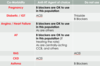
What is the treatment and prognosis of erythema multiforme?
For the majority of cases, no treatment is required as the rash settles by itself over several weeks without complications.
Treatment directed to any possible cause may be required such as oral aciclovir (not topical) for HSV or antibiotics (e.g. erythromycin) for Mycoplasma pneumoniae.
Supportive care:
- oral antihistamines or topical steroids for itch
- mouthwashes containing local anaesthetic and antiseptic reduce pain and secondary infection in patients with involvement of the oral mucosa
Prognosis
Erythema multiforme usually resolves spontaneously without scarring over 2-3 weeks for the EM minor form, and up to 6 weeks for EM major. However it often recurs.
What is the management of impetigo?
In non-remote community settings:
Suspect S. aureus as the pathogen.
For localised skin sores, use:
mupirocin
For multiple skin sores or recurrent infection, use:
di/flucloxacillin 500 mg
In remote community settings in central and northern Australia
Suspect S. pyogenes as the pathogen.
benzathine penicillin
What is the treatment of lichen planus?
Treatment is not always required, but if so consider referral for potent and ultrapotent topical steroids.
What is this?

Lichen Planus

What is this?
What causes it?
What is the treatment?

Pityriasis rosea
We think there may be a viral cause but no-one knows
It will clear up in 6-12 weeks. Dark discolouration of skin may take longer to resolve. It doesn’t normally reccur but it can.
If itchy you can use topical corticosteroid ointment or calamine lotion
What is this?

Psoriasis - Guttate subtype
What can aggravate psoriasis?
Streptococcal tonsillitis and other infections
Injuries such as cuts, abrasions and sunburn
Obesity
Smoking
Excessive alcohol
Stressful event
Medications such as lithium, beta blockers, antimalarias, NSAIDs
Stopping corticosteroids
What does psoriasis look like?
Red, scaly plaques with well-defined edges and symmetrical distribution – usually not itchy.
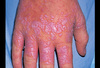
What is this?

Pustular psoriasis
Usually on hands/feet
Often without usual plaque psoriasis
May be painful or “burning”
What are the possible complications of psoriasis you should keep in mind?
No good data on the prevalence of psoriatic arthritis in patients with psoriasis, circa 10%
Patients with psoriasis have 2-3x cardiovascular risk of patients without psoriasis!
What is the management of psoriasis?

What is this?

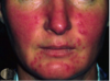
Rosacea is a common persistent eruption of unknown aetiology. It is characterised by central facial erythema, visible blood vessels and acneiform papules and pustules. It is typically chronic and persistent with a fluctuant course.
What is the management of rosacea?
General management
- Apply cool packs if severe
- Minimise factors that cause flushing or irritation (see aetiology)
- Sun protection plus sun avoidance measures are essential
- Use an emollient soap-free cleanser, combined with a low-irritant sunscreen to reduce irritation
- Some people may use a green-tinted foundation to mask erythrotelangiectatic features
Topical therapy – for mild erythema and inflammatory lesions
- 2% sulphur in aqueous cream tds OR
- Metronidazole gel bd OR
- Azelic acid gel OR
- Clindamycin 1% solution bd OR
- Erythomycin 2% gel bd
Long-term maintenance with topical metronidazole is often used to control rosacea and prolong remissions.
Systemic antibiotics – for more severe cases or when topical therapy unsuccessful
- Doxycycline 50 to 100mg daily
- Erythromycin 250 to 500 mg twice daily
- Minocycline 50 to 100mg daily
What is the typical presentation of scabies?
Where does it typically occur (on the body)?
The prominent clinical feature of scabies is itching. It is often severe and usually worse at night. The pruritus is the result of a delayed type-IV hypersensitivity reaction to the mite, mite feces, and mite eggs.

What is the management of scabies?
Permethrin 5% cream (adult and child 6 months or older)
Apply topically to dry skin from the neck down, paying particular attention to hands and genitalia. Apply under the nails using a nailbrush. Leave on for a minimum of 8 hours (usually overnight) and reapply to hands if washed. Application time may be increased to 24 hours if there is a history of treatment failure. Repeat treatment in 7 days
What is the prognosis of Bell’s palsy?
Resolution in 85% of patients within 4-6 weeks.
Generally good and patients may spontaneously recover even without medication.
Related to severity of lesion and more favourable if some recovery or progress seen within 3 weeks of onset.
What are the 3 main things you want to rule out in Bell’s palsy?
Space occupying lesion –> facial twitch or spasm
CVA –> other signs of stroke, not limited to CN VII
Ramsay Hunt Syndrome –> look for vesicles on ear
Otitis Media –> otoscopy
What is the Mx of Bell’s Palsy?
Basics
Eye care for patients who have impaired eye closure
Use of short term (10 days) oral corticosteroids (60 mg/ day) within 72 hours after the onset of symptoms.
Antiviral agents if viral etiology is suspected but only in conjunction with corticosteroids.- contentious
Place and person
GP managed
Referral to neurologist in cases of new or worsening symptoms or incomplete recovery after 3 months.
Investigate and confirm diagnosis
Assess and exclude other possible identifiable causes
Non-invasive management
Psychological counselling and physiotherapy
Definitive management
Specialists may do surgical nerve decompression if fails to recover.
Electrical nerve stimulation to promote motor recovery.
Both have limited evidence
Long term
Follow up 1-2 weeks after onset to monitor effect of medications and eye care.
What are common causes of gradual vision loss?
Cataracts
Age related macular degeneration
Chronic open angle glaucoma
Diabetic retinopathy
What are common causes of sudden onset vision loss?
Branch or central retinal vein occlusion
Branch or central retinal artery occlusion
CVA/TIA
Giant Cell Arteritis
Retinal detachment
Optic neuritis
Migraine
What is the Mx of chalazion/Meibomian cyst?
Benign and self limiting
Apply heat and massage twice a day
Avoid antibiotic ointments
Incision and curettage is second line treatment
Prevention by managing any associated blepharitis
What is the Mx of blepharitis?
No easy cure
Lid hygiene –> warm compresses for 5-10mins bd
If severe try oral tetracycline for 2-3 months
What is the clinical presentation of infective conjunctivitis?
Key features
Gritty red eye
Purulent discharge
Clear cornea
History
Purulent discharge which causes the eyelashes to stick together in the morning
Starts on one eye and spreads to the other (usually)
Hx of contact with a person that has similar symptoms
Examination
Bilateral mucupurulent dischange with uniform engorgement of all of the conjunctivial blood vessels
Non-specific papillary response (larger fleshy swellings on the inside of the eyelid)
Fluorescein staining is negative
What is the Mx of infective conjunctivitis?
Limit the spread by avoiding close contact with others
Use of separate towels and good ocular hygiene
Many cases resolve spontaneously within 5 days; however, symptoms can last up to 14 days if untreated. A ‘delayed prescription’ approach is appropriate.
Mild cases
Saline irrigation of the eyelids and conjunctiva
Antiseptic drop such as propamidine isethionate 0.1%
More severe cases
Chloramphenicol 0.5% eye drops
1-2 hourly for 2 days
Decrease to 4 times daily for another 7 days
Chloramphenicol 1% eye ointment at night
Specific organisms
Pseudomonas use topical gentamicin and tobramycinN. gonorrhea use appropriate systemic antibiotics
When a patient complains of “dizziness” what do you think of that they could actually be experiencing?

What signs/symptoms differentiate central from peripheral vertigo?

What is the Mx of Meniere’s disease
Low salt diet + diuretic
Vestibular rehab
Prochlorperazine maleate (stemetil): 5-10 mg orally every 6-8 hours when required
Do not use stemetil long term –> will stop the brain accommodating to a labyrinthine upset
What is the clinical picture of vestibular neuronitis?
Severe vertigo that lasts for days
Acute inflammation of the vestibular nerve
Cause unknown
Young or middle aged adults
Incapacitating sustained (non-positional) vertigo
Sudden onset
Very unwell and lie still in bed
Nausea and vomiting
No tinnitus or deafness
What is the Mx of vestibular neuronitis?
Reassurance and explanation
Stemetil should be given only in the first few days
After 2-5 days steady resolution usually occurs over a period of 6 to 12 weeks
What are the top 3 DDx for vertigo?
BPPV
Meniere’s disease
Vestibular neuronitis
What pathogen causes syphillus?
Treponema pallidum
What is the epidemiology of syphilis?
Rare in Australia
High-risk sexual activity and previous STIs increase the risk
What are the stages of syphilis?
Early syphilis
primary (chancre), secondary (rash or condylomata lata) or latent syphilis (asymptomatic) of less than two years duration exist based on serology results
Late latent syphilis
Latent syphilis has existed for two or more years or of indeterminate duration, in the absence of neurosyphilis and other symptoms and signs of disease
Tertiary syphilis
where cardiovascular involvement and neurosyphilis is present.

What are the symptoms of secondary syphilis?
Secondary syphilis is caused by haematogenous spread of infection. This leads to a widespread vasculitis.
- Non-itchy, reddish/brown skin rash + mucous membrane lesions.
- Systemic symptoms inc fever, pharyngitis, headache and arthralgia
- condylomata lata (clusters of soft, moist lumps in skin folds of the anogenital area)

What is the natural history of syphilis?

What is this?

Condylomata lata, is a cutaneous condition characterized by wart like lesions on the genitals. They are generally symptoms of the secondary phase of syphilis, caused by the spirochete, Treponema pallidum.
What is tertiary syphilis?
One third of people who have latent syphilis will go on to develop tertiary syphilis - approximately 3 to 15 years after the initial infection. There are three different forms:
gummatous syphilis (15%) - soft tumours on bone/liver/skin neurosyphilis (6.5%) - dementia/paresis/seizures/apathy cardiovascular syphilis (10%) - aortic aneurysms
What is the management syphilis?
Public health
- Syphilis infection (Group C disease) must be notified in writing within five days of diagnosis.
- Advice to cease sexual activity until treated
Contact tracing
- For primary syphilis, Trace sexual contacts for the last 3 months. Such contacts should be treated as for the case, even if their serology is negative.
- For secondary syphilis, this period should be extended to 6 months
- For early latent syphilis, to twelve months.
- For late latent syphilis, any sexual partners and also children of infected women should be evaluated.
Early syphilis (less than 2 years’ duration)
benzathine penicillin 1.8 g IM, as a single dose
Late latent syphilis (asymptomatic syphilis of longer than 2 years’ duration, or of unknown duration.)
benzathine penicillin 1.8 g IM, once weekly for 3 weeks
Tertiary syphilis (syphilis of longer than 2 years’ duration, or of unknown duration, with cardiovascular, central nervous system or skin and bone involvement.)
Expert advice is essential.
benzylpenicillin 1.8 g IV, 4-hourly for 15 days.
What is the clinical picture of initial HSV1 infection?
What is the clinical picture of recurrent HSV1 infection?
HSV1 – First infection
- high fever
- sore throat
- pharyngeal oedema
- Generalised muscle pain
- Rigors
- Cervical lymphadenopathy
- Sometimes splenomegaly
Recurrences
- 6-48 hours of pain/burning/tingling
- Vesicles then form, crust and heal within 10 days
- Systemic manifestations rare
A patient comes to you with this. What do you counsel them about?
How do you treat it?

All patients
- Symptomatic simple analgesia
- Consider 2% topical lidocaine
- Pt education
- Avoid sexual contact when lesions present
- 100% use of condoms
- Lifelong infection, could have contracted long ago
- Lack of serious sequelae for most people
Minor episode (oral)
Aciclovir 5% cream topically, 5 times per day (every 4 hours while awake) for 4 days at the first sign of recurrence
Severe episode
Aciclovir 400 mg orally, 5 times daily for 7 days
Long term suppressive treatment
Aciclovir 400 mg orally, 12-hourly for up to 6 months
Disseminated visceral involvement: pneumonitis, hepatitis, or CNS involvement (meningitis or encephalitis)
Admit to hospital
IV acyclovir
What types of HPV cause genital warts and what types cause cervical cancer?

Apart from HPV infection, what are the other risk factors for cervical cancer?
Smoking
Sexual activity (lifetime number of partners)
HIV and immunosuppression
OCP use for >5years
A patient presents with urethral discharge after unprotected sex, what are your differentials?
Whilst chlamydia is usually asymptomatic, it is much more common than gonorrhea so in a patient with urethral discharge chlamydia is still more common than gonorrheoa!
It could also be HSV
What is the clinical picture of gonorrhoea?
Infections of the cervix, anus and throat usually cause no symptoms.
Men
- Urethral discharge 2 – 10 days after unprotected sex
- Dysuria
- Men who have sex with men
- Hx of STD
- Multiple sexual partners
- Inconsistent condom use
Women
Most women who are infected with gonorrhoea have no symptoms or vague symptoms that mimic common UTI.
Women with gonnorrhoea are just as at risk of developing serious complications from the infections regardless of the presence or severity of symptoms.
The initial symptoms or signs that women MAY experience, include:
- Dysuria
- Vaginal discharge
- Vaginal bleeding
- Dyspareunia
What are the sexual history systems review questions?
The 5 P’s
- Partners
- How many?
- Male/female/both?
- Regular or random?
- Practices
- Frottage/oral/vaginal/anal?
- Drugs used?
- Previous STIs
- Prevention of STIs
- Prevention of Pregnancy
What is the management of gonorrheoa?
Public health
- Gonococcal infection (Group C disease) must be notified in writing within five days of diagnosis.
- Advice to cease sexual activity until treated
- Contact tracing
Sexual partners of individuals with gonorrhoea should be examined and investigated then treated empirically.
Definitive management
Ceftriaxone: 250mg IM as a single dose
It is recommended that all patients with a suspected or confirmed diagnosis of gonorrhoea be treated for Chlamydia if it has not been excluded. As a practical matter, Chlamydia is treated routinely in patients with gonorrhoea.
Azithromycin: 1 g orally as a single dose
Long term
Repeat diagnostic method if want to see if infection has cleared.
What are the complications of untreated chlamydia?
PID
Infertility
Epididymitis
Reactive arthritis
What is the clinical presentation of chlamydia?
Normally asymptomatic but may have Cervical/Urethral discharge
Age 25 years
Multiple sex partner/new sex partner
History or prior STD
Abnormal vaginal bleeding
Dysuria
What are the investigations for chlamydia?
First pass urine – NAAT (nucleic acid amplification test)
Culture of urethral discharge
Also test for gonorrhea, syphilis, HIV and Hep B
What is the management of chlamydia?
Public health
- Chlamydia infection (Group C disease) must be notified in writing within five days of diagnosis.
- Advice to cease sexual activity until treated
Contact tracing
Sexual partners of individuals with chlamydia should be examined and investigated then treated empirically.
Definitive management
Azythromycin 1g as a single dose OR
Doxycycline 100mg BD for 7 days
Long term
patients should be re-tested 3-4 months after the initiation of antibiotics
What is the clinical picture of bacterial vaginosis?
50% asymptomatic
White-to-grey discharge adherent to the vaginal mucosa
Dysuria
Vaginal pruritis
Previous episodes
Dyspareunia
Vulvodynia
What are the diagnostic criteria for bacterial vaginosis?
Amsel criteria

What is the management of bacterial vaginosis?
Treatment of sexual partners is not required
For symptomatic patients, use:
metronidazole 400 mg orally, 12-hourly for 5 days
OR
metronidazole 0.75% vaginal gel, 1 applicator full intravaginally, at bedtime for 5 nights
What are the oestrogenic side effects?
Mastalgia
Nausea
Fluid retention
Abdominal bloating
Headaches
Chloasma
What are the progestogenic side effects?

What are some of the downsides/cons of depo-provera?
Risk of decreased bone density with prolonged use >5 years
Weight gain up to 2 kg per year
Delay in return to fertility up to 9 month
What effect does the implanon have on menses?
1/3 amenorrhoea
1/3 intermittent bleeding
1/3 heavy bleeding –> consider removal
What effect does the Mirena have on menses?
Intermenstrual bleeding first 6 months then at least 90% reduction in blood loss and many have amenorrhoea.
What effect does depo-provera have on menses?
Amenorrhoea occurs usually by 3rd injection
Small % have heavy bleeding
Apart from it’s contraceptive effects, what are the positives (or other uses) of the OCP?
Improved acne
Reduced endometriosis and fibroids risk
Reduced menorrhagia, dysmenorrhea, PMT
Reduced ovarian and endometrial cancer risk
At what age would you switch a women from the COCP to the POP?
35 years if smoker
At menopause if non smoker
What are the options for emergency contraception and how long after unprotected sex can you use them?
You have up to 72 hours after unprotected sex to take the ECP.
You have up to five days after ovulation to have an IUD fitted.
The ECP is most effective at preventing pregnancy (95% effective) if taken within 24 hours of unprotected sex. Within 25 to 48 hours, the effectiveness falls to 85%, and within 49 to 72 hours it is only 58% effective.
What are the causes of conductive hearing loss?
Foreign body
Earwax
Otitis externa
Otitis media
Cholesteatoma
Perforated membrane
Trauma
Eustachian tube dysfunction
Otosclerosis
What are the causes of sensorineural hearing loss?
Cochlear –> CN VIII –> Auditory cortex
Presbycusis
Noice induced
Menierres
Acoustic neuroma
Stroke
What is the management of sudden sensorineural hearing loss?
Do Rinne test
Medical emergency and needs high dose steroids early (Prednisolone 1mg/kg up to 60mg daily)
Refer to specialist
Overall recovery rates are 50-65%
If it recovers, usually within 2-4 weeks
In some patients, the hearing loss is permanent
What is this?

LUDWIGS ANGINA - MEDICAL EMERGENCY
Mortality = 8%
A serious, potentially life-threatening cellulitis of the floor of the mouth, usually occurring in adults with concomitant dental infections and if left untreated, may obstruct the airways, necessitating tracheotomy.
What is the management of Ludwig’s angina?
Basics
- Secure the airway!
- Prepare and be ready for a difficult airway — expect that the patient will require a surgical airway.
- Prevent the development of septic shock and multi-organ failure — give antibiotics early.
Place and person
- Notify ENT, anaesthetics and ICU.
- Admit to ICU
Investigate and confirm diagnosis
- Do not delay treatment whilst waiting for investigations.
Definitive management
- Antibiotics
- Metronidazole 500mg IV every 12 hours AND
- Benzylpenicillin 1.2g IV every 6 hours
- Steroids:
- Dexamethasone IV
- Intensive care
- 1:1 nursing
- Continuous RR, ETCO2, invasive BP and ECG monitoring
- Fluid resuscitation
- Indwelling catheter
*For patients with non-immediate hypersensitivity to penicillin: Cephazolin 1g IV every 8 hours.
For patients with immediate hypersensitivity to penicillin: clindamycin 450 mg IV every 8 hours
What is otitis externa?
What causes it?
What bugs are typically implicated in the infection?
Defined as rapid onset diffuse inflammation of the external ear canal, which may also involve the pinna or tympanic membrane. It is a form of cellulitis that involves the skin and subdermis of the external auditory canal with acute inflammation and variable oedema. Acute diffuse otitis externa (swimmer’s ear) often occurs following skin maceration of the external ear canal from water exposure.
Pseudomonas aeruginosa and/or Staphylococcus aureus are commonly isolated from cultures.
What is the clinical presentation of otitis externa?
RAPID ONSET

Clinical features
- Itching at first
- Pain (mild to intense)
- Fullness in ear canal
- Scant discharge
- Hearing loss
Signs
- Oedema (mild to extensive)
- Tenderness on moving auricle or jaw
- Erythema
- Otorrhoea (offensive if coliform)
- Pale cream ‘wet blotting paper’ debri – C. albicans
- Black spores of Aspergillus nigra
What is the management of otitis externa?
Basics
Simple analgesia
Place and person
N/A
Ix
Culture swab
Management
- The external ear canal must be kept as dry as possible for >2 weeks after treatment
- Remove discharge or other debris from the ear canal by dry aural toilet, not by syringing with water.
- For bacterial infection:
- Dexamethasone 0.05% + framycetin 0.5% + gramicidin 0.005% ear drops
- 3 drops in affected ear tds, for 3-7 days
- Dexamethasone 0.05% + framycetin 0.5% + gramicidin 0.005% ear drops
- For fungal infection
- Triamcinolone acetonide 0.1% + neomycin sulfate 0.25% + gramicidin 0.025% + nystatin
- 3 drops 3tds for 3-7 days
- Triamcinolone acetonide 0.1% + neomycin sulfate 0.25% + gramicidin 0.025% + nystatin
- The tragus should be pumped for 30seconds after instillation by pressing on it repeatedly, within the limitation of any pain.
- Systemic antibiotic therapy provides no additional benefit to topical therapy, unless there is fever, spread of inflammation to the pinna, or folliculitis.
What is the prognosis of acute otitis media?
AOM can be either viral or bacterial (or mixed) in origin, but regardless of cause, it is usually self-limiting (60% of children treated with placebo became pain-free in 24 hours, and spontaneous resolution of AOM occurs in approximately 80% of children).
What is acute otitis media most often related to?
Viral URTI that will self-resolve
What is this?

Acute otitis media
Perforation of the tympanic membrane with otorrhoea
What is the management of acute otitis media?
Generally, conservative management with simple analgesia
Adults with otitis media –> conservative management
Indications for Amoxycillin
- < 6 months old
- Sick child with fever
- Vomiting
- Red-yellow bulging TM
- Discharge
- Loss of TM landmarks
- Persistent fever and pain after 3 days of conservative approach
- Bilateral AOM
What are the differentiating features of safe and unsafe tympanic perforation from chronic otitis media?

What causes tonsilitis?
rhinovirus, coronavirus, adenovirus
What are the 3 stages of pertussis?
Catarrhal stage (7-14 days)
- Anorexia
- Rhinnorrhoea
- Conjunctivitis/lacrimation
- Dry cough
Paroxysmal stage (about 4 weeks)
https://www.youtube.com/watch?v=AIVt3e5EVtc
- Paroxysms of severe coughing with inspiratory ‘whoop’
- Vomiting (after coughing)
- Coughing mainly at night
- Lymphocytosis
Convalescent stage
Chronic cough which may last for weeks
What is the management of pertussis?
Basics
ABC
Place and person
If younger than 6 months – hospitalization
School exclusion until at least 5 days of antibiotic use
Investigate and confirm diagnosis
Nasopharygeal aspirate
FBE, IgA serology
CXR – to exclude pneumonia
Non-invasive management
Good ventilation
Avoid emotional excitement or distress
Avoid overfeeding during paroxysmal stage
Definitive management
Antibiotics minimize transmission but do not effect course of disease
Azithromycin 500 mg 5 days OR
Trimethorpim and sulfamethoxazole 7 days OR
Clarithromycin 7 days
Prophylaxis
Same treatment as above for household and other close contacts if commenced within 3 weeks of onset of cough in the patient
Prevention
Acellular pertussis is part of the immunization schedule
Initial protection
2, 4 and 6months
Booster (dTpa)
4 years
10-15 years
Parents or household contacts of newborns
What is the triad of pyelonephritis?
Fever, flank pain, nausea
What are risk factors for pyelonephritis?
- Frequent sexual intercourse
- Urinary tract infections (UTI)
- Diabetes Mellitus
- Stress incontinence
- Foreign body in urinary tract (calculus, catheter)
- Anatomical/functional urinary abnormality
- Immunosuppressive states (HIV, transplantation, chemotherapy, corticosteroid use)
- Pregnancy
- Hospital acquired infection
- History of urinary tract infection in childhood
What are the Ix for suspected pyelonephritis?
Bedside tests
Urine dipstick
–> Pyuria (pus in the urine) is always present in pyelonephritis, bacteriuria, haematuria
Bloods and urine
Urine culture – WBC casts is diagnostic
Blood cultures
FBE – leuckocytosis
ESR/CRP
Imaging -Not needed to confirm diagnosis but may identify cause
Renal ultrasound/CT may aid in diagnosis of hydronephrosis from a stone or obstruction
In what setting do staghorn calculi generally form?
Staghorn calculi are composed of struvite (magnesium ammonium phosphate) and are usually seen in the setting of infection with urease producing bacteria (e.g. Proteus, Klebsiella, Pseudomonas and Enterobacter). Urease hydrolyses urea to ammonium and increase in the urinary pH.
What are the Ix for suspected urolithiasis?
Bedside Tests
Urine dipstick test - may be +ve for leukocytes, nitrates, blood
Bloods and urine
Urine Microscopy - may be +ve for WBC, RBC and Bacteria
Serum calcium, phosphate, uric acid, bicarbonate
UEC
Imaging
Plain abdominal xray - approx 80% of stones contain calcium which can be seen on xray
CTKUB
99% of stones are visible using this method
For Monash exams the INITIAL AND GOLD STANDARD Ix for urolithiasis is CTKUB not AXR
U/S
Can show some stones and uteric dilatation
Useful in pregnant women, young women or unstable patients in whom exposure to radiation is undesirable
What Ix would you order for someone with recurrent kidney stones?
Stone analysis
To determine the chemical composition of the stone
The stone is either extracted from surgery or passed by the patient
Parathyroid levels
As a possible cause of calcium-containing stones
Hyperparathyroidism –> hypercalcaemia à calcium stones
Urine microscopy
Amino acid / cysteine levels
As a possible cause of cysteine stones
24 hour urine monitoring
should be ordered once the patient is stone free
measure volume, pH, creatinine, sodium, calcium, oxalate, uric acid, citrate
Helps in determining underlying metabolic cause or aetiology for nephrolithiasis
How big can a kidney stone be for medical management?
What is the medical management
<6mm
D/C home on “push fluids” (drinking a lot orally to try to pass the stone)
Medical expulsive therapy (MET) may also help to pass the stone
Tamsulosin (alpha blocker –> smooth muscle relaxation)
Nifedipine (calcium channel blocker –> smooth muscle relaxation)
What are the surgical mx options for kidney stones?
How big does the stone have to be to warrant surgical mx?
>6mm
Extracorporeal shock wave lithotripsy (ESWL)
Ureteroscopy
Percutaneous nephrolithotomy (PCNL)
What is the (full) Mx of carpal tunnel syndrome?
Basics
Education, conservative treatment
Place and person
GP managed, if symptoms persist or worsen refer to orthopaedic/ hand surgeon
Investigate and confirm diagnosis
Nerve conduction study
Non-invasive management
Weight loss (if overweight) Wrist splinting esp at night Ultrasound physiotherapy tx Oral glucocorticoids (short term use only!) Corticosteroid injections into the carpal tunnel region
Definitive management
Surgical decompression
- Open
- Endoscopic
Wide variation in success rates and complications for both
Where do symptoms occur in carpal tunnel syndrome?

What are the indications for surgical management of a fracture?
NO CAST
Non-union
Open fracture
Compromise neurovascular
Articular fracture
Salter harris III IV V
Trauma
If a patient has an egg allergy what vaccine can’t they receive?
influenza, yellow fever and Q fever vaccines
Which vaccine has been related to encephalopathy?
TDAP - pertussis component
tetanus and diphtheria and bordatella pertussis
What are the core features of EBV/infectious mononucleosis?
Fever, pharyngitis, lymphadenopathy (94%, usually posterior cervical chain) and fatigue
Sometimes splenomegaly
What is the prognosis of EBV?
What complications can occur?
Prognosis is typically good for this self-limiting infection. The vast majority of patients recover uneventfully and develop immunity to control the latent infection. Usually, most symptoms will resolve in one to two weeks, although fatigue and malaise may persist for months.
Death can occur rarely from airway obstruction, splenic rupture, neurological complications, haemorrhage (thrombocytopaenia) or secondary infection.
What is this?
What is the treatment?
What are the potential complications?

Measles
There is no specific treatment of measles except for supportive care, but the disease can be prevented by a live virus vaccine. Immediately notify DoH, isolate for >4 days after rash onset.
Complications of measles occur more often in immunocompromised and poorly nourished individuals and include pneumonia, laryngotracheitis, otitis media, and encephalitis.
What is the clinical presentation of measles?
Similarly to malaria, the most important clue is travel to measles-endemic area or exposure to individual with measles in an unvaccinated person.
Other key features include:
Fever
Cough
Coryza
Conjunctivitis
Koplik’s spots
Maculopapular rash

How can you differentiate between measles and rubella?
Measles has Koplick spots
Investigations:
Measles IgM and IgG serology
What is mumps?
What is the clinical presentation?
What is the management?
What are the complications?

Mumps is an acute, self-limited, viral syndrome. It is spread by respiratory droplets, and, before the advent of the mumps vaccine, it affected school-aged children commonly.
Classically, mumps causes swelling of the parotid glands; the involvement of other salivary glands, meninges, gonads, and pancreas is also common.
Management
- 5 days of isolation at home
- Supportive care
- Mumps (Group B disease) must be notified in writing within five days of diagnosis.
- School exclusion: exclude for nine days or until swelling goes down, whichever is sooner.
Complications
The more serious complications of mumps, such as meningitis, encephalitis, and orchitis, may occur in the absence of parotitis, which can delay accurate diagnosis of the clinical syndrome.
What is the full management of osteoporosis?
Basic
Education and reassurance
Place and person
GP managed
Investigate and confirm diagnosis
- FBE
- UEC (rule out renal osteodystrophy)
- LFTs (rule out chronic liver dz)
- CMP (Hypocalcaemia? Primary hyperparathyroidism (high Ca, low phosphate))
- Parathyroid hormone levels
- Vit D levels
- TSH (hyperthyroidism?)
- Serum and urine electrophoretogram and immunoelectrophoretogram (multiple myeloma)
- Anti-TTG and IgA (coeliac)
- DEXA scan
Non invasive
- Quit smoking, reduce alcohol
- Calcium and vitamin D
- Weight bearing exercise
- Physio and OT to prevent falls
Definitive
Indications for pharmacotherapy:
- Minimal trauma fracture
- >70 yrs and T score -3.0 or lower
- T score less than -1.5 and prolonged steroids
Options:
- Bisphosphonates:
- Aledronate, oral, weekly, up to 10 years
- Zoledronic acid, IV, yearly for 3 years
- SERM – Raloxifene
- Monoclonal antibody – Denosumab
- Teriparatide (PTH analogue)
- Strontium ranelate
What do you need to know about bisphosphonates:
- Significantly reduces the risk of # and complications of OP
- Do not eat or drink before taking the drug
- Take once a week, standing up with a full glass of water.
- Take the tablet whole, do not crush or chew.
- Remain standing up for 30mins after taking.
- Do not eat, drink or lie down for 30 mins after taking
- Risks
- Oesophageal irritation
- Upper GI ulcers
- Musculoskeletal aches and pains
- Osteonecrosis of the jaw (rare)
- Atypical femoral stress fractures (rare)
- Delays bone healing if there is a #
When you discover hypertension, what should you do before prescribing an antihypertensive?
1) Is the HTN primary or secondary?
ABCDE
- Apnea, Aldosteronism
- Bruits / Bad Kidneys
- Catecholamines, Cushings, Coartation, Calcaemia
- Drugs
- Corticosteroids
- OCP
- Decongestants (pseudoephedrine)
- ETOH
- NSAIDs
- Oestrogen
- Psych drugs – MAOi, Lithium, Clonidine
- Endocrine (hyperthyroidism)
2) How bad is has it been? - assess for end organ damage
- Heart
- LVH (ECG, TTE/TOE)
- Kidneys
- Albumin:creatnine
- Protein:creatnine
- eGFR
- Eyes
- Fundoscopy
- Vascular
- Auscultate for bruits
- U/S or angiogram
3) How bad might it be in the future? - assess overall CV risk
What are the relative and absolute contraindications to consider before prescribing a hormonal contraceptive?
HOMESICK
Headache / Hypertension
Obesity
Medications (some antivirals/ABx)
Embolism / Thrombus / Clotting disorders
Stroke
IHD
Cancer (Breast, Endometrial)
Kids (ie. parity) / Breastfeeding
A 60yo patient presents with back pain. You know that non-specific musculoskeletal back pain is the right diagnosis is >85% of cases, but what conditions do you want to rule out and what do you ask on history to do that?
- Could it be cancer?
- Pain not relived by bed rest
- Weight loss
- Age
- Hx or FHx of prostate, breast of lung ca
- Could it be cauda equina?
- Bladder/bowel symptoms
- Saddle parasthesia
- Bilateral parasthesia in limbs
- Could it be spinal stenosis?
- Pain in legs when walking but relieved by sitting
- Age >65
- No pain when seated
- Could it be AnkSpond
- Morning stiffness
- Improves with exercise
- Onset <40years
- Slow onset
- Could it be a fracture?
- Point tenderness
-
Trauma
- Age >50
- Osteoporosis or use of corticosteroids
Signs and symptoms of neurocompressive lower back pain
- Pins & needles and/numbness
- Muscle weakness
- Positive straight leg raise and/or slump test
- Sharp, lancinating leg pain
- Leg pain > LBP
- High irritability and flares of pain and activity limitation
What four things do you do to manage non-specific back pain?
- Weight loss + exercise
- Analgesia
- Physio referral
- Monitor
What are the Ottowa ankle rules?

What are the causes of chronic diarhoea?
Persistent Crapping Is Irritating and Highling Distressing say Lamenting Patients
- Parasitic infection –> giardia, cryptosporidium
- Coeliac
- IBS
- IBD (UC/Crohns)
- Hyperthyroid
- DM
- Lactose intolerance/FODMAPS
- Pancreatic insufficiency (CF/chronic pancreatitis)

What are the complications of hypertension?
“SACKED”
Stroke
Aortic Aneurism
CAD
Kidney disease
Eye disease (retinopathy)
Dilated left ventricle
You diagnose a patient with essential hypertension, they ask what the cause of this condition is, what do you say?
No “cause”
But strong associations with:
- ETOH
- BMI
- Salt intake
- Age
- Genetics
What are the secondary causes of hypertension?
Secondary Causes of Hypertension = READ
R- Renal (GN, PCKD, renal artery stenosis)
E - Endocrine (Cushings, Addisons, Phaeochromocytoma, Hypercalcaemia, Hyperthyroidism)
A - Arteries & Apnea (Renal artery stenosis, aortic coarctation)
D - Drugs
Drugs which may cause hypertension = CODE NO + psych drugs
C- Corticosteroids
O - OCP
D - Decongestants (Pseudoephidrine)
E - ETOH
N - NSAIDs
O - Oestrogen
P - Psych drugs = MAOi, Li, Clonidine
What are the side effects of ACEi?
COUGH
Cough (increase in bradykinin)
Oedema (increase in bradykinin)
Unborn baby effects (tetratogen)
GFR decrease (up to 30%)
Hyperkalemia and hypotension
What are the most common side effects of calcium channel blockers?
Reflex tachycardia
Flushing
Headache
Oedema
Hypotension
A patient has a BP reading of 145/89. You decide you need to confirm if this is hypertension. How would you do this?
- Ambulatory BP monitoring
- Home blood pressure monitoring
- Return to clinic for measurement by practice nurse
- Measurement at local pharmacy
What antihypertensives would you use and not use in patients with the following comorbidities?
- Pregnancy
- Diabetic / IGT
- Angina / Heart Failure
- AF
- RAS
- CKD
- Asthma

What second line antihypertensives do you know?
-
Potassium Sparing Diuretics
- Spironolactone and amiloride
-
Alpha Blockers
- Prazosin or Terazosin
-
Centrally acting antiadrenergic drugs
- Methyldopa, moxonidine, clonidine
Ashley is a 17-year-old high achiever. Her academic and physical performances have dropped lately; she is irritable and teary.
She eats a normal diet, with red meat twice a week. Her menarche at 13 years was normal, and she has a BMI of 16kg/m2. She performs in ballet events and is a state level swimmer. Her menstrual periods are light, with one- to two-day flows every second month.
Her GP investigates, finding a normal blood count, with iron studies called normal. Ferritin level is 18μg/L.
Ashley’s family and school are distressed by her fall-off. What can her GP suggest to improve her moods, physical and intellectual performance?
Non-anaemic tissue iron deficiency is a significant cause of symptoms in all ages. The laboratory states her ferritin is normal, using ‘community reference ranges’. This does not mean Ashley is not iron deficient — she definitely is.
Elite athletes and dancers often become iron deficient through damage to red cells in their feet. Symptomatic tissue iron deficiency can result. Ashley’s ballet training is probably causing loss of iron as haemoglobinuria, similar to “march haemoglobinuria” in military recruits.
Children and young adolescents absorb iron well from the gut. If Ashley consumes iron-rich food, oral iron supplementation should suffice. Do not use intramuscular iron. The iron can move back along the needle track into the pigment layers. Ugly dark smudges, which are permanent, will likely result.
A patient who has previously had many instances of Fe deficiency anaemia due to her poor diet and veganism presents to you. She said she feels that she is probably anaemic again but has just started taking Fe supplements she got over the counter. What do you say?
Firstly, all oral iron supplements require 2-3 months for full effect.
BUT, any product with iron salts added can be legally labelled an ‘iron supplement’. Most iron supplements available over-the-counter in Australia have tiny amounts of iron, eg, 5mg in Iron Melts tablets.
At least 100mg of inorganic iron is needed per day to achieve the mass effect to push it across gut mucosal barriers. Thus, the vast majority of OTC iron supplements are almost useless.
What is a safe antihypertensive to use in renal disease?
Calcium channel blockers
What is this?
What causes it?
What is the management?

What is pyogenic granuloma?
Pyogenic granuloma is a relatively common skin growth that presents as a shiny red mass. The surface has a raspberry-like or raw minced meat appearance. Although they are benign (non-cancerous), pyogenic granulomas can cause problems of discomfort and profuse bleeding.
What causes pyogenic granuloma?
The cause of pyogenic granuloma is unknown. The following factors have been identified as having a possible role to play in their development:
- Trauma: some cases develop at the site of a recent minor injury, such as a pinprick.
- Infection: Staphylococcus aureus is frequently present in the lesion
- Hormonal influences: they occur in up to 5% of pregnancies and are rarely associated with oral contraceptives.
- Drug-induced; multiple lesions sometimes develop in patients on systemic retinoids (acitretin or isotretinoin) or protease inhibitors
- Viral infection is possible but not proven
- Underlying microscopic blood vessel malformations
Management
They are usually removed surgically. Histology is essential to confirm the diagnosis, and to rule out a form of skin cancer such as amelanotic (non-pigmented) melanoma. A lobular collection of blood vessels within inflamed tissue is typical of pyogenic granuloma.

What are these?
What is the management?

Dermatofibroma
Benign so removal for cosmesis is only treatment
A 36-year-old female with a diagnosis of coeliac disease develops intensely itchy vesicles and urticarial papules over her buttocks.
What is the diagnosis?

Dermatitis herpetiformis usually presents in young adults in the third or fourth decade of life and is twice as common in males as in females. Most patients have a gluten enteropathy, as in coeliac disease.
Dermatitis herpetiformis is an ‘immunobullous’ condition, which means it is a blistering condition caused by an abnormal immunological reaction. Like other forms of coeliac disease, it involves IgA antibodies and intolerance to the gliaden fraction of gluten found in wheat, rye and barley.
What is this?
What is the management?

Actinic keratoses alternatively known as solar keratoses, are pre-malignant skin lesions. They are seen on sun-exposed sites and are most common in those with fair skin.
They appear as multiple flat or thickened, scaly or warty, skin coloured or reddened lesions. A keratosis may develop into a cutaneous horn.
Management = cryotherapy, 5-Fluorouracil cream or excision
A 35-year-old man presents to his GP complaining of progressive bilateral hearing loss over the last year. Rinne’s test is negative and on examination the tympanic membrane is normal. His father lost his hearing at a similar age and required a hearing aid.
This man has otosclerosis. He has a progressive conductive deafness due to fixation of the stapes in the oval window. It is inherited as a Mendelian dominant and is cured by an operation called stapedectomy.

Which of these clinical features support the diagnosis of acute rheumatic fever carditis in a child?
Ejection systolic murmur at left sternal edge
Bradycardia
Pericardial rub
Short PR interval
Reduced respiratory rate?
Pericardial rub
Acute rheumatic fever is a post-infective condition caused by pathogenic antibodies. Susceptible patients produce antibodies that cross-react with cardiac tissue 2-4 weeks after pharyngeal infection with Lancefield group A beta-haemolytic streptococcus. The effect is a systemic inflammatory condition including pancarditis (pericarditis, myocarditis and endocarditis), arthritis and intra-dermal inflammation.
The pancarditis causes a sustained tachycardia that is particularly prominent at night with loss of normal nocturnal bradycardia and sinus arrhythmia. Conduction abnormalities, including prolonged PR interval, are reported and are included in the diagnostic criteria. Pericarditis may be found clinically with a pericardial rub. In addition patients may have features typical of congestive cardiac failure, including cardiomegaly.
Several murmurs are recognised:
- Aortic regurgitation causing an early diastolic murmur best heard at the left sternal border in expiration with the patient leaning forward
- Mitral regurgitation causing a pansystolic murmur best heard at the apex in expiration radiating into the axilla
- The Carey Coombs murmur, which is a mid-diastolic murmur best heard at the apex. This is not due to mitral stenosis but endocarditis of the mitral valves causes thickening and turbulent blood flow that produces the murmur.
What is the likely bug in this case?
A 55-year-old lady presents with a non-offensive, white vaginal discharge. She also describes polyuria for the last three months with nocturia. Her BMI is 36, and the pH of the vaginal discharge is 4.5.
This woman is showing symptoms of candidiasis. She has a high BMI and polyuria indicating that she may suffer with diabetes which puts a person at a greater risk of developing infection with Candida albicans.
What is the likely bug in this case?
A 26-year-old lady presents to antenatal clinic with a thin vaginal discharge. The discharge has a pH of 4.7 and microscopy reveals epithelial cells covered with bacteria.
Gardnerella infection
This woman is suffering with a condition known as bacterial vaginosis. The exact cause is unknown but typically a bacterial imbalance causes the symptoms. The epithelial cells covered by bacteria are known as ‘clue cells’ and are diagnostic when seen on microscopy. Metronidazole is the treatment of choice.
A 45-year-old woman is referred with a lesion on her cheek. On examination you see it has an irregular surface with smooth sides, central umbilication and a crusty core.
What is the diagnosis and management?

Keratoacanthomas are benign lesions that are often mistaken for BCC/SCC. They occur as a result of hyperplasia of hair follicles and are most common in sun exposed areas. They tend to regress spontaneously over a period of six to 12 months without the need for any treatment.However,some experts consider them to be a variant of SCC,so they are often excised as thy cannot be reliably distinguished clinically.
Keratoacanthomas should be treated for several reasons.
- To obtain pathology: keratoacanthoma can be difficult to distinguish from invasive squamous cell carcinoma.
- To be rid of an unsightly, tender or worrisome lesion
- To minimise the scar, which can be more unsightly if the lesion resolves on its own.
Treatment requires destruction of the lesion. Options include:
- Cryotherapy
- Curettage and cautery or another form of electrosurgery
- Excision
- Radiotherapy
When you are referring for a colonoscopy what do you want to know?
What is the colonoscopists adenoma detection rate?
This varies wildly and you want it to be as high as possible. Be happy with more than 35%
Anthony is a 23-year-old gym junkie who has scaly pale patches on his back and chest that are mildly itchy. He states he’s had them for a while but they get better in the winter.
What is the diagnosis?
What causes this condition?
What is the treatment?

PITYRIASIS VERSICOLOR
Common yeast infection of the skin caused by Malassezia
Overgrowth of normal skin flora
Usually young adults, men more than women and/or those who perspire heavily
Not infectious
Mildly itchy
Clear in winter, recur in summer
Treatment
Topical antifungal cream/shampoo (e.g. Pevaryl®) + minimise sweat
A 31-year-old plumber complains of constant dandruff and a red, scaly, rash over his face. He states that it’s not really itchy but feels “greasy”.
What is the diagnosis?
What is the treatment?

SEBORRHEIC DERMATITIS
- Chronic or relapsing form of eczema that affects scalp/face
- Children – cradle cap
- Adults – dandruff
- Associated with proliferation of commensal Malassezia yeast
- Clinical features
- Ill-defined localised scaly patches or diffuse scale in the scalp
- Salmon-pink, thin, scaly and ill- defined plaques on the face (including nasolabial folds)
- Minimal itch
- Treatment
Keratolytics (e.g. salicylic acid), topical anti-fungals (e.g. Pevaryl®)
A 19-year-old woman comes to you requesting an STI screen. She recently had unprotected sex and now has several fluid-filled vesicles “down there”. She also has small target lesions on her palms and soles with central darkening.
What is the diagnosis?
What is the cause?
What is the management?

ERYTHEMA MULTIFORME
- Hypersensitivity reaction triggered by infection
- 80% HSV but also M. pneumoniae
- Clinical features
- Eruption within 24 hour period
- Target lesions with central darkening
- Start on hands and feet – spread along limbs towards trunk
- Mucous membrane involvement 2-4 days later (if present)
Mx
Rash settles within weeks without Cx
Oral aciclovir (HSV)
Symptomatic – antihistamines, mouth wash for mucosal lesions, etc.
What is this?
What is the management?

Bowen’s Disease –> Intra-epidermal SCC (‘in situ’)
Clinical features
Irregular, flat, red and scaly patch/plaque
Commonly sun-exposed areas
Mx
Cryotherapy
Shave or curettage
5-fluorouracil (Efudix®)
Topical imiquimod (Aldara®)
PDT (photodynamic therapy)

What is this?
What causes it?
What is the management?

Keratocanthoma
Arises from hair follicle skin cells for unknown reasons
Clinical features
Rapidly growing eruption for a few months
Then shrinks and resolves itself
Mx
Self-resolves
Can shave if bothering/cosmetic
A patient presents with some vision loss. What systems questions do you ask them?
- Pain?
- Discharge?
- Floaters/flashes?
- Redness?
- Trauma?
- Abnormalities of eyelids/orbit?
- Contact lenses?
- Glare/dazzle night driving
- Double vision?
What are the risk factors for cataracts?
- A family history of the eye condition
- Diabetes
- An injury to the eye
- Exposed their eyes to sunlight without protection over a long period
- Smoked for a period of time
What are the forms of diabetic retinopathy and what buzz words are associated with each?

What do hard exudates look like on fundoscopy?
What do blot haemorrhages look like on fundoscopy?

What does neovascularisation look like on fundoscopy?

Joan is a 55yo with T1DM who hates coming to the doctor. Her last review was 3 years ago, but her husband has nagged her to come for a checkup today. After taking your history what examination do you perform?
- General
- mental state
- central adiposity or LOW
- Vitals
- check for postural drop of autonomic neuropathy
- Legs
- Inspection
- Hair loss
- Skin atrophy
- Ulcers
- Superficial skin infections
- Necrobiosis lidoidica diabeticorum
- Chacot’s foot or knee
- Bunions/lateral deviation of MTP joints
- Proximal muscle wasting
- Palpation
- Temperature
- Pulses
- Femoral bruit
- Glove and stocking sensation with microfilament
- Loss of tone/power
- Inspection
- Arms
- Superficial skin infection
- Tendon xanthomata
- Acanthosis nigricans in the armpits, back of neck
- Eyes
- H test -> look for CN III opthalmoplegia
- Fundoscopy (non-proliferative or proliferative diabetic retinopathy)
- Neck
- Carotid artery bruits
- Acanthosis nigricans
- Abdomen
- Hepatomegaly from fatty infiltration
- Injection sites
- Full cardiac exam
- Urine dipstick
- Random BGL
In a patient with moderate hypertension and Raynaud’s disease what medication would you prescribe?
Peripheral (dihydropyradine) calcium channel blocker
What commonly used medication can induce Vit B12 deficiency?
Metformin
hypersegmented neutrophils is the buzzword for….
Vit B12 deficiency
What blood test is used to screen for and monitor sarcoidosis?
The angiotensin-converting enzyme (ACE) test is primarily ordered to help diagnose and monitor sarcoidosis. It is often ordered as part of an investigation into the cause of a group of troubling chronic symptoms that are possibly due to sarcoidosis.
What medication can you use for antihistamine-refractory itch in palliative care patients?
Sertaline/SSRIs
Amy complains of pain over the right forefoot made worse on wearing tight heeled shoes. X-ray shows that the 2nd metatarsal head is widened, irregular and flattened at the articular surface.
What is the diagnosis?
Frieberg’s osteochondrosis
Epidemiology
It is commoner in women aged 10-18 (male to female ratio of 1:3).
Aetiology
Physical stress causes repeated microfractures where the middle of the metatarsal meets the growth plate. These restrict circulation to the end of the metatarsal, causing the necrosis. It is an uncommon condition, occurring most often in young women, athletes, and those with abnormally long metatarsals.
Clinical presentation
Clinically they present with pain (sometimes a painful limp), swelling and tenderness.
Management
Initial treatment is generally 4-6 weeks of limited activity, often with crutches or orthotics. In rare cases, surgery is necessary to reduce the bone mass of the metatarsal.
What is Kohler osteochondrosis? Who does it affect and how is it managed?
Köhler bone disease is an osteochondrosis of the navicular bone in the foot. Mid-foot pain and a limp are the most common presenting symptoms. There is point tenderness over the navicular bone on examination.
Patients often present between two and eight years of age, and boys are three to five times more likely to be affected.
The etiology is unknown, and there is usually no history of previous trauma.
The diagnosis is mainly clinical; however, plain radiographs usually demonstrate navicular sclerosis, flattening, and fragmentation.
It is a self-limited condition and symptoms will eventually resolve.

What is Scheuermann’s disease?
A self-limiting skeletal disorder of childhood. It is also known as Scheuermann’s kyphosis (since it results in kyphosis). Scheuermann’s disease describes a condition where the vertebrae grow unevenly with respect to the sagittal plane; that is, the posterior angle is often greater than the anterior. This uneven growth results in the signature “wedging” shape of the vertebrae, causing kyphosis.

What is sesamoiditis?
What is the treatment?
Sesamoiditis
Sesamoiditis is a common ailment of the plantar forefoot, causing pain in the ball of the foot specifically under the big toe joint. The sesamoid bones are very small bones which are located under the big toe joint within the tendons that run to the big toe. They are similar to the knee cap, acting to increase the leverage of the tendons that control the big toe. Due to their location and function they are subjected to massive pressure and forces every time the big toe is used to push the foot forward.
Treatment is conservative

What is metatarsalgia?
What is the management?
Metatarsalgia is a broad term often used by health practitioners to describe pain and inflammation experienced in the forefoot.
Symptoms
This may often include inflammation of the capsule surrounding one or more of the joints in the ball of the foot (capsulitis) or inflammation of the lubricating fluid within the joint or tendons surrounding the joints (synovitis/bursitis). Symptoms may range from a full or inflamed feeling , a bruised tenderness or a burning or throbbing feeling around the bony part of the ball of the foot.
Management is conservative

What is this and what is the management?
How else can this condition sometimes present?
Iritis (anterior uveitis)

Topical steroid drops reduce the inflammation –> requires urgent referral!
Topical cyclopentolate drops dilate the pupil and break down posterior synechiae –> Dilating the pupil also relieves the pain associated with ciliary spasm
How else can it present?
Iris can adhere to the anterior surface of the lens (posterior synechiae) giving an irregular shaped pupil
What is this and what is the management?

Corneal ulcer
Treated with intensive topical antibiotics, ciprofloxacin
Stop use of contacts until ulcer has been healed for at least 2/52
REFER!
A patient with hayfever has incredibly watery eyes, what can you offer them?
olopatadine eye drops
This is a topical antihistamine
What topical eye drop medication can you use for glaucoma?
Pilocarpine –> parasympathomimetic –> meiosis
Latanoprost –> prostaglandin analogue
Timoptol –> topical eye drop beta blocker
How would you go about a smoking cessation counselling OSCE?
- Assess current smoking
- amount per day
- how many years
- Assess motivation and nicotine dependence
- How soon after waking do you smoke?
- Do you want to quit? If so, when? Now? In 6 months?
- Have you tried to quit before? Why did it fail?
- Do you get cravings/withdrawal symptoms if you don’t smoke?
- Benefits of quitting
- 11mins of life for every cigarrete
- Feel better
- Less coughing/wheezing/infections
- Strong immune system
- Reduced risk of cancer, heart disease, strokes, lung cancer
- SAVE MONEY
- Risks of continuing
- Lung function decline
- COPD
- Lung cancer
- Heart disease, strokes etc
- Set a date
- Refer to QUIT line
- What will be the barriers/challenges for you? How can we address them?
- Patches, verenacline (champix), bupropion (zyban)
- Follow up a week after quit date
What are the basic differentials for cognitive decline in the elderly?
5Ds
Dementia
- Frontrotemporal dementia
- Lewy-Body Dementia
- Alzheimer’s disease
- Parksinon’s related
- Alcohol related
- Vascular dementia
Depression
Delirium
Diseases (Parkinson’s / Huntington’s)
Disguises (reversible causes of reduced congition).
- brain tumor
- sub dural haemorrhage
- normal pressure hydrocephalus
An elderly woman presents with cognitive decline and you need to rule out organic causes before diagnosing her with dementia (major neurocognitive disorder). You know you need to perform an “old people screen”. What tests does this include?
- FBE
- UEC / CMP
- LFT
- BGL
- TFTs
- B12 and folate levels
- Syphillis serology
- CXR
- CTB / MRI brain
- Geriatric Depression Scale
What are your top differentials for fever in a returned traveller?
Malaria
Dengue
Typhoid
Hepatitis A
Pneumonia
Gastro
HIV
STI
What history questions would you ask a febrile returned traveller?
AIRWISE BAT
A – Activities? (adventure, sports, surgery)
I – Immunisations and malaria prophylaxis?
R – Rural/jungle visit?
W – Water (drinking and swimming)
I – IVDU
S – Sex
E – What did you eat, street food or hotel?
B – bites and stings
A – Accomodation (luxury/backpacker)
T – Tattoos/piercings
Check for respiratory, GIT, urinary, neuro symptoms

What investigations would you order for a febrile returned traveller?
Bedside
BGL, urine dipstick
Bloods and urine
FBE, UEC, LFTs, Coags
Blood film
Thick and thin films
Blood cultures
Serology for:
- Hep A, B, C
- Dengue
- HIV ELISA
Urine MCS
Stool MCS
Imaging
CXR (Dengue haemorrhagic fever)
What history questions would you ask in a smoking cessation station?
- How many cigs per day?
- How many years?
- How long after you wake up do you smoke?
- How do you feel about becoming a non-smoker?
- Previous quitting attempts?
- Is your partner a smoker / non smoker / trying to quit?
- Do you get any symptoms if you go longer than usual between cigs?
- Do you smoke out of habit (stress, triggers) or dependence (withdrawal symptoms) or both?
What is the first line pharmacotherapy for smoking cessation?
How does it work?
What is it’s major side effect?
Varenicline (Champix)
Nicotinic receptor partial agonist
Nausea is the most common side effect
There is concern about neuropsychiatric adverse effects and people taking varenicline should be monitored for unusual mood changes, depression, behaviour disturbance and suicidal thoughts.
What things would you consider in the risk assessment of an older adult with dementia?
MONEY can’t buy MEDALS or the independence of DRIVING
Money – Finances, can they pay bills, manage their money?
M – Mobility and falls assessment
E – Eating, can they buy food, cook it and eat it?
D – Danger – leave oven on, forget to turn on the cold water?
A – Aggressive when they get confused or hallucinate?
L – Lost (walking, supermarket, disoriented within home)
S – Sexual disinhibition?
Driving – Driving okay? Check whether they remember the road rules, get lost or confused
