Gas exchange and smoking Flashcards

You may prefer our related Brainscape-certified flashcards:
1
Q

Where is ciliated epithelium found in the gas exchange system?

A

Lining the trachea, bronchi and larger bronchioles. The epithelium of the trachea, bronchi and larger bronchioles also contain goblet cells.

2
Q

What function does ciliated epithelium have?

A

The epithelial cells are covered with cilia, which beat back and forth to sweep the mucus and trapped particles and pathogens up the airways towards the mouth where they are swallowed. This action helps prevent the particles and pathogens entering the lungs and causing infections. Any bacteria that are swallowed will be killed by stomach acid.

3
Q

Where are goblet cells found in the gas exchange system?

A

Trachea, bronchi and larger bronchioles

4
Q

What function do goblet cells have?

A

Secrete mucus onto the surface of the ciliated epithelium

5
Q

Where are mucus glands found in the gas exchange system?

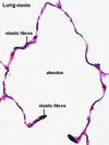
A

Below the epithelium of the trachea and bronchi

6
Q

What function do mucous glands have?

A

Secrete mucus into ducts that open through the ciliated epithelium in order to trap particles and pathogens.

7
Q

Where is cartilage found in the gas exchange system?

A

In the wall of the trachea (in C-shaped rings) and in the wall of the bronchi (as irregular blocks)

8
Q

What function does cartilage have in the gas exchange system?

A

Gives upport to the walls of the trachea and bronchi. During inhalation the pressure inside the airways falls, and the cartilage prevents them from collapsing, holding open the airways to allow easy flow of air.

9
Q

Where is smooth muscle found in the gas exchange system?

A

In the walls of the trachea, bronchi and bronchioles

10
Q

What function does smooth muscle have in the gas exchange system?

A

Smooth muscles cells undergo contraction and relaxation which alters the diameter of the respiratory airways (trachea, bronchi and bronchioles). This is particularly important in bronchioles, which widen during exercise to allow more air to enter the alveoli.

11
Q

Where is squamous epithelium found in the gas exchange system?

A

Alveoli

12
Q

What function does squamous epithelium have in the gas exchange system?

A

It is thin to give a short diffusion pathway for gas exchange. Alveoli provide a large surface area

13
Q

Where are capillaries found in the gas exchange system?

A

In all parts of the gas exchange system - many around the alveoli

14
Q

What function do capillaries have in the gas exchange system?

A

They provide a large surface area for exchange between blood and air in the alveoli

15
Q

What is ventilation or mechanical respiration?

A

Breathing air in and out of the lungs

16
Q

What is cellular respiration?

A

The chemical processes that occur inside cells to transfer energy from molecules, such as glucose and fat, to ATP.

17
Q

What is gas exchange?

A

Diffusion of oxygen and carbon dioxide between air in the alveoli and the blood.

18
Q

What structures form the airways of the gas exchange system?

A

The trachea, which branches into two bronchi which branch into numerous bronchioles

19
Q

How does the structure of bronchi differ from that of the trachea?

A

They have a similar structure, but the cartilage is in blocks in the walls of the bronchi instead of in rings as it is in the trachea. Bronchioles have no cartilage.

20
Q

What are these cells?

A

Ciliated epithelium

21
Q

What are these cells?

A

Goblet cells

22
Q

What are these structures?

A

Mucous glands

23
Q

What is this tissue?

A

Cartilage

24
Q

What is this tissue?

A

Smooth muscle

25
Q

Look carefully at the micrograph

A

Elastin fibers

26
Q

What are these cells?

A

Squamous epithelium of alveoli

27
Q

Study the attached micrograph

A
28
Q

Draw a plan diagram of an alveolus

A
29
Q

What is the gas exchange surface of the lung?

A

The squamous epithelium of the alveoli

30
Q
A
31
Q

Describe the process of gas exchange between aor in the alveoli and the blood

A

Alveloli are tiny air-filled sacs, adapted for the efficient exchange of gases by diffusion between the air and blood capillaries. This requires (1) a short diffusion distance (less than 0.3 µm) (2) a large surface area (3) a steep concentration gradient. Breathing ventilates the alveoli, maintianing near constant concentrations of oxygen and carbon dioxide in alveolar air. Blood flows through capillaries in the lungs, bringing constant supply of deoxygenated blood. Ventilation and blood flow combine to maintain a steep concentration gradient for oxygen between the blood and alveolar air. The same is also true for carbon dioxide, although the concentration gradient is not as steep as it is for oxygen.

32
Q

In the lungs, which gases are diffused from the blood?

A

Carbon dioxide

33
Q

In the lungs, which gases are diffused into the blood?

A

Oxygen

34
Q
A
35
Q

What functions do the airways have in mechnical respiration/breathing?

A

The airways allow the uninterrupted flow of air into and out of the lungs. To do this they must be kept open and be able to respond to demands for an increased oxygen supply by widening when necessary.

The airways also protect the gas exchange surface. The lining of the airways is formed by ciliated epithelium that contains goblet cells. Beneath the epithelium there are mucous glands. Goblet cells and mucous glands seceret mucous onto the surface of the epithelium to trap any small particles - including pathogens, such as bacteria, viruses and fungal spores - that have passes he hairs in the nose.

36
Q

On what two systems does tobacco smoke have an effect?

A

The gas exchange and the cardiovascular systems

37
Q

Are the effects of tobacco short term or long term?

A

Both short term and long term

38
Q
A
39
Q

What are the important constituents of cigarette smoke?

A

(1) Tar (a thick, black, oily liquid that contains carcinogens and settles in the bronchi and bronchioles)
(2) Carbon monoxide (a gas that combines with haemoglobin)
(3) nicotine (the drug in tobacco that is absorbed into the blood)

40
Q

What are carcinogens?

A

Cancer-causing chemicals e.g. benzpyrene

41
Q

When you see a question on smoking DO NOT automatically assume it is asking about the effects on the lungs (gas exchnage system), it may be asking about the effects on the cardiovascular system.

A
42
Q

What is chronic bronchitis?

A

Long term disease involving the inflammation of the walls of the bronchi and other air passages so that breathing becomes difficult. It often leads to emphysema.

43
Q

What is emphysema?

A

Lung disease in which the walls of the alveoli are destroyed. Thsi leads to the formation of larger air sacs and a decrease in the surface area fot gas exchange, making breathing difficult. Often leads to heart failure.

44
Q

What is chronic obstructive pulmonary disease?

A

The long-term lung diseases chronic bronchitis and emphysema

45
Q

What are the effects of tar on the gas exchange system?

A

(a. ) Tar is an irritant, causing inflammation of the epithelium lining the tract.
(b. ) The goblet cells and the mucous glands secrete more mucus which accumulates in the bronchi
(c) The tar causes the cilia to be inhibited and so mucus is not moved upwards, resulting in a suitable environment for pathgens to grow.
(d) Long-term smoking causes progressive chnages in the linings of the airways, particularly in the bronchi, leading to chronic bronchitis.
(e) Smoking stimulates macrophages in the alveoli, which in turn release chemicals that attract neutrophils
(f) Proteases, particulalry elastase, are secreted by these neutrophils and macrophages.
(g) Proteases are enzymes that digest lung tissue and are responsible for the damage seen in emphysema

46
Q

How does smoking cause lung cancer?

A

The carcinogens in the tobacco smoke interact with DNA in bronchial epithelial cells causing mutations. Cells become tumerous if the genes that control the cell cycle and mitosis mutate. The mutations increase the chnace that cells lining the bronchus will start to divide uncontrollably by motiosis. A cell that starts to divide in this way may form a group of cells known as a tumour. These cells may invade healthy lung tissue and block the airways or blood vessels. One of the symptoms of lung cancer is coughing up blood.

47
Q

What does the word chronic mean?

A

Chronic means long-term. Although many think it means severe or harmful. The word acute means short-term.

48
Q

What is carboxyhaemoglobin?

A

The irreversible combination of carbon monoxide with haemoglobin.

49
Q

What are the short term effects of nicotine and carbon monoxide on the cardiovascular system?

A

(a) Carbon monoxide diffuses into red blood cells where it combines irreversibly with haemoglobin to form carboxyhaemoglobin so less haemoglobin is avaliable to take up oxygen.
(b) Nicotine is rapidly absorbed and acts at synapses, stimulating the nervous system to the heart and promoting the secretion of adrenaline from the adrenal glands.
(c) This increases heart rate and blood pressure.
(d) Nicotine also stimulates neurones taht supply arterioles.
(e) The neurones stimulate smooth muscle to contract so reducing blood flow to capillaries.
(f) Both carbon monoxide and nicotine can damage the endothelium that lines blood vessles.
(g) the damage to blood vessels leads to inflammation increasing the risks of atheroma (build-up of fatty deposits in the walls of arteries, especially coronoary arteries) and thrombosis (formation of blood clots).

50
Q

Describe the structure of the tissue that forms the lining of the bronchus.

A

The lining is made from ciliated epithelium. This has many cells with cilia facing into the lumen. In between these cells are goblet cells that make mucus and realease it onto the surface.

51
Q

Explain how the distribution of cartilage in the lining of the bronchus differs from the distribution in the trachea.

A

The cartilage in the trachea forms C-shaped rings that are arranged in intervals down the length of the organ. There are no rings in the bronchus, just blocks of cartilage in the walls, not arranged in rings.

52
Q

During exercise, changes occur in the bronchi and in the arteries that supply leg muscles. Outline the changes that occur in both of these structures.

A

During exercise, the bronchial lumen widens/expands; the smooth muscle relaxes; the cartilage, moves further apart; there is reduced resistance to air flow, increasing the volume of air flowing in and out of the lungs. During exercise, the arteries lumen widens as blood pressure increases; the elastic tissue is stretched more; the smooth muscle relaxes; and this results in an increase in blood flow.

53
Q

Which cells are found in the wall of the trachea?

A

Cartilage cells (chondrocytes), ciliated epithelium, goblet cells.

54
Q

Which components of tobacco smoke can cross the gas exchange surface?

A

Carbon monoxide and nicotine

55
Q

Which feature of the mammalian gas exchange surface is an adapatation for reducing the diffiusion distance between air and blood?

A

Squamous epithelial cells

56
Q

State four structural functions of the trachea that are adaptations for its function. Explain how each feature is an adaptation.

A

The trachea has :

(1) C-shaped/incomplete, rings of cartilage that keep the lumen open to allow air to pass freely to and from the gas exchange surface
(2) smooth muscle in the wall of the trachea allows expansion of the lumen when the muscle relaxes to allow more air to move in and out of the lungs
(3) goblet cells/mucous glands, secrete mucus that is sticky and traps, pathogens/dust
(4) cilia beat back and forwards to move mucus, up the trachea/away from the alveoli

57
Q

Outline the effects of tobacco smoke on the lining of the bronchus.

A

Tar from tobacco smoke is an irritant, causing inflammation of the epithelial lining of the respiratory tract. The smoke causes the goblet cells and mucous glands secrete more mucus which accumulates in the bronchi. The smoke inhibits the cilia and so mucus is not moved upwards, away from the alveoli. This results in a suitable environment for pathogens to grow. This causes progressive changes in the linings of the airway, leading to chronic bronchitis. The carcinogens in tobacco smoke interact with the DNA of the bronchial epithelial cells causing mutations and increase the chances that cells in the lining of the bronchus will start to divide uncontrollably, forming a group of cells known as a tumour. These cells may invade healthy lung tissue and block the bronchal airway and/or the broncial blood vessels. This may result in coughing up of blood.

58
Q

The blood of smokers often has 10% less oxygen in it compared with the blood of nonsmokers. Explain what causes this reduction in transport of oxygen by the blood.

A

(1) One of the constituents of cigarette smoke is carbon monoxide. This diffuses into red blood cells where it combines irreversibly with haemoglobin to form carboxyhaemoglobin so less haemoglobin is avaliable to take up oxygen.
(2) Smoking stimulates macrophages in the alveoli, which in turn release chemicals that attract neutrophils. Proteases, particularly elastase, are secreted by these neutrophils and macrophages. Proteases are enzymes which digest lung tissue thereby reducing the number of alveoli by creating large air sacs and thereby reducing the gas exchange surface area thereby reducing the volume of oxygen which can diffuse into the blood.
(3) Smoking is an irritant which casues inflammation of the walls of the bronchi and other air passages which reduces the volume of air which is ventilated with each breath, reducing the amount of oxygen within the alveolar air, reducing the amount of oxygen avaliable for diffusion into the blood.

59
Q

People with emphysema often have low oxygen concentrations in their blood. Explain how this happens.

A

Emphysema is a lung disease in which the walls of alveloli are destroyed. This is as a result of tobacco smoking which stimulates macrophages in the alveoli, which in turn stimulates the release of chemicals which attract neutrophils. Proteases, particlaurly elastase, are secreted by these neutrophils and macrophages. Proteases are the enzymes responsible for digesting lung tissue, causing the walls of the alveoli to be destroyed. This leads to the formation of large air sacs and a decrease in the surface area for gas exchange reducing the diffusion of oxygen into the blood.

60
Q

What cells are labelled A-D and what is their function?

A

A - macrophage. They are responsible for removing particles such as dust or microorganisms from the respiratory surfaces by phagocytosis. Alveolar macrophages are frequently seen to contain granules of exogenous material such as particulate carbon that they have picked up from respiratory surfaces.

B - squamous epithelial cell. They provide a short diffusion distance and forms wall of alveolus

C - Red blood cell. They transport oxygen and carbon dioxide.

D - Endothelial cell. They provide a short diffusion distance and form the wall of the capillary.

61
Q

Where does exchange of oxygen and carbon dioxide occue on the bocy?

A

The alveoli of the lungs

62
Q

How does the exchange of gases occur in the lung?

A

Exchange occurs by diffusion between the air in the alveoli and blood in capillaries surrounding each alveolus.

63
Q

What structures does air pass through to reach the alveoli?

A

Nose and/or mouth, trachea, bronchi and bronchioles.

64
Q

Study the attached trnsverse section of the trachea - practice drawing it!

A
65
Q

Study the attached transverse section of a bronchus. Make sure you can draw it.

A
66
Q

Study the attached transverse section of a bronchiole. Make sure you can draw it.

A
67
Q

Study the atached plan diargam of ciliated epithelium. Make sure you can draw it.

A
68
Q

Where are elastic fibers found in the respiratory system?

A

Elastic fibers are present in the walls of all the bronchial tubes and between the alveoli. During inhalation, the elastic fibers stretch to allow the airways and alveoli to expand. During exhalation they recoil, helping to decrease the volume of the alveoli and push the air out of the lungs.

69
Q

Describe the arrangement and function of cartilage in the trachea.

A

Cartilage is present in the trachea in C-shaped rings. It supports the trachea wall and prevents its collapse during inhalation.

70
Q

Explain the role of goblet cells in the gas exchange system.

A

Goblet cells secrete mucus, which traps particles and pathogens. The mucus is removed by the action of cilia, preventing these impurities from entering the lungs.

71
Q

Explain the function of elastic fibers in the tissue between the alveoli.

A

The elastic fibers stretch during inhalation, allowing the alveloi to expand. During exhalation they recoil, helping to decrease the volume of the alveoli and push the air out of the lungs.

72
Q

Explain the process of gas exchange in the alveoli

A

(1) The walls of each alveolus are composed of a single layer of flat squamous epithelial cells.
(2) There is a network of blood capillaries in contact with the alveolus, and these also have walls made of a single layer of endothelial cells.
(3) The distance between the air in the alveolus and the blood inside the capillaries is very short (less than 1µm).
(4) This short distance speeds up the rate of diffusion of gases.
(5) Compared with the blood in the capillaries, alveolar air has a higher concentration of oxygen and a lower concentration of carbon dioxide.
(6) Oxygen therefore diffuses from the air in the alveolus, across the walls of the alveolus and capillary and enters the blood.
(7) Carbon dioxide diffuses in the opposite direction.
(8) The diffusion gradient is maintained by:
- blood flow past the alveolus, which brings deoxygenated blood from the pulmonary artyery and takes away the oxygenated blood through the pulmonary vein.
- ventilation of the lungs, which replaces alveolar air with air from outside the body.

73
Q

What is tissue X and cell Y in the attached micrograph?

A

X - ciliated epithelium

Y- red blood cell / erythrocyte

74
Q

State three ways in which the lining of the trachea, bronchi and bronchioles provides protection against the entry of bacterial pathogens.

A

(1) The epithelial cells are covered with cilia, which beat back and forth to sweep the mucus and trapped pathgens and particles towards the mouth, where they are swallowed. This action helps to prevent the particles and entering the lungs and causing infection. Any bacteria that are swallowed will be destroyed by the stomach acid.
(2) Within the ciliated epithelium there are goblet cells which secrete mucus to trap particles and pathogens. This acts as a barrier to entry to the lungs.
(3) Capillaries bring phagocytes to engulf bacteria

75
Q

Study the attached picture. State what is happening at stage J and explain the role of the

A