fractures Flashcards
what coud you use when describing location of a fracture/
Which bone?
Thirds (long bones)
Proximal, middle, distal third
Anatomic orientation
E.g. proximal, distal, medial,
lateral, anterior, posterior
Anatomic landmarks
E.g. head, neck, body /
shaft, base, condyle
Segment (long bones)
Epiphysis, physis,
metaphysis, diaphysis
What type of fracture is this?

transverse fracture
- occurs with pure bending force where the cortex on one side fails in compression and the cortex on the other side in tension
- usually don’t shorten (ubless completely displaced) but may angulate or result in rotational malialignment

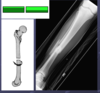
what type of fracture is this?

oblique
- occur with shearing force (e.g. fall from height, deceleration)
- can be fixed with interfragmentary screw
- tend to shorten and may also angulate
what type of fracture is this?

Segmental
- occur when bone is fracture in two seperate places
- very unstable and require stabilisation with long rods or plates
what type of fracture is this?

linear/ longitudinal/ splint
what type of fracture is this?

Comminuted (>3 pieces)
- generally reflection of higher energy injury (or poor bone quality)
- there may be substantial soft tissue swelling and periosteal damage with reduced blood supply to the fracture site which may impair healing
- very unstable and tend to be stablised surgically
what type of fracture is this?

impaction/ compression
what type of fracture is this?

Avulsion
describing a fracture
can be described according to the site of the fracture, whether its position is satisfactory or not and its stability (likelihood of displacing) which is related to the fracture pattern and degree of initial displacement
A fracture of long bone can be described according to the site of the bone involved in terms of proximal, middle or distal third. It can also be described according to type of bone involved (diaphyseal, metaphyseal or epiphyseal)
A fracture at the end of a long bone (metaphyseal/epiphyseal) can be intra articular (extending into joint) or extra-articular. Intra-articular fractures have a greater risk of stiffness, pain, and post-traumatic OA.
Fracture displacement depends on the degree of translation, angulation and rotation
describe translation
Sometimes confusingly called ‘displacement’
Extent to which Fx fragments are not axially aligned
Convention: describe displacement of distal fragment relative to proximal
Describe in % of bone width / direction (100% generally referred to as “off-ended” fracture)
** translation of distal fragment can be described as anterior or posterior displacement and medially or laterally translated

describe angulation
The extent to which Fx fragments are not anatomically aligned in a angular fashion
Convention: describe angulation in the direction that the distal end of the bone is pointing to relative to where it should be
Describe in degrees

describe rotation
Extent to which Fx fragments are rotated relative to each other
Convention: describe which direction the distal fragment is rotated relative to the proximal portion of the bone
other signs of fracture on Xray
periosteal reaction
callus
fat pad sign - means there is intra-articular effusion. post injury = blood;
posterior fat pad sign always abnormal 9anterior can be normal
management on subcapital and transcervical displaced fracture in the elderly
Unipolar hemiarthroplasty
Involves an open exposure of the hip joint - Anterolateral / Posterior • Resection and replacement of the native femoral head • Large metal head articulates with native acetabulum • Possible drawer backs: Dislocation risk, infection, loosening
management on subcapital and transcervical displaced fracture in the slightly fitter
Bipolar hemiarthroplasty
Involves an open exposure of the hip joint
Resection and replacement of the native femoral head
22mm metal head articulated with polyethylene liner, which is encased in a large metal head liner, which articulates with native acetabulum
Advantages: ? ↑ROM; ↓Acetabular erosion • Disadvantages: Dislocation risk, infection, loosening
subcapital and transcervical displaced fracture in the biologically fit and young
Total hip arthroplasty •
Advantages: ? ↑ROM; addresses deformity/pre- existing arthritis; longevity compared to hemiarthroplasty •
Disadvantages: Dislocation risk, infection, loosening
undisplaced or stable impacted fractures
Cannulated screws
Suitable in #s with an intact chondral buttress, such as high transcervical or subcapital #s •
Limited approach required (? + capsulotomy) • Low profile and bone preserving •
Compression at the fracture site •
Biomechanical advantages of 3 screws •
Importance of placement / configuration
basicervical fracture management
Biological and mechanical transition between intracapsular fractures and intertrochanteric fractures •
distal to capsule; vascular supply survives; decreased rate of AVN → FIXATION •
Bony neck cortices not intact → not favourable to cannulated screws •
Dynamic hip screws better resist bending forces
intertrochanteric fracture
Zone of transition between the femoral neck and shaft
Extracapsular, therefore, blood supply to femoral head unaffected and AVN risk ↓
Bony neck cortices not intact → not favourable to cannulated screws
Dynamic hip screws better resist bending forces
intertrochanteric fracture management
Dynamic hip screw •
Involves a limited approach to lateral proximal femur, fracture site not opened •
On-table reduction on trauma table •
Guide wire passed using fixed angle guide •
Large bore cannulated, partially threaded screw passed +/- de-rotation wire/screw • Importance of screw positioning (TAD) •
De-rotation plate allows compression at fracture site, increasing healing, decreasing non-union
describe secondary fracture healing process

name 5 fracture patterns

name 3 fracture patterns

treatment options for fracture
Non-surgical – boot, cast, splint, traction, etc •
Percutaneous wires •
External fixator • I
ntramedullary fixation •
Open reduction and internal fixation (ORIF) •
Arthroplasty •
Excison/amputation
limping child

what is buckle fracture
Compressive force in children

what is Greenstick fracture
Force to one side of bone may cause break in only one cortex in children

Plastic deformation
In very young children, neither cortex may break

salter harris classification
higher grade fractures are more likely to cause growth disturbance
I. Fracture passes transversely through physis separating epiphysis from metaphysis
II Transversely through physis but exits through metaphysis – Triangular fragment
III. Crosses physis and exits through epiphysis at joint space
IV. Extends upwards from the joint line, through the physis and out the metaphysis
V. Crush injury to growth plate

most common elbow fracture in children
supracondylar
Weakest part of the elbow joint where humerus flattens and flares
– Most are extension type (98%)
- Classified by Gartland – I, IIa, IIb, III
Potential for vascular compromise
– Check pulse!!! Reduce fracture if pulse compromised
– Check nerve function in hand
elbow landmarks

Gartland classification for supracondylar fractures
Gartland Classification (Extension Type): I – Undisplaced (treat conservatively) IIa – Displaced posteriorly but intact posterior periosteal hinge and anterior humeral line transects capitellum (normally treat conservatively) IIb - Displaced posteriorly but intact posterior periosteal hinge. Anterior humeral line does not transect capitellum (needs MUA +/- wires) III - Displaced posteriorly. No posterior periosteal hinge (needs MUA +/- wires)
toddlers fracture
Children younger than 2 years old learning to walk
No specific injury notable most of the time
Child refuses to bear weight on leg – Examine hip, thigh and knee to r/o other causes of limping
Often undisplaced spiral fracture of tibia with no fibular fracture
Initial x-ray often normal- diagnosis on f/u films with lucent line or periosteal reaction
factors suggesting NAI
Majority of fractures in child < 1 year are from abuse
Greater risk of abuse:
– first-born
– premature infants
– stepchildren
– children with learning or physical disabilities
Most common sites: femur, humerus, tibia
Unexplained fractures in different stages of healing
Femoral fracture in child < 1 year
Scapular fracture in child
Epiphyseal and metaphyseal fractures of the long bones
NAI bone fracture investigations
Clinical signs of fracture
Localised bony (marked) tenderness - not diffuse mild tenderness
Swelling
deformity
Crepitus - from bone ends grating with an unstable fracture
*not all MSK injuries require X-ray to exclude fratures. Most are done on clincial judgement. Guidelibes e.g. ottawa for ankle injury exist. General rule - if a patient cannot weight bear, X-ray.
Assessment of injured limb
- open or closed
- distal neurovascular status
- compartment syndrome?
- status of the skin and soft tissue envelope
Investigations of fracture
- X-ray - usually AP and lateral. TWO VIEWS ALWAYS REQUIRED.
- CT can be used for complex fractures and can help determine the degree of articular damage and help surgical planning for complex intra-articular fractures
- MRI can be used for occult fractures where there is clinical suspicion but normal X-ray
- Technetium bone scans can be useful to detect stress fractures (e.g. hip, femur, tibia, fibula, 2nd metatarsal) as these fail to show up on x-ray until hard callus begins to form
initial management of a long bone fracture
Assessment & analgesia
splintage/immobilization of the limb (temporary plaster slab, sling, orthosis, thomas splint if femoral shaft)
investigations
If fracture is grossly displaced, if there is obvious dislocation, if there is risk of skin damage from excessive pressure, reduction of the fracture shoul dbe perfoemed before waiting for x-rays. xray post reduction should still demonstrate ay fracture adequately.
definitive fracture management of undisplaced, minimally displaced, and minimally angulated fractures which are considered to be stable
non-operatively with a period of splintage or immobilization and then rehabilitation
definitive fracture management of displaced or angulated fractures where position is unacceptable
require reduction under anesthetic. They may perform closed reduction and cast application with serial X-rays to ensure no loss of position.
definitive management of unstable injuries
surgical stabilisation which mau involve the use of small percutaneous pins (K-wires) for small fragments, cerclage wires, screws, plates & screws, intramedullary nails or external fixation
definitive management of unstable extra-articular diaphyseal fractures
open reduction and internal fixation (ORIF) using plates and screws with the aim of anatomic reduction and rigid fixation leading to primary bone healing
It may be preferable to avoid ORIF particularly where the soft tissues are too swollen, where the blood supply to the fracture site is tenuous (high energy), where ORIF may cause extensive blood loss (eg femoral shaft) or plate fixation may be prominent (eg tibia). In this case closed reduction and indirect internal fixation with an intramedullary nail with dissection distant to the fracture site may be used with the aim of a functional reduction and stable fixation allowing micromotion required for secondary bone healing.
Another alternative for extra‐articular diaphyseal fractures is external fixation which again aims for secondary bone healing however carries the risk of pin site infection and loosening.
definitive management of displaced intra-articular fractures
anatomic reductoin and rigid fixation by ORIF using wires, screws and plates.
Fractures involving a joint with predictable poor outcome may be treated with joint replacement or arthodesis
Complications of fractures
Early local: compartment syndrome, vascular injury with ischaemia, nerve compression or injury, and skin necrosis
Early systemic: hypovolaemia, fat embolism, shock, ARDS, acute renal failure, SIRS, multi-organ dysfunction syndrome, death
Late local: stiffnessm loss of function, chronic regional pain syndrome, infection, non-union, malunion, Volkmann’s ischaemic contacture, post traumatic Oa and DVT
late systemic: PE (tends to be several days to weeks but can occur much sooner)
open fracture managment
ABx
prompt surgery - debridement + internal or external fixation (list 3 reasons why)
May just need closed primarily if not contamined and skin and muscle is viable and can be closed without tension (resulting in skin necrosis and wound breakdown)
dislocation and instability management
Any dislocation should be reduced as soon as possible. Most dislocations can be reduced by closed manipulation under sedation and analgesia or occasionally general or regional anaesthetic. Delayed presentation of a dislocation (eg in alcoholics) increases the risk of requiring an open reduction and recurrent insatbility.
Dislocations may occur after significant trauma however people with hypermobility (including Ehlers Danlos and Marfan’s) may sustain a dislocation with a seemingly innocuous injury and some can voluntarily dislocate joints (eg shoulder).
Dislocations can occur with associated injuries including tendon tears, nerve injury, vascular injury and compartment syndrome. Recurrent dislocation may require soft tissue repair / reconstruction or occasionally bony surgical prcedures.
Fractures can occur with dislocations (known as fracture‐dislocation) and these may reduce with closed reduction however ORIF may be required if reduction cannot be achieved, if a bony fragment prevents congruent reduction or if the joint is very unstable.
complete spinal cord injury
Complete spinal cord injury results in no sensory or voluntary motor function below the level of the injury (reflexes should return). The level of the injury is determined by the most distal spinal level with partial function (after spinal shock has resolved) as determined by the presence of dermatomal sensation and myotomal skeletal muscle voluntary contraction. The prognosis for recovery from complete cord injuries is poor.
incomplete spinal cord injury
With incomplete spinal injuries, some neurologic function (sensory and/or motor) is present distal to the level of injury. In general, the greater the function present,the faster the recovery is and the better the prognosis. Sacral sparing with preservation of perianal sensation, voluntary anal sphincter contraction and big toe flexion(FHL muscle, S1/2) indicates some continuity of the corticospinal (motor) and spinothalamic (course touch, pain, temperature) tracts. The presence of sacral sparingindicates an incomplete cord injury with a better prognosis than a complete injury.
