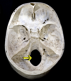
Foramen - extra testing Flashcards

1
Q

Contents of Foramen Ovale

A

Contents: OVALE

  • Otic ganglion (inferior)
  • V3 cranial nerve (mandibular branch of the trigeminal nerve)
  • Accessory meningeal artery
  • Lesser petrosal nerve
  • Emissary veins
2
Q

Contents of Foramen Spinosum

A

Contents: MMA fighters break your SPINE

  • Middle Meningeal Artery
  • Middle Meningeal Vein
  • Meningeal branch of CN V3/Nervus Spinosus
3
Q

Contents of Foramen Rotundum

A

Contents:

  • Maxillary branch of trigeminal nerve (CN V2)
  • Artery of foramen rotundum
  • Emissary veins
4
Q

Contents of Foramen Lacerum

A

ICA Nerve/Artery of Pterygoid canal

5
Q

Contents of Jugular Foramen

A

Contents: If someone hits you with a glass JUG call 9-11

  • Glossopharyngeal nerve (CN IX)
  • Vagus nerve (CN X)
  • Accessory nerve (CN XI)
  • Jugular bulb
  • Inferior petrosal and sigmoid sinuses
6
Q

Contents of Foramen Magnum

A

Contents: Special Meninges Make A Special Vertical Sheath

  • Spinal cord
  • Meninges
  • Meningeal lympthatics
  • Accessory nerve
  • Sympathetic plexus on vertebral arteries
  • Vertebral arteries
  • Spinal branch of vertebral arteries
7
Q

Contents of Stylomastoid Foramen

A

Stylomastoid Artery

Facial Nerve

8
Q

Contents of Superior Orbital Fissure

A

Contents: Lazy French Tarts Sit Nakedly In Sexual Anticipation

  • Lacrimal nerve
  • Frontal nerve (branch of opthalmic nerve of trigeminal nerve (CN V))
  • Trochlear nerve (CN IV)
  • Superior opthalmic vein
  • Nasociliary nerve (branch of opthalmic nerve (CN V1))
  • Inferior division of the oculomotor nerve (CN III)
  • Superior dividion of the oculomotor nerve (CN III)
  • Abducens nerve (CN VI)
  • (+ A branch of the inferior opthalmic vein)
9
Q

CN I - Name + Exit Point

A

Ophthalmic

Cribiform Plate

10
Q

CN II - Name + Exit Point

A

Optic

Optic Canal

11
Q

CN III - Name + Exit Point

A

Oculomotor

Superior Orbital Fissure

12
Q

CN IV - Name + Exit Point

A

Trochlear

Superior Orbital Fissure

13
Q

CN V - Name + Exit Point

A

Trigeminal ‘Standing Room Only’

V1 - Sup Orbital Fissure

V2 - Foramen Rotundum

V3 - Foramen Ovale

14
Q

CN VI - Name + Exit Point

A

Abducens

Superior Orbital Fissure

15
Q

CN VII - Name + Exit Point

A

Facial Nerve

Interal Acoustic Meatus -> Foramen Spinosum

16
Q

CN VIII - Name + Exit Point

A

Vestibulocochlear

IAM - Coke down (inf)

17
Q

CN IX - Name + Exit Point

A

Glossopharyngeal

Jugular Foramen

18
Q

CN X - Name + Exit Point

A

Vagus

Jugular Foramen

19
Q

CN XI - Name + Exit Point

A

Accessory

Jugular Foramen

20
Q

CN XII - Name + Exit Point

A

Hypoglossal

Hypoglossal Canal

21
Q

Femoral artery

A

arises after inguinal ligament

enters femoral triangle

Profunda femoris

medial and lateral circumflex femoral artery

22
Q

branches of internal iliac artery in lower limbs

A

I Love Going Places In My Very Own Underwear

I: iliolumbar artery

L: lateral sacral artery

G: gluteal (superior and inferior) arteries

P: (internal) pudendal artery

I: inferior vesical (vaginal in females) artery

M: middle rectal artery

V: vaginal artery (females only)

O: obturator artery

U: umbilical artery and uterine artery (females only)

The first three branches in the mnemonic (iliolumbar, lateral sacral and superior gluteal) are branches of the posterior division of the internal iliac artery, the remaining branches are of the anterior division.

23
Q

popliteal artery

A

arises after adductor hiatus (small gap in adductor magnus which allows vessels to pass posterior at the limb)

Branches at knee then:

anterior tibial artery (dorsalis pedis)
Tibioperoneal trunk (posterior tibial and peroneal)
24
Q

Veins in the legs - deep

A

Anterior tibial vein

Posterior tibial vein

popliteal vein -

femoral vein

25
Superficial vein of legs
Great saphenous vein - medial aspect of leg, anterior to medial malleolus Small saphenous vein - lateral aspect of leg - posterior to lateral malleolus, joins the popliteal vein
26
What travels through the cribiform foramina in the cribiform plate?
Location: Anterior cranial fossa Cranial bone: Ethmoid bone Contents: * Olfactory nerve (CN I) * Anterior ethmoidal nerves
27
What travels through the optic canal?
Location: Middle cranial fossa Cranial bone: Sphenoid bone Contents: * Optic nerve (CN II) * Opthalmic artery (located inferolaterally within the canal)
28
What travels through the superior orbital fissure?
Location: Middle cranial fossa Cranial bone: Sphenoid bone Contents: **L**azy **F**rench **T**arts **S**it **N**akedly **I**n **S**exual **A**nticipation * Lacrimal nerve * Frontal nerve (branch of opthalmic nerve of trigeminal nerve (CN V)) * Trochlear nerve (CN IV) * Superior opthalmic vein * Nasociliary nerve (branch of opthalmic nerve (CN V1)) * Inferior division of the oculomotor nerve (CN III) * Superior dividion of the oculomotor nerve (CN III) * Abducens nerve (CN VI) (+ A branch of the inferior opthalmic vein)
29
What travels through the foramen rotundum?
Location: Middle cranial fossa Cranial bone: Sphenoid bone Contents: * Maxillary branch of trigeminal nerve (CN V2) * Artery of foramen rotundum * Emissary veins
30
What travels through the foramen ovale?
Location: Middle cranial fossa Cranial bone: Sphenoid bone Contents: **OVALE** * **O**tic ganglion (inferior) * **V**3 cranial nerve (mandibular branch of the trigeminal nerve) * **A**ccessory meningeal artery * **L**esser petrosal nerve * **E**missary veins
31
What travels through foramen spinosum?
Location: Middle cranial fossa Cranial bone: Sphenoid bone Contents: **MMA fighters break your SPINE** * **M**iddle **M**eningeal **A**rtery * Middle Meningeal Vein * Meningeal branch of CN V3/Nervus Spinosus
32
What travels through the internal acoustic meatus?
Location: Middle cranial fossa Cranial bone: Petrous part of temporal bone Contents: * Facial nerve (CN VII) * Vestibulocochlear nerve (CN VIII) * Vestibular ganglion * Labrynthine artery **EXTRA:** There are 4 regions within the IAM: anterio-superior/anterio-inferior/postero-superior/postero-inferior **...Seven UP, Coke DOWN** * Facial nerve (**CN VII**) located superiorly * Vestibulo**coch**lear nerve located inferiorly
33
What travels through the jugular foramen?
Location: Posterior cranial fossa Cranial bone: * Anterior aspect= Petrous portion of temporal bone * Posterior aspect= Occipital bone Contents: **If someone hits you with a glass JUG call 9-11** * Glossopharyngeal nerve (CN **IX**) * Vagus nerve (CN **X**) * Accessory nerve (CN **XI**) * Jugular bulb * Inferior petrosal and sigmoid sinuses
34
What travels through the hypoglossal canal?
Location: Posterior cranial fossa Cranial bone: Occipital bone Content: * Hypoglossal nerve (CN XII)
35
What travels through the foramen magnum?
Location: Posterior cranial fossa Cranial bone: Occipital bone Contents: **S**pecial **M**eninges **M**ake **A S**pecial **V**ertical **S**heath * Spinal cord * Meninges * Meningeal lympthatics * Accessory nerve * Sympathetic plexus on vertebral arteries * Vertebral arteries * Spinal branch of vertebral arteries
36
Describe the borders of anterior triangle
Midline Inferior border of mandible Medial border of sternocleidomastoid
37
Submental
**Borders:** Inferiorly – hyoid bone. Medially – midline of the neck. Laterally – anterior belly of the digastric **Contents:** Lymph nodes
38
Submandibular
**Borders:** Superiorly – body of the mandible. Anteriorly – anterior belly of the digastric muscle. Posteriorly – posterior belly of the digastric muscle. **Contents:** Submandibular gland, lingual nerve Facial artery and Vein
39
Carotid triangle
**Borders:** Superior – posterior belly of the digastric muscle. Lateral – medial border of the sternocleidomastoid muscle. Inferior – superior belly of the omohyoid muscle. **Contents: Common carotid artery**(which bifurcates within the carotid triangle into the external and internal carotid arteries), **Internal jugular vein Hypoglossal**and**vagus nerves**
40
Muscular triangle borders
**Borders:** Superiorly – hyoid bone. Medially – imaginary midline of the neck. Supero-laterally – superior belly of the omohyoid muscle. Infero-laterally – inferior portion of the sternocleidomastoid muscle. **Contents:** Midline organs: **thyroid, parathyroid, larynx, trachea,** infrahyoid muscle
41
Describe the borders of the posterior triangle
**Borders:** Anterior – posterior border of the sternocleidomastoid. Posterior – anterior border of the trapezius muscle. Inferior – middle 1/3 of the clavicle. Covered by investing layer of fascia and floor formed by prevertebral fascia
42
Content of posterior triangle
**Muscles** - scalene, levator scapulae, splenius capitis, omohyoid (inf belly crosses post triangle in superomedial direction) **Vascular** - **External Jugular Vein** (formed by retromandibular and posterior auricular vein) - drains into the **Subclavian vein** **Nerves** - Spinal accessory nerve, **cervical plexus inc. phrenic nerve**
43
Aorta - Abdominal branches
**I**n **C**ase **M**y **S**tudents **R**eally **L**ove **G**ames **I**'m **M**onopoly **I**nferior phrenic **C**oeliac(T12) **M**iddle adrenal **S**MA(L1) **R**enal **L**umbar **G**onadal (L2), **I**MA(L3), **M**edian sacral (L4),
44
Coeliac trunk
T12 **Left gastric** (oesophageal branches) **Splenic** (L gastroepiploic (greater curve stomach), short gastric arteries **Common hepatic artery** : gives rise to R gastric, R + L hepatic, cystic and sup. and inf. gastroduodenal (head of panc.)
45
Superior mesenteric artery
L1 Major branches: **Inferior pancreaticoduodenal artery** **Jejunal & ileal arteries** (forms the anastamotic arcades) **Right & middle colic** **Ileocolic** (gives rise to branches supply illeum, caecum, colon and appendix) - can ligate appendicular artery in appendix
46
Inferior mesenteric artery
L3, supplies the hindgut, retroperitoneal main branches: **Left colic** (from distal 1/3 of transverse colon) **Sigmoid** **Superior rectal** Anastomosis with middle colic - Marginal artery of Drummond
48
Inferior vena cava
Formed by CIV at L5 Drains - Lumbar veins, right gonadal, right adrenal, right and left renal, inferior phrenic and hepatic veins
52
Venous drainage
IVC Hepatic portal system
53
External Carotid artery
Bifurcates from CCA at C4 **S**he **A**lways **L**ikes **F**riends **O**ver **P**apa, **M**ama and **S**ister * *S**uperior thyroid * *A**scending pharyngeal * *L**ingual * *F**acial * *O**ccipital * *P**osterior auricular * *M**axillary * *S**uperficial temporal
54
Internal carotid artery
Bifurcates from CCA at C4 no extracranial branch enters via carotid canal **C**alming **V**oices **M**ake **I**ntra-**O**perative **S**urgery **P**leasurable **A**nd Almost **M**emorable **C**: caroticotympanic artery (C2) **V**: Vidian artery (C2) **M**: meningohypophyseal trunk (C4) **I**: inferolateral trunk (C4) **O**: ophthalmic artery (C6) **S:** superior hypophyseal artery (C6) **P**: posterior communicating artery (C7) **A:** anterior choroidal artery (C7) **A**: anterior cerebal artery (C7) **M**: middle cerebral artery (C7) - **forms circle Willis**
55
Quadrangular space - content
axillary nerve posterior circumflex humeral artery
56
Quadrangular space - border
superior - teres minor inferior - teres major lateral - humerus medial - long head of triceps
57
Triangular space - borders
superior - teres minor inferior - teres major lateral - long head triceps
58
Triangular interval - borders
Long head of triceps Teres major Humerus
59
Clavipectoral triangle - borders
lateral - deltoid superior - clavicle inferior - pectoralis major
61
Cubital fossa - borders
Lateral - brachioradialis medial - pronator teres superior - imaginary line between epicondyles roof - bicipital aponeurosis floor - brachialis and supinator
62
Cubital fossa - content
Radial nerve Biceps tendon Brachial artery Median nerve
63
Carpal tunnel - borders
Formed by two layers - deep carpal arch and superficial flexor retinaculum Lateral - scaphoid, trapezium Medial - hamate, pisiform
64
Carpal tunnel - content
FDS/FDP/FPL tendons median nerve
65
Anatomical snuffbox - borders
Medial/ulnar - Extensor pollicis longus Lateral - Extensor pollicis brevis, Abductor Pollicis longus Proximal - radius styloid Floor - scaphoid and trapezium
66
Anatomical snuffbox - content
Radial artery Radial nerve cephalic vein
71
Clavipectoral triangle - content
Cephalic vein lateral pectoral nerve medial pectoral nerve
78
Triangular INTERVAL contents
Radial nerve Profunda brachii artery
79
Hesselbachs Triangle and relevance
**Medial** – lateral border of the rectus abdominis muscle. **Latera**l – inferior epigastric vessels. **Inferior** – inguinal ligament. Demarcate an area of potential weakness in the abdominal wall – through which herniation of the abdominal contents can occur.