female reproductive system Flashcards

1
Q

What does menapause begin?

A
  1. the female reproductive system remains incompletely developed until gonadotropic hormones released by anterior pituitary initiate puberty.
  2. the first menstrual flow begins at age 13 and repeated on a 28 day cycle throughout the womans reproductive age, except during prenancy
  3. menopause occurs between ages of 45-55
2
Q

what age does the female reproductive developement finalize and how?

A
  1. the female reproductive system remains incompletely developed until gonadotropic hormones released by anterior pituitary initiate puberty.
  2. the first menstrual flow begins at age 13 and repeated on a 28 day cycle throughout the womans reproductive age, except during prenancy
  3. menopause occurs between ages of 45-55
3
Q

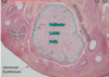
Describe the tissue type and composition of the ovary cortex and medulla

A
  1. ovaries
    1. site of oogenesis
    2. histology
      1. almond shaped
      2. simple cuboidal epithelium (surface or germinal epitehlium)
      3. dense connective tissue capsule: tunica albuginea
      4. cortex
        1. highly cellular (fibroblasts) connective tissue stroma
        2. many ovarian follicles at various stages of devopment suspended in the CT stroma
      5. medulla
        1. contains loose connective tissue and blood vessels entering the organ through hilum from the mesenteries suspending the ovaries
4
Q

What is the ovary covered by?

A
  1. ovaries
    1. site of oogenesis
    2. histology
      1. almond shaped
      2. each ovary is covered by a simple cuboidal epithelium (surface or germinal epitehlium)
      3. dense connective tissue capsule: tunica albuginea
      4. cortex
        1. highly cellular (fibroblasts) connective tissue stroma
        2. many ovarian follicles at various stages of devopment suspended in the CT stroma
      5. medulla
        1. contains loose connective tissue and blood vessels entering the organ through hilum from the mesenteries suspending the ovaries
5
Q

ovaries are the stie of _______

A
  1. ovaries
    1. site of oogenesis
    2. histology
      1. almond shaped
      2. simple cuboidal epithelium (surface or germinal epitehlium)
      3. dense connective tissue capsule: tunica albuginea
      4. cortex
        1. highly cellular (fibroblasts) connective tissue stroma
        2. many ovarian follicles at various stages of devopment suspended in the CT stroma
      5. medulla
        1. contains loose connective tissue and blood vessels entering the organ through hilum from the mesenteries suspending the ovaries
6
Q
  1. these develope around the first month of gestation
A
  1. oogonia and primary oocytes
    1. oogonia
      1. primoridial cells that develop after the first mont hof gestation
      2. these cells undergo several mitotic divisions and are found in the cortex of the developing ovary, where they continue to divide until 6th month of fetal life.
    2. each ovary posses ~3million oogonia, but most undergo atresia
      1. process of degeneration and death
    3. primary oocyte
      1. result from the final mitotic division of the surviving oogonia
      2. begin meiosis while still in the fetus, but are arrested in prophase of first meitotic division
      3. remain arrested in this stage until just before ovulation
      4. young woman has ~400000 follicles, when ovulation begins around puberty
  2. quick math
    1. ovary starts with 300million oogonia
    2. 400k are arrested in P1 as follicles in a young woman who starts puberty
    3. only 500 oocytes are ovulated over a lifetime
    4. meaning that 99% of the oocytes undergo atresia
7
Q
  1. results from the final mitotic division of the surviving oogonia. Describe the process leading up through meiosis
A
  1. oogonia and primary oocytes
    1. oogonia
      1. primoridial cells that develop after the first mont hof gestation
      2. these cells undergo several mitotic divisions and are found in the cortex of the developing ovary, where they continue to divide until 6th month of fetal life.
    2. each ovary posses ~3million oogonia, but most undergo atresia
      1. process of degeneration and death
    3. primary oocyte
      1. result from the final mitotic division of the surviving oogonia
      2. begin meiosis while still in the fetus, but are arrested in prophase of first meitotic division
      3. remain arrested in this stage until just before ovulation
      4. young woman has ~400000 follicles, when ovulation begins around puberty
  2. quick math
    1. ovary starts with 300million oogonia
    2. 400k are arrested in P1 as follicles in a young woman who starts puberty
    3. only 500 oocytes are ovulated over a lifetime
    4. meaning that 99% of the oocytes undergo atresia
8
Q

discuss and label all 5 major steps in follicular development

A
  1. ovarian follicles
    1. primordial follicles
      1. most primitive follicles
      2. each consists of
        1. primary oocyte surrounded by a singla layer of squamous follicular cells
      3. the oocyte is 25micrometes in diameter at this stage, the follicular cells are attached to one another by sedmosomes and havea basal lamina seperating the follicle from surrounding stromal connective tissue
      4. primordial follicles location
        1. found adjacent to the tunica albuginea in the mature ovary
    2. primary follicles unilaminar
      1. develop from the primordial follicle
      2. surrounding cell maturation
        1. the follicular cells surrounding the primary oocyte become cuboidal in shape
        2. a primary follicle is surrounding by a. single layer of cuboidal cells
          1. the primary oocyte grows to 100-150 micrometers, developing a large nucleus
    3. multilaminar primary follicle
      1. a primary follicle with 2 or more layer of granulosa cells
        1. granulosa cells function to assist flow of nutrients and other molecules between the blood and follicular fluid
      2. formation of the zona pellucida
        1. distinct acidphilic refractile layer develops between oocyte and the follicular calls
        2. composed of
          1. glyocproteins secreted by both follicular cells and the oocyte
      3. theca folliculi
        1. organized layer of connective tissue outside of the basal lamina of the primary follicle
        2. orgaizes into 2 distint layers
          1. theca interna
            1. form from the connective tissue immediatly surrounding the basal lamina
          2. theca externa
            1. the conncective tissue layer surrounding the theca interna
    4. secondary follicle( antral follicle)
      1. increase in granulosa cell population, from FSH leads to
        1. cumulus oophus
          1. group of granulosa cells associated with the oocyte
        2. corona radiadata
          1. lies immediatly outside the zona pellucida
          2. first single layer of granulosa cells surrounding the oocyte
          3. remains with oocyte at the time of oculation, unlike other granulosa cells
    5. graafian follicle (mature follicle)
      1. granulosa cell proliferation and liquor folliculi accumulation continues unti mature graafian follicle is formed
      2. 2/5cm
      3. priamry ooctye + cornoa radiata detach from the cumulus oophorus anflota freely in liquor folliculi
      4. theca interna is more pronounced
      5. this is when the secondary oocyte is formed
9
Q

a follicle is found wit ha single layer of squamous follicular cells.

  1. What is the stage
  2. what is the location?
    1. describe the attachment and seperation of them
A
  1. ovarian follicles
    1. primordial follicles
      1. most primitive follicles
      2. each consists of
        1. primary oocyte surrounded by a singla layer of squamous follicular cells
      3. the oocyte is 25micrometes in diameter at this stage, the follicular cells are attached to one another by sedmosomes and havea basal lamina seperating the follicle from surrounding stromal connective tissue
      4. primordial follicles location
        1. found adjacent to the tunica albuginea in the mature ovary
    2. primary follicles unilaminar
      1. develop from the primordial follicle
      2. surrounding cell maturation
        1. the follicular cells surrounding the primary oocyte become cuboidal in shape
        2. a primary follicle is surrounding by a. single layer of cuboidal cells
          1. the primary oocyte grows to 100-150 micrometers, developing a large nucleus
    3. multilaminar primary follicle
      1. a primary follicle with 2 or more layer of granulosa cells
        1. granulosa cells function to assist flow of nutrients and other molecules between the blood and follicular fluid
      2. formation of the zona pellucida
        1. distinct acidphilic refractile layer develops between oocyte and the follicular calls
        2. composed of
          1. glyocproteins secreted by both follicular cells and the oocyte
      3. tehca folliculi
        1. organized layer of connective tissue outside of the basal lamina of the primary follicle
        2. orgaizes into 2 distint layers
          1. theca interna
            1. form from the connective tissue immediatly surrounding the basal lamina
          2. theca externa
            1. the conncective tissue layer surrounding the theca interna
    4. secondary follicle( antral follicle)
      1. increase in granulosa cell population, from FSH leads to
        1. cumulus oophus
          1. group of granulosa cells associated with the oocyte
        2. corona radiadata
          1. lies immediatly outside the zona pellucida
          2. first single layer of granulosa cells surrounding the oocyte
          3. remains with oocyte at the time of oculation, unlike other granulosa cells
    5. graafian follicle (mature follicle)
      1. granulosa cell proliferation and liquor folliculi accumulation continues unti mature graafian follicle is formed
      2. 2/5cm
      3. priamry ooctye + cornoa radiata detach from the cumulus oophorus anflota freely in liquor folliculi
      4. theca interna is more pronounced
      5. this is when the secondary oocyte is formed
10
Q

a follicular cell is surrounded by a single layer of cuboidal follicular cells

  1. what is the stage
A
  1. ovarian follicles
    1. primordial follicles
      1. most primitive follicles
      2. each consists of
        1. primary oocyte surrounded by a singla layer of squamous follicular cells
      3. the oocyte is 25micrometes in diameter at this stage, the follicular cells are attached to one another by sedmosomes and havea basal lamina seperating the follicle from surrounding stromal connective tissue
      4. primordial follicles location
        1. found adjacent to the tunica albuginea in the mature ovary
    2. primary follicles unilaminar
      1. develop from the primordial follicle
      2. surrounding cell maturation
        1. the follicular cells surrounding the primary oocyte become cuboidal in shape
        2. a primary follicle is surrounding by a. single layer of cuboidal cells
          1. the primary oocyte grows to 100-150 micrometers, developing a large nucleus
    3. multilaminar primary follicle
      1. a primary follicle with 2 or more layer of granulosa cells
        1. granulosa cells function to assist flow of nutrients and other molecules between the blood and follicular fluid
      2. formation of the zona pellucida
        1. distinct acidphilic refractile layer develops between oocyte and the follicular calls
        2. composed of
          1. glyocproteins secreted by both follicular cells and the oocyte
      3. tehca folliculi
        1. organized layer of connective tissue outside of the basal lamina of the primary follicle
        2. orgaizes into 2 distint layers
          1. theca interna
            1. form from the connective tissue immediatly surrounding the basal lamina
          2. theca externa
            1. the conncective tissue layer surrounding the theca interna
    4. secondary follicle( antral follicle)
      1. increase in granulosa cell population, from FSH leads to
        1. cumulus oophus
          1. group of granulosa cells associated with the oocyte
        2. corona radiadata
          1. lies immediatly outside the zona pellucida
          2. first single layer of granulosa cells surrounding the oocyte
          3. remains with oocyte at the time of oculation, unlike other granulosa cells
    5. graafian follicle (mature follicle)
      1. granulosa cell proliferation and liquor folliculi accumulation continues unti mature graafian follicle is formed
      2. 2/5cm
      3. priamry ooctye + cornoa radiata detach from the cumulus oophorus anflota freely in liquor folliculi
      4. theca interna is more pronounced
      5. this is when the secondary oocyte is formed
11
Q

a follicle is surrounded by a zona pellucida and several layers of granulosa cells

  1. what atage is it in?
  2. describe the other structures associated with this stage and the physiology
A
  1. ovarian follicles
    1. primordial follicles
      1. most primitive follicles
      2. each consists of
        1. primary oocyte surrounded by a singla layer of squamous follicular cells
      3. the oocyte is 25micrometes in diameter at this stage, the follicular cells are attached to one another by sedmosomes and havea basal lamina seperating the follicle from surrounding stromal connective tissue
      4. primordial follicles location
        1. found adjacent to the tunica albuginea in the mature ovary
    2. primary follicles unilaminar
      1. develop from the primordial follicle
      2. surrounding cell maturation
        1. the follicular cells surrounding the primary oocyte become cuboidal in shape
        2. a primary follicle is surrounding by a. single layer of cuboidal cells
          1. the primary oocyte grows to 100-150 micrometers, developing a large nucleus
    3. multilaminar primary follicle
      1. a primary follicle with 2 or more layer of granulosa cells
        1. granulosa cells function to assist flow of nutrients and other molecules between the blood and follicular fluid
      2. formation of the zona pellucida
        1. distinct acidphilic refractile layer develops between oocyte and the follicular calls
        2. composed of
          1. glyocproteins secreted by both follicular cells and the oocyte
      3. theca folliculi
        1. organized layer of connective tissue outside of the basal lamina of the primary follicle
        2. orgaizes into 2 distint layers
          1. theca interna
            1. form from the connective tissue immediatly surrounding the basal lamina
          2. theca externa
            1. the conncective tissue layer surrounding the theca interna
    4. secondary follicle( antral follicle)
      1. increase in granulosa cell population, from FSH leads to
        1. cumulus oophus
          1. group of granulosa cells associated with the oocyte
        2. corona radiadata
          1. lies immediatly outside the zona pellucida
          2. first single layer of granulosa cells surrounding the oocyte
          3. remains with oocyte at the time of oculation, unlike other granulosa cells
    5. graafian follicle (mature follicle)
      1. granulosa cell proliferation and liquor folliculi accumulation continues unti mature graafian follicle is formed
      2. 2/5cm
      3. priamry ooctye + cornoa radiata detach from the cumulus oophorus anflota freely in liquor folliculi
      4. theca interna is more pronounced
      5. this is when the secondary oocyte is formed
12
Q

a follicle has generated a cumulous oophus.

  1. what stage is it in?
  2. what other structure are present?
    1. list their function
A
  1. ovarian follicles
    1. primordial follicles
      1. most primitive follicles
      2. each consists of
        1. primary oocyte surrounded by a singla layer of squamous follicular cells
      3. the oocyte is 25micrometes in diameter at this stage, the follicular cells are attached to one another by sedmosomes and havea basal lamina seperating the follicle from surrounding stromal connective tissue
      4. primordial follicles location
        1. found adjacent to the tunica albuginea in the mature ovary
    2. primary follicles unilaminar
      1. develop from the primordial follicle
      2. surrounding cell maturation
        1. the follicular cells surrounding the primary oocyte become cuboidal in shape
        2. a primary follicle is surrounding by a. single layer of cuboidal cells
          1. the primary oocyte grows to 100-150 micrometers, developing a large nucleus
    3. multilaminar primary follicle
      1. a primary follicle with 2 or more layer of granulosa cells
        1. granulosa cells function to assist flow of nutrients and other molecules between the blood and follicular fluid
      2. formation of the zona pellucida
        1. distinct acidphilic refractile layer develops between oocyte and the follicular calls
        2. composed of
          1. glyocproteins secreted by both follicular cells and the oocyte
      3. tehca folliculi
        1. organized layer of connective tissue outside of the basal lamina of the primary follicle
        2. orgaizes into 2 distint layers
          1. theca interna
            1. form from the connective tissue immediatly surrounding the basal lamina
          2. theca externa
            1. the conncective tissue layer surrounding the theca interna
    4. secondary follicle( antral follicle)
      1. increase in granulosa cell population, from FSH leads to
        1. cumulus oophus
          1. group of granulosa cells associated with the oocyte
        2. corona radiadata
          1. lies immediatly outside the zona pellucida
          2. first single layer of granulosa cells surrounding the oocyte
          3. remains with oocyte at the time of oculation, unlike other granulosa cells
    5. graafian follicle (mature follicle)
      1. granulosa cell proliferation and liquor folliculi accumulation continues unti mature graafian follicle is formed
      2. 2/5cm
      3. priamry ooctye + cornoa radiata detach from the cumulus oophorus anflota freely in liquor folliculi
      4. theca interna is more pronounced
      5. this is when the secondary oocyte is formed
13
Q

the primary oocyte is found floating in the middle of the antrum.

  1. what stage is it in?
  2. what other structures are present?
    1. list their functions
A
  1. ovarian follicles
    1. primordial follicles
      1. most primitive follicles
      2. each consists of
        1. primary oocyte surrounded by a singla layer of squamous follicular cells
      3. the oocyte is 25micrometes in diameter at this stage, the follicular cells are attached to one another by sedmosomes and havea basal lamina seperating the follicle from surrounding stromal connective tissue
      4. primordial follicles location
        1. found adjacent to the tunica albuginea in the mature ovary
    2. primary follicles unilaminar
      1. develop from the primordial follicle
      2. surrounding cell maturation
        1. the follicular cells surrounding the primary oocyte become cuboidal in shape
        2. a primary follicle is surrounding by a. single layer of cuboidal cells
          1. the primary oocyte grows to 100-150 micrometers, developing a large nucleus
    3. multilaminar primary follicle
      1. a primary follicle with 2 or more layer of granulosa cells
        1. granulosa cells function to assist flow of nutrients and other molecules between the blood and follicular fluid
      2. formation of the zona pellucida
        1. distinct acidphilic refractile layer develops between oocyte and the follicular calls
        2. composed of
          1. glyocproteins secreted by both follicular cells and the oocyte
      3. tehca folliculi
        1. organized layer of connective tissue outside of the basal lamina of the primary follicle
        2. orgaizes into 2 distint layers
          1. theca interna
            1. form from the connective tissue immediatly surrounding the basal lamina
          2. theca externa
            1. the conncective tissue layer surrounding the theca interna
    4. secondary follicle( antral follicle)
      1. increase in granulosa cell population, from FSH leads to
        1. cumulus oophus
          1. group of granulosa cells associated with the oocyte
        2. corona radiadata
          1. lies immediatly outside the zona pellucida
          2. first single layer of granulosa cells surrounding the oocyte
          3. remains with oocyte at the time of oculation, unlike other granulosa cells
    5. graafian follicle (mature follicle)
      1. granulosa cell proliferation and liquor folliculi accumulation continues unti mature graafian follicle is formed
      2. 2/5cm
      3. priamry ooctye + cornoa radiata detach from the cumulus oophorus and float freely in liquor folliculi
      4. theca interna is more pronounced
      5. this is when the secondary oocyte is formed
14
Q

describe the meisosis events in the generation of a corpus luteum. Be location and stage specific

A

formation of the secondary oocyte in the graafian follicle

  1. secondary oocyte
    1. forms when the primary oocyte in the graafian follicle compeltes the first meitotic division
      1. remember all the oogonium are arrested in Prophase1 in the begining of the life cycle for females
    2. secondary oocyte enters the second meitotic division and is arrested in metaphase
    3. after being arrested in M2
      1. the secondary oocyte is oculated WITH THE CORONA RADIATA
    4. the remnants of the graafian follicle generate a corpus luteum
15
Q

a oocyte is found is taken up for collection in the uterine tube

  1. Was meiosis complete? assume no sexual history
  2. what does the graafian follicle become?
A

formation of the secondary oocyte in the graafian follicle

  1. secondary oocyte
    1. forms when the primary oocyte in the graafian follicle compeltes the first meitotic division
      1. remember all the oogonium are arrested in Prophase1 in the begining of the life cycle for females
    2. secondary oocyte enters the second meitotic division and is arrested in metaphase
    3. after being arrested in M2
      1. the secondary oocyte is oculated WITH THE CORONA RADIATA
    4. the remnants of the graafian follicle generate a corpus luteum
16
Q

what type of oocyte is ejected from the ovary and with what structure?

A

formation of the secondary oocyte in the graafian follicle

  1. secondary oocyte
    1. forms when the primary oocyte in the graafian follicle compeltes the first meitotic division
      1. remember all the oogonium are arrested in Prophase1 in the begining of the life cycle for females
    2. secondary oocyte enters the second meitotic division and is arrested in metaphase
    3. after being arrested in M2
      1. the secondary oocyte is oculated WITH THE CORONA RADIATA
    4. the remnants of the graafian follicle generate a corpus luteum
17
Q

what occurs during ovulation, theoretically is occurs how often?

A

ovulation

  1. the release of oocyte from the ovary, occurring at ~14th day of the 28 day menstrual cycle
  2. oculation is a result of several homronally induced factors
    1. increased volume and pressure of liquor folliculi
    2. enzymatic breakdown of the follicular wall
    3. contraction of smooth muscle in the theca externa
    4. stigma
      1. area on the ovary surface that becomes ischemic dur to the pressure of the Graafian follicle against the ovarian wall
      2. the tissue bvecomes necrotic and ruptures ( along with the follicular wall)
      3. the oocyte is released through this opening and is transported into the oviduct.
18
Q

what notable changes results in ovulation

A

ovulation

  1. the release of oocyte from the ovary, occurring at ~14th day of the 28 day menstrual cycle
  2. ovulation is a result of several homronally induced factors
    1. increased volume and pressure of liquor folliculi
    2. enzymatic breakdown of the follicular wall
    3. contraction of smooth muscle in the theca externa
    4. stigma
      1. area on the ovary surface that becomes ischemic dur to the pressure of the Graafian follicle against the ovarian wall
      2. the tissue bvecomes necrotic and ruptures ( along with the follicular wall)
      3. the oocyte is released through this opening and is transported into the oviduct.
19
Q

area on the ovary surface tha becomes ischemic due to pressure of the graafian follicle. what happens to this area?

A

ovulation

  1. the release of oocyte from the ovary, occurring at ~14th day of the 28 day menstrual cycle
  2. oculation is a result of several homronally induced factors
    1. increased volume and pressure of liquor folliculi
    2. enzymatic breakdown of the follicular wall
    3. contraction of smooth muscle in the theca externa
    4. stigma
      1. area on the ovary surface that becomes ischemic dur to the pressure of the Graafian follicle against the ovarian wall
      2. the tissue bvecomes necrotic and ruptures ( along with the follicular wall)
      3. the oocyte is released through this opening and is transported into the oviduct.
20
Q

during ovulation

  1. what increases in volume?
  2. how does the follicular wall break down?
  3. Where does smooth muscle contraction occur?
  4. what occurs in the stigma?
A

ovulation

  1. the release of oocyte from the ovary, occurring at ~14th day of the 28 day menstrual cycle
  2. oculation is a result of several homronally induced factors
    1. increased volume and pressure of liquor folliculi
    2. enzymatic breakdown of the follicular wall
    3. contraction of smooth muscle in the theca externa
    4. stigma
      1. area on the ovary surface that becomes ischemic dur to the pressure of the Graafian follicle against the ovarian wall
      2. the tissue bvecomes necrotic and ruptures ( along with the follicular wall)
      3. the oocyte is released through this opening and is transported into the oviduct.
21
Q

forms from the remnants of collapsed graafian follicle.

  1. describe the cell types and products
A
  1. corpus luteum
    1. formed from the remnants of collapsed graafian follicle after oculation
    2. a blood clot is formed at the rupture site and the modified granulosa and theca interna cells begin secretion of veious hormones
    3. granulosa lutein cells
      1. ~80 of structure
      2. synthesis steroid and highly dense with the cellular machinary to synthesize cholesterol and downstream products
        1. estrogen
        2. progesterone
    4. theca lutein cells
      1. small dark staining cells derived from the theca interna
      2. synthesize
        1. progesterone
        2. androgens
      3. degenerate 14 post ovulation if fertilization does not occur
  2. corpus albicans
    1. dense connective tissue scar formed at the site of an involueted corpus luteum
22
Q

dense connective tissue sxar formed at the site of an involuted corpus luteum

A
  1. corpus luteum
    1. formed from the remnants of collapsed graafian follicle after oculation
    2. a blood clot is formed at the rupture site and the modified granulosa and theca interna cells begin secretion of veious hormones
    3. granulosa lutein cells
      1. ~80 of structure
      2. synthesis steroid and highly dense with the cellular machinary to synthesize cholesterol and downstream products
        1. estrogen
        2. progesterone
    4. theca lutein cells
      1. small dark staining cells derived from the theca interna
      2. synthesize
        1. progesterone
        2. androgens
      3. degenerate 14 post ovulation if fertilization does not occur
  2. corpus albicans
    1. dense connective tissue scar formed at the site of an involuted corpus luteum
23
Q

describe the following with regard to the corpus luteum

  1. synthesize estrogen and progesterone, ~80% of the cells in the corpus luteum
  2. small dark, staining cells synthesize: progesteron and androgens
    1. what occurs in these cells 14 days after ovulation
A
  1. corpus luteum
    1. formed from the remnants of collapsed graafian follicle after oculation
    2. a blood clot is formed at the rupture site and the modified granulosa and theca interna cells begin secretion of veious hormones
    3. granulosa lutein cells
      1. ~80 of structure
      2. synthesis steroid and highly dense with the cellular machinary to synthesize cholesterol and downstream products
        1. estrogen
        2. progesterone
    4. theca lutein cells
      1. small dark staining cells derived from the theca interna
      2. synthesize
        1. progesterone
        2. androgens
      3. degenerate 14 post ovulation if fertilization does not occur
  2. corpus albicans
    1. dense connective tissue scar formed at the site of an involuted corpus luteum
24
Q

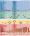
what are the hormones that control the follicle development and ovulation? (3 main ones)

A

hormonal control of follicle development and ovulation is largely dependent on FSH and LH

  1. FSH
    1. the most influential hormone in the days 8-10 of mentrual cycle
    2. stimulates 10-20 primary follicles to develop
    3. origin
      1. released from pituitary pars distalis
      2. binds to receptors on the follicular cell surface
      3. cells enlarge and proliferate in response to FSH stimulation, formining the layers of granulosa cells in the multilaminar primary follicle.
      4. granluosa cells induce production of estrogen leading to secondary follicle development
  2. LH
    1. sythesis and release
      1. pars distalis
      2. stimulated by increased estrogen
      3. occurs around day 14
    2. sharp increase in LH release results from high circulating levels of estrogens
    3. binds to receptors on the theca interna cells
      1. stimulated production of androgens
    4. Androgen produced by the theca interna is converted to estrogen by the granulosa cells
    5. rising estrogen levels suppresses FSH release from pars distalis
      1. directly on the anterior pituitary
      2. and indirectly on the hypothalmus, decreasing GnRH
    6. function
      1. induces the primary oocyte to complete meiosis 1 and proceed to metaphase 2
      2. high levels also induce ovulation
    7. post ovulation the corpus luteum is formed and maintained dur to binding of LH to receptors on theca interna
  3. progesterone
    1. synthesized in both cell lines
      1. theca lutein
      2. granulosa cells
    2. function
      1. inhibits release of LH
        1. directly on the anterior pituitary
        2. indirectly, via on the hypothalmus to decrease the GnRH
    3. fertilization?
      1. no
        1. LH levels fall below levels required to maintain corpus luteum and it will consequently begin to degenerate
        2. occurs ~14days post ovulation
25
the most influential hormone in the hormonal control of follicle developmen and ovulation 1. how many follicles are stimulated? 2. released from and binds to? 3. what does it induce on the follicular cells?
hormonal control of follicle development and ovulation is largely dependent on FSH and LH 1. ***_FSH_*** 1. ***_the most influential hormone in the days 8-10 of mentrual cycle_*** 2. ***_stimulates 10-20 primary follicles to develop_*** 3. ***_origin_*** 1. ***_released from pituitary pars distalis_*** 2. ***_binds to receptors on the follicular cell surface_*** 3. ***_cells enlarge and proliferate in response to FSH stimulation, formining the layers of granulosa cells in the multilaminar primary follicle._*** 4. ***_granluosa cells induce production of estrogen leading to secondary follicle development_*** 2. LH 1. sythesis and release 1. pars distalis 2. stimulated by increased estrogen 3. occurs around day 14 2. sharp increase in LH release results from high circulating levels of estrogens 3. binds to receptors on the theca interna cells 1. stimulated production of androgens 4. Androgen produced by the theca interna is converted to estrogen by the granulosa cells 5. rising estrogen levels suppresses FSH release from pars distalis 1. directly on the anterior pituitary 2. and indirectly on the hypothalmus, decreasing GnRH 6. function 1. induces the primary oocyte to complete meiosis 1 and proceed to metaphase 2 2. high levels also induce ovulation 7. post ovulation the corpus luteum is formed and maintained dur to binding of LH to receptors on theca interna 3. progesterone 1. synthesized in both cell lines 1. theca lutein 2. granulosa cells 2. function 1. inhibits release of LH 1. directly on the anterior pituitary 2. indirectly, via on the hypothalmus to decrease the GnRH 3. fertilization? 1. no 1. LH levels fall below levels required to maintain corpus luteum and it will consequently begin to degenerate 2. occurs ~14days post ovulation
26
stimulated by increased estrogen 1. location of synthesis/action on which cells? 1. what reaction occurs in the other pituitary hormone? 2. day of occurance? 3. function 4. what maintains levels post ovulation?
hormonal control of follicle development and ovulation is largely dependent on FSH and LH 1. FSH 1. the most influential hormone in the days 8-10 of mentrual cycle 2. stimulates 10-20 primary follicles to develop 3. origin 1. released from pituitary pars distalis 2. binds to receptors on the follicular cell surface 3. cells enlarge and proliferate in response to FSH stimulation, formining the layers of granulosa cells in the multilaminar primary follicle. 4. granluosa cells induce production of estrogen leading to secondary follicle development 2. ***_LH_*** 1. ***_sythesis and release_*** 1. ***_pars distalis_*** 2. ***_stimulated by increased estrogen_*** 3. ***_occurs around day 14_*** 2. ***_sharp increase in LH release results from high circulating levels of estrogens_*** 3. ***_binds to receptors on the theca interna cells_*** 1. ***_stimulated production of androgens_*** 4. ***_Androgen produced by the theca interna is converted to estrogen by the granulosa cells_*** 5. ***_rising estrogen levels suppresses FSH release from pars distalis_*** 1. ***_directly on the anterior pituitary_*** 2. ***_and indirectly on the hypothalmus, decreasing GnRH_*** 6. ***_function_*** 1. ***_induces the primary oocyte to complete meiosis 1 and proceed to metaphase 2_*** 2. ***_high levels also induce ovulation_*** 7. ***_post ovulation the corpus luteum is formed and maintained dur to binding of LH to receptors on theca interna_*** 3. progesterone 1. synthesized in both cell lines 1. theca lutein 2. granulosa cells 2. function 1. inhibits release of LH 1. directly on the anterior pituitary 2. indirectly, via on the hypothalmus to decrease the GnRH 3. fertilization? 1. no 1. LH levels fall below levels required to maintain corpus luteum and it will consequently begin to degenerate 2. occurs ~14days post ovulation
27
theca lutein cells and granulosa cells generate this post ovulation. 1. function? 2. what occurs w/o fertilization?
hormonal control of follicle development and ovulation is largely dependent on FSH and LH 1. FSH 1. the most influential hormone in the days 8-10 of mentrual cycle 2. stimulates 10-20 primary follicles to develop 3. origin 1. released from pituitary pars distalis 2. binds to receptors on the follicular cell surface 3. cells enlarge and proliferate in response to FSH stimulation, formining the layers of granulosa cells in the multilaminar primary follicle. 4. granluosa cells induce production of estrogen leading to secondary follicle development 2. LH 1. sythesis and release 1. pars distalis 2. stimulated by increased estrogen 3. occurs around day 14 2. sharp increase in LH release results from high circulating levels of estrogens 3. binds to receptors on the theca interna cells 1. stimulated production of androgens 4. Androgen produced by the theca interna is converted to estrogen by the granulosa cells 5. rising estrogen levels suppresses FSH release from pars distalis 1. directly on the anterior pituitary 2. and indirectly on the hypothalmus, decreasing GnRH 6. function 1. induces the primary oocyte to complete meiosis 1 and proceed to metaphase 2 2. high levels also induce ovulation 7. post ovulation the corpus luteum is formed and maintained dur to binding of LH to receptors on theca interna 3. ***_progesterone_*** 1. ***_synthesized in both cell lines_*** 1. ***_theca lutein_*** 2. ***_granulosa cells_*** 2. ***_function_*** 1. ***_inhibits release of LH_*** 1. ***_directly on the anterior pituitary_*** 2. ***_indirectly, via on the hypothalmus to decrease the GnRH_*** 3. ***_fertilization?_*** 1. ***_no_*** 1. ***_LH levels fall below levels required to maintain corpus luteum and it will consequently begin to degenerate_*** 2. ***_occurs ~14days post ovulation_***
28
list the three layers and important concepts of the uterine tubes.
oviducts=fallopian tubes=uterine tubes 1. histological layers 1. ***_mucosa_*** 1. thrown into ***_longitudincal folds_*** and is lined by 1. simple columnar epithlium 2. ***_two cells types = epithelium_*** 1. ***_ciliated cells_*** 1. beat cilia in a wave towards the uterus, thus propelling the contents,ovum, in that direction 2. ***_peg cells_*** 1. non ciliated 2. secretory 1. provide nutrient to ovum and spermatozoa in case of fertilization to the embryo 2. ***_muscularis_*** 1. ***_the smooth muscle is arranged as inner circular layer and outer longitudinal layer_*** 3. ***_serosa_*** 1. ***_simpole squamous epithelium (mesothelium) covering with a layer of highly vascular loose connective tissue_*** 1. lies adjacent to the smooth muscle layer 2. ***_many autonomic nerve fibers are present here._*** 2. four anatomical regions 1. infundibulum 1. funnel shaped open ended region of the oviduct adjacent to the ovary 2. the ovulated secondary oocyte is released near the fimbriae of the infundibulum 3. fimbriae 1. finger like infundibular extensions that draw the oocyte int othe oviduct via an active "beating" movement 2. ampulla 1. long-tube like region where fertilization normally takes place 2. the ampulla extends from infundibulum to the isthmus and accounts for 2/3 the length of the oviduct 3. isthmus 1. the narrowed medial region of the oviduct that goes into the 4th region, the intramural region 4. intramural region 1. 1 cm in length 2. opens into the wall of the uterus
29
1. thrown into longitudincal folds and is lined by 1. two cells types
oviducts=fallopian tubes=uterine tubes 1. histological layers 1. ***_mucosa_*** 1. ***_thrown into longitudincal folds and is lined by_*** 1. ***_simple columnar epithlium_*** 2. ***_two cells types = epithelium_*** 1. ***_ciliated cells_*** 1. ***_beat cilia in a wave towards the uterus, thus propelling the contents,ovum, in that direction_*** 2. ***_peg cells_*** 1. ***_non ciliated_*** 2. ***_secretory_*** 1. ***_provide nutrient to ovum and spermatozoa in case of fertilization to the embryo_*** 2. muscularis 1. the smooth muscle is arranged as inner circular layer and outer longitudinal layer 3. serosa 1. simpole squamous epithelium (mesothelium) covering with a layer of highly vascular loose connective tissue 1. lies adjacent to the smooth muscle layer 2. many autonomic nerve fibers are present here. 2. four anatomical regions 1. infundibulum 1. funnel shaped open ended region of the oviduct adjacent to the ovary 2. the ovulated secondary oocyte is released near the fimbriae of the infundibulum 3. fimbriae 1. finger like infundibular extensions that draw the oocyte int othe oviduct via an active "beating" movement 2. ampulla 1. long-tube like region where fertilization normally takes place 2. the ampulla extends from infundibulum to the isthmus and accounts for 2/3 the length of the oviduct 3. isthmus 1. the narrowed medial region of the oviduct that goes into the 4th region, the intramural region 4. intramural region 1. 1 cm in length 2. opens into the wall of the uterus
30
***_the smooth muscle is arranged as inner circular layer and outer longitudinal layer in the uterine tube_***
oviducts=fallopian tubes=uterine tubes 1. histological layers 1. mucosa 1. thrown into longitudincal folds and is lined by 1. simple columnar epithlium 2. two cells types = epithelium 1. ciliated cells 1. beat cilia in a wave towards the uterus, thus propelling the contents,ovum, in that direction 2. peg cells 1. non ciliated 2. secretory 1. provide nutrient to ovum and spermatozoa in case of fertilization to the embryo 2. ***_muscularis_*** 1. ***_the smooth muscle is arranged as inner circular layer and outer longitudinal layer_*** 3. serosa 1. simpole squamous epithelium (mesothelium) covering with a layer of highly vascular loose connective tissue 1. lies adjacent to the smooth muscle layer 2. many autonomic nerve fibers are present here. 2. four anatomical regions 1. infundibulum 1. funnel shaped open ended region of the oviduct adjacent to the ovary 2. the ovulated secondary oocyte is released near the fimbriae of the infundibulum 3. fimbriae 1. finger like infundibular extensions that draw the oocyte int othe oviduct via an active "beating" movement 2. ampulla 1. long-tube like region where fertilization normally takes place 2. the ampulla extends from infundibulum to the isthmus and accounts for 2/3 the length of the oviduct 3. isthmus 1. the narrowed medial region of the oviduct that goes into the 4th region, the intramural region 4. intramural region 1. 1 cm in length 2. opens into the wall of the uterus
31
many autonomic nerve fibers are present here in this layer of the uterine tube 1. what is the structure?
oviducts=fallopian tubes=uterine tubes 1. histological layers 1. mucosa 1. thrown into longitudincal folds and is lined by 1. simple columnar epithlium 2. two cells types = epithelium 1. ciliated cells 1. beat cilia in a wave towards the uterus, thus propelling the contents,ovum, in that direction 2. peg cells 1. non ciliated 2. secretory 1. provide nutrient to ovum and spermatozoa in case of fertilization to the embryo 2. muscularis 1. the smooth muscle is arranged as inner circular layer and outer longitudinal layer 3. ***_serosa_*** 1. ***_simpole squamous epithelium (mesothelium) covering with a layer of highly vascular loose connective tissue_*** 1. ***_lies adjacent to the smooth muscle layer_*** 2. ***_many autonomic nerve fibers are present here._*** 2. four anatomical regions 1. infundibulum 1. funnel shaped open ended region of the oviduct adjacent to the ovary 2. the ovulated secondary oocyte is released near the fimbriae of the infundibulum 3. fimbriae 1. finger like infundibular extensions that draw the oocyte int othe oviduct via an active "beating" movement 2. ampulla 1. long-tube like region where fertilization normally takes place 2. the ampulla extends from infundibulum to the isthmus and accounts for 2/3 the length of the oviduct 3. isthmus 1. the narrowed medial region of the oviduct that goes into the 4th region, the intramural region 4. intramural region 1. 1 cm in length 2. opens into the wall of the uterus
32
list and describe the four sections of the uterine tube
oviducts=fallopian tubes=uterine tubes 1. histological layers 1. mucosa 1. thrown into longitudincal folds and is lined by 1. simple columnar epithlium 2. two cells types = epithelium 1. ciliated cells 1. beat cilia in a wave towards the uterus, thus propelling the contents,ovum, in that direction 2. peg cells 1. non ciliated 2. secretory 1. provide nutrient to ovum and spermatozoa in case of fertilization to the embryo 2. muscularis 1. the smooth muscle is arranged as inner circular layer and outer longitudinal layer 3. serosa 1. simpole squamous epithelium (mesothelium) covering with a layer of highly vascular loose connective tissue 1. lies adjacent to the smooth muscle layer 2. many autonomic nerve fibers are present here. 2. f***_our anatomical regions_*** 1. ***_infundibulum_*** 1. ***_funnel shaped open ended region of the oviduct adjacent to the ovary_*** 2. ***_the ovulated secondary oocyte is released near the fimbriae of the infundibulum_*** 3. ***_fimbriae_*** 1. ***_finger like infundibular extensions that draw the oocyte int othe oviduct via an active "beating" movement_*** 2. ***_ampulla_*** 1. ***_long-tube like region where fertilization normally takes place_*** 2. ***_the ampulla extends from infundibulum to the isthmus and accounts for 2/3 the length of the oviduct_*** 3. ***_isthmus_*** 1. ***_the narrowed medial region of the oviduct that goes into the 4th region, the intramural region_*** 4. ***_intramural region_*** 1. ***_1 cm in length_*** 2. ***_opens into the wall of the uterus_***
33
funnel shaped open ended region of the oviduct adjacent to the ovary. Contains fimbriae
oviducts=fallopian tubes=uterine tubes 1. histological layers 1. mucosa 1. thrown into longitudincal folds and is lined by 1. simple columnar epithlium 2. two cells types = epithelium 1. ciliated cells 1. beat cilia in a wave towards the uterus, thus propelling the contents,ovum, in that direction 2. peg cells 1. non ciliated 2. secretory 1. provide nutrient to ovum and spermatozoa in case of fertilization to the embryo 2. muscularis 1. the smooth muscle is arranged as inner circular layer and outer longitudinal layer 3. serosa 1. simpole squamous epithelium (mesothelium) covering with a layer of highly vascular loose connective tissue 1. lies adjacent to the smooth muscle layer 2. many autonomic nerve fibers are present here. 2. four anatomical regions 1. ***_infundibulum_*** 1. ***_funnel shaped open ended region of the oviduct adjacent to the ovary_*** 2. ***_the ovulated secondary oocyte is released near the fimbriae of the infundibulum_*** 3. ***_fimbriae_*** 1. ***_finger like infundibular extensions that draw the oocyte int othe oviduct via an active "beating" movement_*** 2. ampulla 1. long-tube like region where fertilization normally takes place 2. the ampulla extends from infundibulum to the isthmus and accounts for 2/3 the length of the oviduct 3. isthmus 1. the narrowed medial region of the oviduct that goes into the 4th region, the intramural region 4. intramural region 1. 1 cm in length 2. opens into the wall of the uterus
34
long-tube like region where fertilization normally takes place
oviducts=fallopian tubes=uterine tubes 1. histological layers 1. mucosa 1. thrown into longitudincal folds and is lined by 1. simple columnar epithlium 2. two cells types = epithelium 1. ciliated cells 1. beat cilia in a wave towards the uterus, thus propelling the contents,ovum, in that direction 2. peg cells 1. non ciliated 2. secretory 1. provide nutrient to ovum and spermatozoa in case of fertilization to the embryo 2. muscularis 1. the smooth muscle is arranged as inner circular layer and outer longitudinal layer 3. serosa 1. simpole squamous epithelium (mesothelium) covering with a layer of highly vascular loose connective tissue 1. lies adjacent to the smooth muscle layer 2. many autonomic nerve fibers are present here. 2. four anatomical regions 1. infundibulum 1. funnel shaped open ended region of the oviduct adjacent to the ovary 2. the ovulated secondary oocyte is released near the fimbriae of the infundibulum 3. fimbriae 1. finger like infundibular extensions that draw the oocyte int othe oviduct via an active "beating" movement 2. ***_ampulla_*** 1. ***_long-tube like region where fertilization normally takes place_*** 2. ***_the ampulla extends from infundibulum to the isthmus and accounts for 2/3 the length of the oviduct_*** 3. isthmus 1. the narrowed medial region of the oviduct that goes into the 4th region, the intramural region 4. intramural region 1. 1 cm in length 2. opens into the wall of the uterus
35
the narrowed medial region of the oviduct that goes into the 4th region, the intramural region
oviducts=fallopian tubes=uterine tubes 1. histological layers 1. mucosa 1. thrown into longitudincal folds and is lined by 1. simple columnar epithlium 2. two cells types = epithelium 1. ciliated cells 1. beat cilia in a wave towards the uterus, thus propelling the contents,ovum, in that direction 2. peg cells 1. non ciliated 2. secretory 1. provide nutrient to ovum and spermatozoa in case of fertilization to the embryo 2. muscularis 1. the smooth muscle is arranged as inner circular layer and outer longitudinal layer 3. serosa 1. simpole squamous epithelium (mesothelium) covering with a layer of highly vascular loose connective tissue 1. lies adjacent to the smooth muscle layer 2. many autonomic nerve fibers are present here. 2. four anatomical regions 1. infundibulum 1. funnel shaped open ended region of the oviduct adjacent to the ovary 2. the ovulated secondary oocyte is released near the fimbriae of the infundibulum 3. fimbriae 1. finger like infundibular extensions that draw the oocyte int othe oviduct via an active "beating" movement 2. ampulla 1. long-tube like region where fertilization normally takes place 2. the ampulla extends from infundibulum to the isthmus and accounts for 2/3 the length of the oviduct 3. ***_isthmus_*** 1. ***_the narrowed medial region of the oviduct that goes into the 4th region, the intramural region_*** 4. intramural region 1. 1 cm in length 2. opens into the wall of the uterus
36
region that opens into the wall of the uterus
oviducts=fallopian tubes=uterine tubes 1. histological layers 1. mucosa 1. thrown into longitudincal folds and is lined by 1. simple columnar epithlium 2. two cells types = epithelium 1. ciliated cells 1. beat cilia in a wave towards the uterus, thus propelling the contents,ovum, in that direction 2. peg cells 1. non ciliated 2. secretory 1. provide nutrient to ovum and spermatozoa in case of fertilization to the embryo 2. muscularis 1. the smooth muscle is arranged as inner circular layer and outer longitudinal layer 3. serosa 1. simpole squamous epithelium (mesothelium) covering with a layer of highly vascular loose connective tissue 1. lies adjacent to the smooth muscle layer 2. many autonomic nerve fibers are present here. 2. four anatomical regions 1. infundibulum 1. funnel shaped open ended region of the oviduct adjacent to the ovary 2. the ovulated secondary oocyte is released near the fimbriae of the infundibulum 3. fimbriae 1. finger like infundibular extensions that draw the oocyte int othe oviduct via an active "beating" movement 2. ampulla 1. long-tube like region where fertilization normally takes place 2. the ampulla extends from infundibulum to the isthmus and accounts for 2/3 the length of the oviduct 3. isthmus 1. the narrowed medial region of the oviduct that goes into the 4th region, the intramural region 4. intramural region 1. 1 cm in length 2. opens into the wall of the uterus
37
This portion of the uterus does not undergo degeneration and proliferation during the menstrual cycle. 1. what occurs mid cycle in this area? 2. what is the structure connecting to the vagina? 3. list the change of epithelium heading into the vagina
uterus 1. structure 1. body 2. fundus 3. ***_cervix_*** 1. ***_endometrium of the cervix does not undergo the phases of degeneraion and proliferation_*** 2. ***_extensive glands present in the cervical endometrium do exhibit functional changes_*** 1. ***_mid cycle the glands secrete 10x's more mucus for transportation of spermatozoa_*** 3. ***_endocervical canal_*** 1. ***_portion of the uterus that links the uterus to the upperportion of the vagina_*** 4. ***_epithelium transistions to stratified squamous non-keratinized epithelium_*** 2. histology 1. endometrium 1. mucosal lining of the uterus 1. simple columnar ciliated mixed with non ciliated secretory cells 2. lamina propria 1. dense irregular collagenous connective tissue 2. with many cells and reicular fibers 3. branched tubular glands extend from the epithilium to lamina propria 1. these are lined by simple columnar nonciliated secretory cells 4. undergoes extensive changes during the menstrual cycle 2. myometrium 1. thick layer of smooth muscle in the wall of the uterus 2. consist of 1. highly vascularized middle circular layer between less vascularized longitudinal layers 2. 3. perimetrium 1. the outer most layer 2. is a serous membrane continuous with the peritoneum
38
List the anatomical regions of the uterus
***_uterus_*** 1. ***_structure_*** 1. ***_body_*** 2. ***_fundus_*** 3. ***_cervix_*** 1. endometrium of the cervix does not undergo the phases of degeneraion and proliferation 2. extensive glands present in the cervical endometrium do exhibit functional changes 3. mid cycle the glands secrete 10x's more mucus for transportation of spermatozoa 4. endocervial canal 1. portion of the uterus that links the uterus to the upperportion of the vagina 5. epithelium transistions to stratified squamous non-keratinized epithelium 2. histology 1. endometrium 1. mucosal lining of the uterus 1. simple columnar ciliated mixed with non ciliated secretory cells 2. lamina propria 1. dense irregular collagenous connective tissue 2. with many cells and reicular fibers 3. branched tubular glands extend from the epithilium to lamina propria 1. these are lined by simple columnar nonciliated secretory cells 4. undergoes extensive changes during the menstrual cycle 2. myometrium 1. thick layer of smooth muscle in the wall of the uterus 2. consist of 1. highly vascularized middle circular layer between less vascularized longitudinal layers 2. 3. perimetrium 1. the outer most layer 2. is a serous membrane continuous with the peritoneum
39
list the histological arrangment of the uterus
uterus 1. structure 1. body 2. fundus 3. cervix 2. histology 1. ***_endometrium_*** 1. mucosal lining of the uterus 1. simple columnar ciliated mixed with non ciliated secretory cells 2. lamina propria 1. dense irregular collagenous connective tissue 2. with many cells and reicular fibers 3. branched tubular glands extend from the epithilium to lamina propria 1. these are lined by simple columnar nonciliated secretory cells 4. undergoes extensive changes during the menstrual cycle 2. ***_myometrium_*** 1. thick layer of smooth muscle in the wall of the uterus 2. consist of 1. highly vascularized middle circular layer between less vascularized longitudinal layers 2. 3. ***_perimetrium_*** 1. the outer most layer 2. is a serous membrane continuous with the peritoneum
40
mucosal lining of the uterus 1. describe cell type and lamina propria 2. what does this layer undergo during menstrual cycle?
uterus 1. structure 1. body 2. fundus 3. cervix 2. histology 1. ***_endometrium_*** 1. ***_mucosal lining of the uterus_*** 1. ***_simple columnar ciliated mixed with non ciliated secretory cells_*** 2. ***_lamina propria_*** 1. ***_dense irregular collagenous connective tissue_*** 2. ***_with many cells and reicular fibers_*** 3. ***_branched tubular glands extend from the epithilium to lamina propria_*** 1. ***_these are lined by simple columnar nonciliated secretory cells_*** 4. ***_undergoes extensive changes during the menstrual cycle_*** 2. myometrium 1. thick layer of smooth muscle in the wall of the uterus 2. consist of 1. highly vascularized middle circular layer between less vascularized longitudinal layers 2. 3. perimetrium 1. the outer most layer 2. is a serous membrane continuous with the peritoneum
41
thick layer of smooth muscle in the wall of the uterus 1. describe the layers and vasuculature
uterus 1. structure 1. body 2. fundus 3. cervix 2. histology 1. endometrium 1. mucosal lining of the uterus 1. simple columnar ciliated mixed with non ciliated secretory cells 2. lamina propria 1. dense irregular collagenous connective tissue 2. with many cells and reicular fibers 3. branched tubular glands extend from the epithilium to lamina propria 1. these are lined by simple columnar nonciliated secretory cells 4. undergoes extensive changes during the menstrual cycle 2. ***_myometrium_*** 1. ***_thick layer of smooth muscle in the wall of the uterus_*** 2. ***_consist of_*** 1. ***_highly vascularized middle circular layer between less vascularized longitudinal layers_*** 2. 3. perimetrium 1. the outer most layer 2. is a serous membrane continuous with the peritoneum
42
serous membrane of the uterus that is continuous with \_\_\_\_
uterus 1. structure 1. body 2. fundus 3. cervix 2. histology 1. endometrium 1. mucosal lining of the uterus 1. simple columnar ciliated mixed with non ciliated secretory cells 2. lamina propria 1. dense irregular collagenous connective tissue 2. with many cells and reicular fibers 3. branched tubular glands extend from the epithilium to lamina propria 1. these are lined by simple columnar nonciliated secretory cells 4. undergoes extensive changes during the menstrual cycle 2. myometrium 1. thick layer of smooth muscle in the wall of the uterus 2. consist of 1. highly vascularized middle circular layer between less vascularized longitudinal layers 2. 3. ***_perimetrium_*** 1. ***_the outer most layer_*** 2. ***_is a serous membrane continuous with the peritoneum_***
43
what are the two importan layers of the uterus? which has phases and what are they?
1. ***_functional layer_*** = stratum functionalis 1. lies adjacent to the lumen of the uterus 2. thickest part of the endometrium 3. spiral arteries are highly coiled arteries that supply the functional layer 4. undergoes continuous changes during cycle 1. ***_menstrual_*** 1. no fertilization or implantation occur, corpus luteum regresses 2. ~day 8-10 circulating levels of progesterone + estrogen decrease, causing mensturation. 3. progesterone decreasing causes 1. spasm of muscle contraction in spiral arteries 2. increased synthesis by arterial cells of prostaglandins 1. produces strong vasoconstriction and local hypoxia 3. NO AFFECT ON BASAL LAYER,spiral arteries her are not affected by these activites 2. ***_proliferative=follicular=estrogenic phase_*** 1. after the menstrual phase, the uterine mucosa is really thin. end of this phase leaves the endometrium at 2-3mm 2. coincides with the rapid growth of a small group of ovarian follicles growing as vesicular follicles 3. development of their theca interna, these follicles acrively secrete estrogen and increase its plasma concentration 4. estrogen affects 1. induces regeneration of the functional layer of the endometrium, lost during menstruation 2. cells in the basal ends of the glands proliferate, migrate and form new epithelial covering over the surface exposed during menstruation 3. spiral arteris lengthen 3. ***_secretory = luteal_*** 1. starts as a result of the progesterone secreted by the corpus luteum 2. progesterone affects 1. stimulates epithelial cells of the uterine glands that formed during the proliferative phase and these cells begin to secrete and accumulate GLYCOGEN 2. dilation of the glandular lumen 3. superficial blood vessels noe include thin-walled, blood filled LACUNAE 4. inhibits contraction of teh myometrium that might interfere with embryo impolantation 5. the endometrium reaches its maximum thickness as a result of accumulation of secretions and edema in the stroma 2. ***_basal layer_*** 1. retained during menstruation and gives rise to the new functional layer in the next menstrual cycle
44
thickest part of the endometrium what occurs here if there is no fertilization
1. ***_functional layer = stratum functionalis_*** 1. ***_lies adjacent to the lumen of the uterus_*** 2. ***_thickest part of the endometrium_*** 3. ***_spiral arteries are highly coiled arteries that supply the functional layer_*** 4. ***_undergoes continuous changes during cycle_*** 1. menstrual 1. no fertilization or implantation occur, corpus luteum regresses 2. ~day 8-10 circulating levels of progesterone + estrogen decrease, causing mensturation. 3. progesterone decreasing causes 1. spasm of muscle contraction in spiral arteries 2. increased synthesis by arterial cells of prostaglandins 1. produces strong vasoconstriction and local hypoxia 3. NO AFFECT ON BASAL LAYER,spiral arteries her are not affected by these activites 2. proliferative=follicular=estrogenic phase 1. after the menstrual phase, the uterine mucosa is really thin. end of this phase leaves the endometrium at 2-3mm 2. coincides with the rapid growth of a small group of ovarian follicles growing as vesicular follicles 3. development of their theca interna, these follicles acrively secrete estrogen and increase its plasma concentration 4. estrogen affects 1. induces regeneration of the functional layer of the endometrium, lost during menstruation 2. cells in the basal ends of the glands proliferate, migrate and form new epithelial covering over the surface exposed during menstruation 3. spiral arteris lengthen 3. secretory = luteal 1. starts as a result of the progesterone secreted by the corpus luteum 2. progesterone affects 1. stimulates epithelial cells of the uterine glands that formed during the proliferative phase and these cells begin to secrete and accumulate GLYCOGEN 2. dilation of the glandular lumen 3. superficial blood vessels noe include thin-walled, blood filled LACUNAE 4. inhibits contraction of teh myometrium that might interfere with embryo impolantation 5. the endometrium reaches its maximum thickness as a result of accumulation of secretions and edema in the stroma 2. basal layer 1. retained during menstruation and gives rise to the new functional layer in the next menstrual cycle
45
in this phase the progesterone decreases. 1. what does this cause?
1. functional layer = stratum functionalis 1. lies adjacent to the lumen of the uterus 2. thickest part of the endometrium 3. spiral arteries are highly coiled arteries that supply the functional layer 4. undergoes continuous changes during cycle 1. ***_menstrual_*** 1. ***_no fertilization or implantation occur, corpus luteum regresses_*** 2. ***_~day 8-10 circulating levels of progesterone + estrogen decrease, causing mensturation._*** 3. ***_progesterone decreasing causes_*** 1. ***_spasm of muscle contraction in spiral arteries_*** 2. ***_increased synthesis by arterial cells of prostaglandins_*** 1. ***_produces strong vasoconstriction and local hypoxia_*** 3. ***_NO AFFECT ON BASAL LAYER,spiral arteries her are not affected by these activites_*** 2. proliferative=follicular=estrogenic phase 1. after the menstrual phase, the uterine mucosa is really thin. end of this phase leaves the endometrium at 2-3mm 2. coincides with the rapid growth of a small group of ovarian follicles growing as vesicular follicles 3. development of their theca interna, these follicles acrively secrete estrogen and increase its plasma concentration 4. estrogen affects 1. induces regeneration of the functional layer of the endometrium, lost during menstruation 2. cells in the basal ends of the glands proliferate, migrate and form new epithelial covering over the surface exposed during menstruation 3. spiral arteris lengthen 3. secretory = luteal 1. starts as a result of the progesterone secreted by the corpus luteum 2. progesterone affects 1. stimulates epithelial cells of the uterine glands that formed during the proliferative phase and these cells begin to secrete and accumulate GLYCOGEN 2. dilation of the glandular lumen 3. superficial blood vessels noe include thin-walled, blood filled LACUNAE 4. inhibits contraction of teh myometrium that might interfere with embryo impolantation 5. the endometrium reaches its maximum thickness as a result of accumulation of secretions and edema in the stroma 2. basal layer 1. retained during menstruation and gives rise to the new functional layer in the next menstrual cycle
46
this phase has a release of a hormone inducing release of glycogen from glands and formation of lacunae 1- describe this phase and the important structures
1. functional layer = stratum functionalis 1. lies adjacent to the lumen of the uterus 2. thickest part of the endometrium 3. spiral arteries are highly coiled arteries that supply the functional layer 4. undergoes continuous changes during cycle 1. menstrual 1. no fertilization or implantation occur, corpus luteum regresses 2. ~day 8-10 circulating levels of progesterone + estrogen decrease, causing mensturation. 3. progesterone decreasing causes 1. spasm of muscle contraction in spiral arteries 2. increased synthesis by arterial cells of prostaglandins 1. produces strong vasoconstriction and local hypoxia 3. NO AFFECT ON BASAL LAYER,spiral arteries her are not affected by these activites 2. proliferative=follicular=estrogenic phase 1. after the menstrual phase, the uterine mucosa is really thin. end of this phase leaves the endometrium at 2-3mm 2. coincides with the rapid growth of a small group of ovarian follicles growing as vesicular follicles 3. development of their theca interna, these follicles acrively secrete estrogen and increase its plasma concentration 4. estrogen affects 1. induces regeneration of the functional layer of the endometrium, lost during menstruation 2. cells in the basal ends of the glands proliferate, migrate and form new epithelial covering over the surface exposed during menstruation 3. spiral arteris lengthen 3. ***_secretory = luteal_*** 1. ***_starts as a result of the progesterone secreted by the corpus luteum_*** 2. ***_progesterone affects_*** 1. ***_stimulates epithelial cells of the uterine glands that formed during the proliferative phase and these cells begin to secrete and accumulate GLYCOGEN_*** 2. ***_dilation of the glandular lumen_*** 3. ***_superficial blood vessels noe include thin-walled, blood filled LACUNAE_*** 4. ***_inhibits contraction of teh myometrium that might interfere with embryo impolantation_*** 5. ***_the endometrium reaches its maximum thickness as a result of accumulation of secretions and edema in the stroma_*** 2. basal layer 1. retained during menstruation and gives rise to the new functional layer in the next menstrual cycle
47
this phase has a release of a hormone inducing regeneration of the functional layer lost during menstruation.
1. functional layer = stratum functionalis 1. lies adjacent to the lumen of the uterus 2. thickest part of the endometrium 3. spiral arteries are highly coiled arteries that supply the functional layer 4. undergoes continuous changes during cycle 1. menstrual 1. no fertilization or implantation occur, corpus luteum regresses 2. ~day 8-10 circulating levels of progesterone + estrogen decrease, causing mensturation. 3. progesterone decreasing causes 1. spasm of muscle contraction in spiral arteries 2. increased synthesis by arterial cells of prostaglandins 1. produces strong vasoconstriction and local hypoxia 3. NO AFFECT ON BASAL LAYER,spiral arteries her are not affected by these activites 2. ***_proliferative=follicular=estrogenic phase_*** 1. ***_after the menstrual phase, the uterine mucosa is really thin. end of this phase leaves the endometrium at 2-3mm_*** 2. ***_coincides with the rapid growth of a small group of ovarian follicles growing as vesicular follicles_*** 3. ***_development of their theca interna, these follicles acrively secrete estrogen and increase its plasma concentration_*** 4. ***_estrogen affects_*** 1. ***_induces regeneration of the functional layer of the endometrium, lost during menstruation_*** 2. ***_cells in the basal ends of the glands proliferate, migrate and form new epithelial covering over the surface exposed during menstruation_*** 3. ***_spiral arteris lengthen_*** 3. secretory = luteal 1. starts as a result of the progesterone secreted by the corpus luteum 2. progesterone affects 1. stimulates epithelial cells of the uterine glands that formed during the proliferative phase and these cells begin to secrete and accumulate GLYCOGEN 2. dilation of the glandular lumen 3. superficial blood vessels noe include thin-walled, blood filled LACUNAE 4. inhibits contraction of teh myometrium that might interfere with embryo impolantation 5. the endometrium reaches its maximum thickness as a result of accumulation of secretions and edema in the stroma 2. basal layer 1. retained during menstruation and gives rise to the new functional layer in the next menstrual cycle
48
the only location of stratified squamous non-keratonized epithilium in the female reproductive tract
vagina 1. fibromuscular tubular structure extending from the cervix to the vestibule of the external genitalia 2. epithelium 1. stratified squamous non keratinized 3. lamina propria 1. loos fibrous elastic tissue 2. glands NOT presentin the mucosa 1. mucus secreted by the cervial glands is responsible fro lubricating the mucosal surface 4. muscularis 1. smooth muscles longitudinally 2. skeletal at the exgternal opening 5. adventitia- surrounds the vagina
49
describe the following of the vagina 1. epithelium 2. lamina propria 3. muscularis 4. type of glands in vagina
vagina 1. fibromuscular tubular structure extending from the cervix to the vestibule of the external genitalia 2. ***_epithelium_*** 1. stratified squamous non keratinized 3. ***_lamina propria_*** 1. loos fibrous elastic tissue 2. ***_glands NOT presentin the mucosa_*** 1. ***_mucus secreted by the cervial glands is responsible fro lubricating the mucosal surface_*** 4. ***_muscularis_*** 1. smooth muscles longitudinally 2. skeletal at the exgternal opening 5. ***_adventitia_***- surrounds the vagina
50
discuss the hormones involved with breast tissue, general
mammary glands 1. structure 1. modified apocrine sweat glands 2. developmentally stimulated by sex hormones 3. compound tubuloalveolar glands, composed of 15-20 lobes 4. lactiferous duct that extends to the nipple drains each lobe 5. stratified cuboidal epithelium 6. tissue around gland 1. dense irregular connective tissue and adipose tissue 7. plasma cells 1. occur in large nuumbers in the connective tissue 2. IgA Ab enter the secretions of mammary glands 3. allow for passive immunity to neonate 2. male and female are comparable through puberty, with sex hormones inititiating further development 3. ***_hormones_*** 1. ***_ovaries_*** 1. ***_estrogen and progensterone_*** 2. ***_pars distalis (anterior pituitary)_*** 1. ***_prolactin_*** 2. ***_somatotropin_*** 3. ***_adrenal glands_*** 1. ***_glucocorticoids_*** 4. glandular mass decreases with age
51
describe the histology of the mammary glands 1. what makes the 15-20 lobes?
mammary glands 1. ***_structure_*** 1. ***_modified apocrine sweat glands_*** 2. ***_developmentally stimulated by sex hormones_*** 3. ***_compound tubuloalveolar glands, composed of 15-20 lobes_*** 4. ***_lactiferous duct that extends to the nipple drains each lobe_*** 5. ***_stratified cuboidal epithelium_*** 6. ***_tissue around gland_*** 1. ***_dense irregular connective tissue and adipose tissue_*** 7. ***_plasma cells_*** 1. ***_occur in large nuumbers in the connective tissue_*** 2. ***_IgA Ab enter the secretions of mammary glands_*** 3. ***_allow for passive immunity to neonate_*** 2. male and female are comparable through puberty, with sex hormones inititiating further development 3. hormones 1. ovaries 1. estrogen and progensterone 2. pars distalis (anterior pituitary) 1. prolactin 2. somatotropin 3. adrenal glands 1. glucocorticoids 4. glandular mass decreases with age
52
describe the following for mammary glands during prengnancy 1. hormones that increase development of the mamary glands 2. growth in ______ tissue compared the surrounding connective tissue 3. partially surround each alveolus. How do they assist with milk ejection?
***_mammary glands during pregnancy_*** 1. ***_increased levels of estrogen and progesterone during pregnancy activate further development of mammary glands_*** 2. ***_duct system branches and grows resulting in increased glanduular tissue relative to surrounding connective tissue_*** 3. ***_changes to tissue_*** 1. ***_alveoli develop in the lobuules at the ends of terminal interlobalar ducts_*** 2. ***_alveolous is round group of cuboidal cells that will secrete milk during lactation_*** 3. ***_myoepthilial cells_*** 1. ***_paritally surround each alveolus and assist in milk ejection_*** 4. lactatin mammary gland 1. during pregnancy extensive development of the mammary gland gradually begin to produce COLOSTRUM for few days after childbirth 1. this contains high levels of proteins Ab 2. milk replaces colostrum with in a fewdays after birth and it consists of lipids, carbohydrates(lactose), proteins (casein), minerals and electrolytes 3. secretory cells release the 1. lipid component as an APOCRINE secretion and 2. protein componenent as a MEROCRINE secretion 5. milk production accelerates after child birth due to 1. circulating levels of progesteron and estrogen being reduced after birth, 1. they both are inhibitors of milk production 2. prolactin from pars distalis 1. increases milk production 3. ocytocin from pars nervosa 1. initiation of milk ejection reflex by contraction of myoepithelial cells surrounding alveoli
53
formation of this substance during first few days after childbirth
mammary glands during pregnancy 1. increased levels of estrogen and progesterone during pregnancy activate further development of mammary glands 2. duct system branches and grows resulting in increased glanduular tissue relative to surrounding connective tissue 3. changes to tissue 1. alveoli develop in the lobuules at the ends of terminal interlobalar ducts 2. alveolous is round group of cuboidal cells that will secrete milk during lactation 3. myoepthilial cells 1. paritally surround each alveolus and assist in milk ejection 4. ***_lactatin mammary gland_*** 1. ***_during pregnancy extensive development of the mammary gland gradually begin to produce COLOSTRUM for few days after childbirth_*** 1. ***_this contains high levels of proteins Ab_*** 2. milk replaces colostrum with in a fewdays after birth and it consists of lipids, carbohydrates(lactose), proteins (casein), minerals and electrolytes 3. secretory cells release the 1. lipid component as an APOCRINE secretion and 2. protein componenent as a MEROCRINE secretion 5. milk production accelerates after child birth due to 1. circulating levels of progesteron and estrogen being reduced after birth, 1. they both are inhibitors of milk production 2. prolactin from pars distalis 1. increases milk production 3. ocytocin from pars nervosa 1. initiation of milk ejection reflex by contraction of myoepithelial cells surrounding alveoli
54
compare the secretion methods of proteins and lipids in breast milk
mammary glands during pregnancy 1. increased levels of estrogen and progesterone during pregnancy activate further development of mammary glands 2. duct system branches and grows resulting in increased glanduular tissue relative to surrounding connective tissue 3. changes to tissue 1. alveoli develop in the lobuules at the ends of terminal interlobalar ducts 2. alveolous is round group of cuboidal cells that will secrete milk during lactation 3. myoepthilial cells 1. paritally surround each alveolus and assist in milk ejection 4. ***_lactatin mammary gland_*** 1. ***_during pregnancy extensive development of the mammary gland gradually begin to produce COLOSTRUM for few days after childbirth_*** 1. ***_this contains high levels of proteins Ab_*** 2. ***_milk replaces colostrum with in a fewdays after birth and it consists of lipids, carbohydrates(lactose), proteins (casein), minerals and electrolytes_*** 3. ***_secretory cells release the_*** 1. ***_lipid component as an APOCRINE secretion and_*** 2. ***_protein componenent as a MEROCRINE secretion_*** 5. milk production accelerates after child birth due to 1. circulating levels of progesteron and estrogen being reduced after birth, 1. they both are inhibitors of milk production 2. prolactin from pars distalis 1. increases milk production 3. ocytocin from pars nervosa 1. initiation of milk ejection reflex by contraction of myoepithelial cells surrounding alveoli
55
what is colostrum replaced with in a few days after birth?
mammary glands during pregnancy 1. increased levels of estrogen and progesterone during pregnancy activate further development of mammary glands 2. duct system branches and grows resulting in increased glanduular tissue relative to surrounding connective tissue 3. changes to tissue 1. alveoli develop in the lobuules at the ends of terminal interlobalar ducts 2. alveolous is round group of cuboidal cells that will secrete milk during lactation 3. myoepthilial cells 1. paritally surround each alveolus and assist in milk ejection 4. lactating mammary gland 1. during pregnancy extensive development of the mammary gland gradually begin to produce COLOSTRUM for few days after childbirth 1. this contains high levels of proteins Ab 2. ***_milk replaces colostrum with in a fewdays after birth and it consists of lipids, carbohydrates(lactose), proteins (casein), minerals and electrolytes_*** 3. secretory cells release the 1. lipid component as an APOCRINE secretion and 2. protein componenent as a MEROCRINE secretion 5. milk production accelerates after child birth due to 1. circulating levels of progesteron and estrogen being reduced after birth, 1. they both are inhibitors of milk production 2. prolactin from pars distalis 1. increases milk production 3. ocytocin from pars nervosa 1. initiation of milk ejection reflex by contraction of myoepithelial cells surrounding alveoli
56
Breast feeding leads to which of the following 1. increase in estrogen and progesterone 2. increse in estrogen and decrease in progesterone 3. decrease in estrogen and increase in progesterone 4. decrese in estrogen and progesterone
mammary glands during pregnancy 1. increased levels of estrogen and progesterone during pregnancy activate further development of mammary glands 2. duct system branches and grows resulting in increased glanduular tissue relative to surrounding connective tissue 3. changes to tissue 1. alveoli develop in the lobuules at the ends of terminal interlobalar ducts 2. alveolous is round group of cuboidal cells that will secrete milk during lactation 3. myoepthilial cells 1. paritally surround each alveolus and assist in milk ejection 4. lactatin mammary gland 1. during pregnancy extensive development of the mammary gland gradually begin to produce COLOSTRUM for few days after childbirth 1. this contains high levels of proteins Ab 2. milk replaces colostrum with in a fewdays after birth and it consists of lipids, carbohydrates(lactose), proteins (casein), minerals and electrolytes 3. secretory cells release the 1. lipid component as an APOCRINE secretion and 2. protein componenent as a MEROCRINE secretion 5. ***_milk production accelerates after child birth due to_*** 1. ***_circulating levels of progesteron and estrogen being reduced after birth,_*** 1. ***_they both are inhibitors of milk production_*** 2. prolactin from pars distalis 1. increases milk production 3. ocytocin from pars nervosa 1. initiation of milk ejection reflex by contraction of myoepithelial cells surrounding alveoli
57
what three things occur to accelerate milk production after child birth?
mammary glands during pregnancy 1. increased levels of estrogen and progesterone during pregnancy activate further development of mammary glands 2. duct system branches and grows resulting in increased glanduular tissue relative to surrounding connective tissue 3. changes to tissue 1. alveoli develop in the lobuules at the ends of terminal interlobalar ducts 2. alveolous is round group of cuboidal cells that will secrete milk during lactation 3. myoepthilial cells 1. paritally surround each alveolus and assist in milk ejection 4. lactatin mammary gland 1. during pregnancy extensive development of the mammary gland gradually begin to produce COLOSTRUM for few days after childbirth 1. this contains high levels of proteins Ab 2. milk replaces colostrum with in a fewdays after birth and it consists of lipids, carbohydrates(lactose), proteins (casein), minerals and electrolytes 3. secretory cells release the 1. lipid component as an APOCRINE secretion and 2. protein componenent as a MEROCRINE secretion 5. ***_milk production accelerates after child birth due to_*** 1. ***_circulating levels of progesteron and estrogen being reduced after birth,_*** 1. ***_they both are inhibitors of milk production_*** 2. ***_prolactin from pars distalis_*** 1. ***_increases milk production_*** 3. ***_ocytocin from pars nervosa_*** 1. ***_initiation of milk ejection reflex by contraction of myoepithelial cells surrounding alveoli_***
58
two hormones released from the pars nervosa have what role in milk produdtion and delivery to the child?
mammary glands during pregnancy 1. increased levels of estrogen and progesterone during pregnancy activate further development of mammary glands 2. duct system branches and grows resulting in increased glanduular tissue relative to surrounding connective tissue 3. changes to tissue 1. alveoli develop in the lobuules at the ends of terminal interlobalar ducts 2. alveolous is round group of cuboidal cells that will secrete milk during lactation 3. myoepthilial cells 1. paritally surround each alveolus and assist in milk ejection 4. lactatin mammary gland 1. during pregnancy extensive development of the mammary gland gradually begin to produce COLOSTRUM for few days after childbirth 1. this contains high levels of proteins Ab 2. milk replaces colostrum with in a fewdays after birth and it consists of lipids, carbohydrates(lactose), proteins (casein), minerals and electrolytes 3. secretory cells release the 1. lipid component as an APOCRINE secretion and 2. protein componenent as a MEROCRINE secretion 5. milk production accelerates after child birth due to 1. circulating levels of progesteron and estrogen being reduced after birth, 1. they both are inhibitors of milk production 2. ***_prolactin from pars distalis_*** 1. ***_increases milk production_*** 3. ***_ocytocin from pars nervosa_*** 1. ***_initiation of milk ejection reflex by contraction of myoepithelial cells surrounding alveoli_***