Female Pelvic Viscera Flashcards
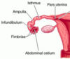
Label all:


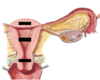
Adnexa of uterus =
- ovary + uterine tube
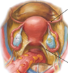
Label all:
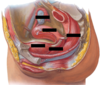

Label:

bladder
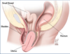
Location of uterus in relation to the bladder in females:
- uterus lays on top of urinary bladder.
- urge to urinate is entirely pressure driven.
- increased urge to urinate during pregnancy due to inreased uterine weight on the bladder.
Vestibule of vagina:
- area between labia minora.
- urethra and vaginal canal open into vestibule.
Path of vagina to abdominal cavity:
cause of pelvic inflammatory disease
- opening of vagina
- up vagina
- into cervix
- into uterus
- through and out fallopian tubes
Vagina blood supply:
- pelvic portion: vaginal artery; branch of uterine artery/internal iliac.
- perineal portion: internal pudendal artery.
Nervous innervation to pelvic vagina:
- Autonomics: uterovaginal plexus
- Pain: pelvic splanchnic nerves (parasympathetic; S2-S4 lower limb pain)
Uterovaginal plexus is a sub-plexus of:
inferior hypogastric plexus

Nervous innervation to perineal vagina:
Pudendal nerve (motor and sensory)
Vaginal fornices:
- recesses created by cervix entering vagina.
- exploited for examination and surgical access.

The anterior fornix can be used to palpate:
bladder
The lateral fornices can be used to palpate:
ovaries, oviduct, ureters
The posterior fornix can be used to palpate:
uterus, rectum, rectouterine pouch (pouch of Douglas)
Through what vaginal pouch can you enter the rectouterine pouch (pouch of Douglas), and therefore the abdominal/pelvic cavity?
posterior fornix
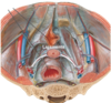
Label all:


Label all:
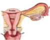
The two openings of the cervix:
- internal os = between cervix and uterus.
- external os = between cervix and vagina.
What is the normal anatomical position of the uterus:
anteverted and anteflexed.
- anteverted = angle cervix enters vagina.
- anteflexed = angle between cervix and uterus body.

Anteflexion of uterus:
- angle between cervix and uterus body.
- uterus tipped forward as it rests on the urinary bladder.

Anteversion of uterus:
- angle cervix enters vagina.
- protects/covers external os of cervix.

As the urinary bladder fills in females, what will occur to the uterus?
- retroflexion: uterus body and fundus tips posteriorly.
- retroflex uterus = retrovert cervix angle into vagina.
Label all:


What occurs to the uterine artery as it enter and traverese the uterine wall?
- uterine artery coils back on itself
- when uterus expands during pregnancy, uterine artery can expand with it.

Infundibulum:
- distal-most portion of uterine tube
- the opening into peritoneal cavity

The little fingers on the end of the uterine tube surrounding the infundibulum:
fimbriae
Fertilization typically takes place in what part of the uterine tube?
ampulla

Label all:


Label all:

What ligaments form the white lines in the image?
- transverse cervical (cardinal) ligaments.
- carry uterine arteries.
- primary support of cervix at pericervical ring.
Label all:


Cause of uterine prolapse (2):
- pericervical ring dilated or stretched.
- connective tissue structures attaching to pericervical ring damaged.
external os of the cervix protrudes first.
Radical hysterectomy:
- removal of entire uterus, adnexa, and upper part of vagina.

Total hysterectomy:
- removal of uterus fundus, body, and cervix.
- adnexa and upper part of the vagina remain.

Partial/sub-total hysterectomy:
- removal of uterine body and fundus.
- pericervial ring and adnexa stay.

