Eye Anatomy Flashcards

1
Q

Label the orbit bones

A
2
Q

Label the fissures/ canal/ formina of the orbit

A
3
Q

What is contained within the optic canal?

A

Optic nerve

Ophthalmic artery

4
Q

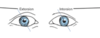
where do eyelids meet?

A

Medial and laterla Canthus

5
Q

Label this

What is the orbital septum?

A

orbital septum- connecitve tissue that attaches to orbit- extension of the periosteum

6
Q

Describe the Muscle which is superficial to the fibrous framework of the orbit ?

what is there function?

A

Orbicularis oculi muscle——> movements of the eyelid (close)

orbital; part- forcefully

Palpebral part- gently

7
Q

what Nerve innervated the orbicularis occuli?

A

CN VII facial nerve

8
Q

Label this

A
9
Q

What is the role of Levator palpebrae?

innervated by

A

innervated by CNIII- oculomotor

10
Q

what is the role of the superiro trasal msucle

A
11
Q

Label this?

A
12
Q

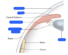
Describe the outer coat of the eye?

A

cornea

Sclera

limbus

13
Q

Describe the middle cosat of the eye (uvea/ vascular layer)

A

iris

ciliary body (ciliary muscles/ciliary processes)

choriod

14
Q

Describe the inner coat of the eye?

A

retinal pigment epithelium

retina

light sensitive tissue that lines back of the eye

15
Q

what is the role of the cornea?

A
16
Q

What makes up 85% of the outer (fiborous) coat of the eye and provides attachments for extraocular muscles?

A

Sclera

17
Q

What is this?

A

Keratoconus

non inflamamotry eye condition- cornea thins and progressive becomes cone like bulge

18
Q

what is the uveal layer

A

uveal or vascular

Middle coat of the eye

19
Q

what is the choriod

A

Most posterior and highly vasuclarised area of the posteriro eye

20
Q

what are the Ciliary processes

A

connected to the lens of the eyeball by the zonula fubres (suspensory ligament of the lens)

21
Q

describe the action and innevration of the ciliary msucle

A

parasympathetic control and CNIII m(oculomotor)

contracts and pulls the ciliary proccess forward-

suspensory ligament relax

lens fattens , more relaced

22
Q

label this

A
23
Q

what is the iris?

A

the iris is an extension of the ciliary body that cotnrols pupil size

this is the coloured area of the eye

24
Q

what is the role of the spincter pupillae?

A

Parasympathetic

constrict the pupil

CNIII (oculomotor)

(circular fibres)

25
Describe the role of the Dilator pupiullae what condition causes paralysis
radiuclar fibres sympathetic dilates the pupil (horners syndrome -due to lesion of the sympathetic fibres cuase parlaysis of the pupillary dilator iris and miosis)\_
26
what is the function of the Lens? how do the ciliary body alter its ability?
focusing light and producing clear sharp images ciliary muscle contract- fattens lens for close vision ciliary muscle relax- lens flattens for long visison
27
Label
28
describe the chambers of the eye?
iris divides the space between the cornea and lens into 2 chamber * Posterior chamber * Bounded by the iris, ciliary processes, zonular fibres and the lens * Anterior chamber * Bounded by the cornea and the iris * Filled with aqueous humour * Canal of Schlemm * Drainage of aqueous humour *
29
What is the conjuctiva?
thin translucent mucous membrane that lines the eyeball and eyelid (palpebral= eyelid), (bulbar= eye) **bulba conjucitva** **palpebral conjuctivae**
30
what is the angle of difference between the axis of orbit and the axis of the eye ball?
23 degrees eeyeball has to move 23 degrees (laterally) to be in the axis of the orbit
31
what are the extraocular muscles of the eye and what is there innervation! hint **(LR6(SO4)3)**
Lateral rectus- CNVI (abducnes) superiro oblique ( CN IV trochlear) CNIII (occulomotor) * **superior rectus** * **medial rectus** * **inferiro rectus** * **inferiro oblique**
32
Label this and what is the innervation
LR6(SO4)3
33
what are these movements of the eye?
34
what is the role of rotation eye movement (extorsion and intorsion)
help when we turn to maintain binocular vision
35
Role of Medial rectus and innervation
adduction (pulls to midline) CNIII
36
what is the role of laterl rectus and its innervation
abduction (pulls away from the midline) Suppplied by CNVI
37
what are 'yolk muslces' of the eye
medial and lateral rectus are both yolk muscles When we look right Medial rectus of left eye contracts Lateral rectus of right eye contracts **MAKE sure that eyes look in the same direction!**
38
Describe the movement of superiro rectus and its innervation
CNIII **_Elevation_, intorsion and adduction**
39
when the eye in abduction what is the only movement superiro rectus can do?
Elevate!
40
Describe the movemetns and innevation of inferiro rectus?
**Depression, extorsion and adduction!**
41
describe the only movemetn inferiro rectus can do when the eye is abducted?
con only depress the eye
42
which muscle attaches to the Trochlea in the eye?
superiro oblique- acts as a pulley sustem
43
describe the movement of the superiro oblique and its innervation
when the muscle contracts intorsion depression and abduction (think diagonally) CNIV
44
when the eye is adducted describe the only movement superiro obliwue can do?
Depression of the eye
45
describe the movement of the inferior obliwue and its innervation
CNIII extorsion, elevation adn adduction (think diagnoally)
46
Describe the only movement inferiro oblique can perform if the eye is adducted?
Elevation
47
What is the ophtalmic artery a branch of ?
internal carotid artery!
48
Label the circle of willis
49
what artery is a branch of the opthlamic artery enters the optic canal / dsic to supply the inner retina?
Central retinal artery
50
what arteries from the circle of Zinn in the orbit?
ciliary arterys branchs of the opthalmic artery supply the outer retina anatastomose
51
Describe Nerve supply to the eye? special sense? gene sens Motor? Parasympathetic?
52
Which CN supplies sensation over the cornea?
The o**phthalmic branch of the trigeminal nerve** Tested by the corneal reflex **CNV1**
53
Describe sympathetic innervation of muscles of the eye?
Superiro trasal and the dilator pupillae are both controlled by sympathetic innervation Pathology- **horners syndroem** **ptossis and miosis (due to unooposed PS nerve supply)**
54
what is the edinger westphal nucleus involved in?
Parasympathetic control of pupil light reflex - travels with CNIII
55
where do we find the Motor nuclei of CN III, IV, VI
Located medially in the braintsem are the motor nuclie of the cranila nerves (extraocualr movements)
56
CNVI opthalmic branch of the trigeminal supplies the general sensation of the cornea- where is its nucleus located
extends thought the brainstem laterally (blue)
57
Label this sagital T1 weight MRI of orbit
Sagittal rectus Optic nerve Levator Pupillae superiors muscle Orbicularis oculi muscle (right) Eyeball Inferior rectus muscle Inferior oblique muscle of eyeball
58
Describe the route of lacrimiantion?
59
what is the role of aqueous humour where is it produced?
supplies nutrients to the avascular front part of the eye clear produced by ciliary bodies
60
Describe the Flwo fo aqueous humour through the eye?
Active secretion and ultrafiltration by ciliary epithelium into the posteriro cghambver flows in front of lens through the pupil to ansteiro change drain out the angle of the eye between the cornea and the iris through the traebcular netowrk into the **canal of schlemm**
61
what is glaucomma ?
anything that disrupts the ciruclation of fluid can casue pressure in the eye to become too low or too high! can be primary (open or closed angle) or secondary due to steriro, uveitis or truama!
62
what is open angle glaucoma?
outflow of Aqueous humour through the trabeuclar meshwork is reduced causes a graudual reduction of the peripheral vision- until end stage
63
what is a closed angle glaucoma?
iris is forced agsints the traebcular meshwork (tissue in the anteriro chamber) prventing drianage of aquaopus humour **_Opthalamic Emergency can rapdily lead to blindness_**
64
what is a glaucoma?
a blockage of aquosu humour draining from the anteriro comaprtment of the eye fluid increase increase in intraocular pressure damages optic nerve
65
what would we seen upon examination of gluacoma
snellen chart reduction ina cuity visual field intirlla peripheraly then central fundoscopy increased cup: disc ratio