Exam review Flashcards
Neural Crest derived mesenchyme from the first and second pharyngeal arches migrates into the
developing face to give rise to facial bones (as well as anterior portion of the sphenoid and frontal bones) and cartilage (Meckel’s cartilage which becomes mandible, incus, and malleus

What are the contents of the Infratemporal Fossa
- Inferior portion of Temporalis muscle
- Lateral and Medial Pterygoid muscles
- Maxillary artery
- Pterygoid venous plexus
- Nervous structure:
- Inferior Alveolar (V3)
- Lingual (V3)
- Buccal (V3)
- Chorda Tympani (CN VII)
- Otic ganglion
Boundaries of the Infratemporal Fossa
- Lateral
- Ramus of Mandible
- Anterior
- Maxilla
- Medial
- Lateral Pterygoid Plate
- Roof
- Sphenoid
- Posterior
- Tympanic plate and Mastoid and syloid process
- Inferior
- Angle of Mandible

Muscles of Mastication are innervated by
CN V3
what are the 4 paired muscles of mastication
- Temporalis
- Masseter
- Lateral pterygoids
- Medial Pterygoids


What are the functions of the intrinsic muslces of the tongue
- Curls, squeeze, and fold the tongue during chewing and speaking
Extrinsic muscles of the tongue
- originate on other head and neck structures and insert on the tongue “Glossus”
- left and right genioglossus originate on the mandible and protract the tongue
- The left and right syloglossus muscles originate on the styloid processes of the temporal bone.
- elevate and retract the tongue (pull the tongue back into the mouth)
- The left and right hyoglossus muscles originate at the hyoid bone and insert on the sides of the tongue.
- Depress and retract the tongue
- The left and right palatoglossus muscles originate on the soft palate.
- Elevate the posterior portion of the tongue
- What goes through the pterygoid canal
- What nerves join
- What modalities do they carry
- Deep Petrosal nerve and Greater petrosal nerve joint ot form the nerve of the pterygoid canal
- Deep petrosal nerve arises from the internal carotid plexus and conveys postysnaptic sympathetic fibers which join
- The greater petrosal nerve carry the parasympathetic fibers of the facial nerve (CN VII) to the pterygopalatine ganglion. (Pre-synaptic parasympathetic fibers are form the superior cervical ganglion)
Contents of the Pterygopalatine Fossa
- Maxillary Nerve (CN V2)
- Pterygopalatine ganglion
- Third part of Maxillary artery
Focus on the flow of cerebral spinal fluid and where it ends up
- Circulation of CSF
- CSF is produced by the choroid plexus in the ventricles
- CSF flows form the third ventricle through the mesencephalic aquaduct into the fourth ventricle
- As the CSF flows through the subarachnoid space, it removes waste products and provides buoyancy to support the brain
- Excess CSF flows into the arachnoid villi, then drains into the dural venous sinuses. Pressure allows the CSF to be released into the blood without permitting any venous blood to enter the subarachnoid space. The greater pressure on the CSF in the subarachnoid space assures that CSF moves in the venous sinuses
functional regions of the cerebral cortex
- Each hemisphere is subdivided into five funcitonal areas called lobes
- Frontal lobe
- Higher intellectual funcitons (concentration, decision making, planning): personality: verbral communication; voluntary motor control of skeletal muscles
- Cortices and Association areas within lobe:
- Primary motor cortex
- Premotor cortex
- motor speech area (broca’s area)
- Frontla eye fields
- Parietal lobe
- Sensory interpretation of textures and shapes; understanding speech and formulating words to express thoughts and emotions (Wernicke’s area)
- Cortices and Association areas within lobes
- Primary somatosensory cortex
- Somatosensory association area
- Part of Wernicke’s area
- Part of gnostic area
- Temporal Lobe
- Interpretation of auditory and olfactory sensations; storage of audiotyr and olfactory experiences
- Cortices and Association Areas within lobe
- Primary auditory cortex
- Primary olfactory cortex
- Audiotyr association area
- Olfactory association area
- Part of Wernicke’s area
- Part of gnostic area
- Occipital lobe
- Conscious perception of visual stimuli; integration of eye focusing movements; correlation of visual images with previous visual experiences
- Cortices and Association areas within Lobe
- Primary visual cortex
- Visual Association areas
- Insula
- Interpretation of taste; memory
- Cortices and Association areas within lobe
- primary gustatory cortex
What is the difference between nasal conchae and meatus
- Nasal Conchae
- divide nasal cavity into 4 passages
- spheno-ethmoidal recess (opening of sphenoid sinus)
- Superior nasal meatus (opening of ethmoidal sinuses)
- Middle nasal meatus (opening of frontal sinus)
- maxillary sinus also opens into middle nasal meatus in posterior part of semilunar hiatus at the maxillary ostium (below ethmoid bulla)
- Inferior nasal meautus (opening of nasolacrimal duct)
- divide nasal cavity into 4 passages
Excess CSF is removed form the subarachnoid space by
Arachnoid granulation
Gnostic Area
Parietal, occipital, temporal- E.g. Clock indicates 12:30, smell food cooking, friend talks about hunger, you interpret it to be lunch time
Wernicke’s area
left hemisphere- recognize, understand, and comprehend spoken or written language. Works with Broca’s area which is the motor speech area
The nasolacrimal duct drains into the
inferior nasal meatus
The Palatine tonsil is the nasopharynx or orothopharynx
Oropharynx
The malleus and incus originate from ____ while the stapes originates from
Malleus and incus originate from Meckel’s Cartilage and the stapes originates from Reichert’s cartilage
The platysma is innervated by
by the cervical branch of the facial nerve
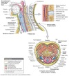
What are the 5 facial layers of the neck
- Superficial fascia
- Investing layer
- Pretracheal layer
- Alar fascia and carotid sheath


The middle ear connets to the ______ via the pharyngotympanic tube (Eustacian Tube)
Nasopharynx


