Exam 4 Wed.3.30.Kidneys Flashcards
What is the Renal capsule?
- surrounds each kidney
- embedded in a mass of fat; protection from trauma

What is Renal fascia?
- surrounds kidney and fat
- attaches kidney to posterior abdominal wall; protection from trauma
*Dr.T emphasized how much it hold kidneys in place and how it may best be appreciated when watching a kidney being taken out of a body

What is Hilum?
- central indentation where blood vessels, nerves, lymphatics and ureter enter and exit
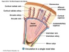
What is the cortex?
- outer region of kidney
- contains glomeruli, most of proximal tubule and some distal tubule
- not the salty region of the kidneys

What is the Medulla?
- inner region of kidney
- contains primarily pyramids, Loop of Henle, and collecting ducts

What are the Pyramids?
contain loop of Henle and collecting ducts

What is the Calyx?
- calices- plural form
- chambers that receive urine from collecting ducts, forming entry into renal pelvis

What is the Lobe?
- structural unit of the kidney
- contains pyramid and overlying cortex ~ 14/kidney

Something to do if you’re mega stressed out or maybe just need a mental break
What is the nephron?
- functional unit of kidney
- 1.2 million per kidney

Tubular structures- What do they do (general)? List the structures (5)
Together function to form urine (end product) via secretion and absorption
–Renal corpuscle- glomerulus and Bowman’s capsule
–Proximal convoluted tubule (PCT)
–Loop of Henle (important in electrolyte and water balance)
–Distal convoluted tubule (DCT)
–Collecting duct (for urine)

What is a Glomerulus?
tuft of capillaries
What is Bowman’s capsule?
surrounds glomerular capillaries
Disadvantage to the “out hole”
- entry point for bacteria
- Women have a shorter urethra than men making them more prone to bladder infections
- 1/3 women get a UTI
Name two (sorta 3) chronic illnesses are highly dependent on kidney function
- Diabetes and hypertension
- Should include metabolic syndrome since it is teh precursor to diabetes
- rely on glucose and electrolyte regulation and balance
Number 1 function of the kidneys
maintain a stable internal environment- homeostasis
What happens to the old kidney with a kidney transplant?
- Docs leave it where they found it. It’s hooked into the aorta and inf. vena cava.
- Won’t remove unless infected
- The new kidney is hooked into inferior vessels near the ilium
Where are the kidneys?
- Tucked under rib cage and musculature for protection
- If kidneys are aggravated, may send pain signals if tapped/palpated over costovertebral angle.
Descriptive word for kidney pain
Colicky (cramping and wave like pain)
List kidney blood vessles in Dr. T’s PP (10)
- Renal arteries
- Interlobar arteries
- Arcuate arteries
- Interlobular
- Afferent arteriole
- Glomerular capillaries
- Efferent arteriole
- Peritubular capillaries
- Vasa recta
- Renal veins
*Bold were vessels emphasized in lecture. She was not as concerned about teh other vessels

Where do Renal arteries branch off from?
abdominal aorta
Where are Interlobar arteries located?
travel down renal columns and between pyramids

*Not emphasized by Dr. T
Where are Arcuate arteries located?
arch over pyramids in cortical medullary junction

*Not emphasized by Dr. T
What are Interlobular vessels connected to?
afferent arteriole

*Not emphasized by Dr. T
What do Afferent arteriole do?
controls blood flow into glomerulus

What do Glomerular capillaries do?
- form a filter
- fist-like structure
What do Efferent arteriole do?
carries blood minus filtrate out of glomerulus

Where are the Peritubular capillaries and what do they do?
- surround tubules and loop of Henle
- involved in reabsorption and secretion
* Not emphasized by Dr. T

What is Vasa recta and what does it do?
- part of peritubular system
- responsible for concentrating urine and blood supply to kidneys
*Not emphasized by Dr. T

What do Renal veins do?
deliver filtered and reabsorbed blood to inferior vena cava

Two facts about blood filtration and the kidneys
- 1000 to 1200 ml or 20 to 25 % of cardiac output per minute
- Body’s entire blood volume passes through kidney in 5 minutes
What is GFR? (2)
- Glomerular Filtration Rate (GFR) – rate of filtration and cleansing of blood
- GFR is related to blood flow and pressure, controlled by neural and hormonal actions
What is the function of a nephron?
include glomerular fitlration rate
include daily urine output volume
- Filter blood/plasma
- Reabsorb water, electrolytes, glucose, proteins, within tubular system and capillaries
- maintain fluid volume, electrolyte balance, pH
- Glomerular filtration rate - 180 L/day filtered, and only produces 1 to 2 L/day of urine output – a 99% reabsorption of water
Auto regulation is an important part of renal blood flow. What are the components of auto regulation? (3)
what is arterial BP range?
- Arterial BP 80 to 180 mmHg
- Macula densa sensing Na+ filtration
- pretty important, controls GFR
- Constriction or dilation of afferent arteriole controls GFR
Neuronal regulation is involved with renal blood flow- How?
–ANS regulation- sympathetic stimulation causes vasoconstriction of afferent arteriole
*If things go wrong here. a person becomes hypertensive.
Hormonal regulation is involved in renal blood flow
How? (2)
- Renin- released in kidney due to decrease BP or Na+ causes retention of Na+ and H2O and increases BP
- ADH- Decreased BP or BV signals hypothalamus signaling posterior pituitary to release ADH resulting in water reabsorption into blood vessels from distal tubules and collecting ducts
ADH increase= retain water
ADH decrease= lose water
What are the effects of exercise on renal blood flow?
- Sodium and water loss- relative decrease in BV and loss of Na+(sweating)
- Increase in BP
- sympathetic stimulation, etc.
What are the effects of increased BP on renal blood flow?
- Renin and ADH decreased, afferent dilation to increase filtration
- can use meds like ACE inhibitors if needed to control BP
Dr. T’s advice about learning this section of the kidney
Be able to draw out the parts and tubular units of the kidney and be able to say what each area does.
Do not need to worry about vascular section as much.
3 parts of glomular filtration membrane
- Inner- has pores
- Middle- selective permeability
- Outer- contains projections called podocytes

What is the main point of having the Podocytes
Back up to say “Keep this” “don’t keep that”
Two parts of the Podocytes of epithelium
- Pedicles- projections which adhere to basement membrane and other podocytes
- Filtration slits- formed between pedicles; covered with anions (negative charge) preventing protein filtration

What is the Glomerular filtrate?
- what passes through the 3 layers membrane
- forms primary urine
What is Bowman’s space?
(different from capsule, but not by much)
area between capillary layers and bowman’s capsule
What are Juxtaglomerular cells?
renin-releasing cells located within afferent arteriole and some in efferent

Where and what are Macula Densa?
- sits between afferent and efferent arteriole
- sodium sensing cells of distal tubule

4 tubes that make up pathway of filtrate
(with details about each)
- Proximal tubule- cuboidal cells with microvilli (brush border); enhances reabsorption
- Loop of Henle- descends into medulla; ascending portion actively transports solutes
- Distal tubule
- Collecting duct- connect nephron to ureter

Why is the medulla salty?
to maximaize water reabsorption

2 cells of the dital tubule and their function
- Principle cells- reabsorb sodium and secrete potassium
- Intercalated cells- reabsorb potassium and bicarbonate and secrete hydrogen
List the hormones of the kidney (5)
- ADH
- Aldosterone
- Atrial Natriuretic Peptide (ANP)
- Urodilatin
- Erythropoietin
(renin?)
Function of ADH
controls H2O reuptake in distal tubules
Function of Aldosterone
reabsorb sodium into vessels from distal tubules and collecting ducts and secrete potassium (K+) from vessels

Function of Atrial Natriuretic Peptide (ANP)
include 2 specific things it does?
diuretic decreases aldosterone by inhibiting renin inhibits Na+ and H20 reabsorption into vessels also dilates vessels

Function of Urodilatin
a natriuretic peptide stimulated by increase BV and BP
Natriuretic:
- Pertaining to or characterized by natriuresis.
- A substance that increases urinary excretion of sodium, usually as a result of decreased tubular reabsorption of sodium ions from glomerular filtrate.
As in Atrial Natriuretic Peptide
Purpose of using Diuretics
- enhance urine flow by decreasing reabsorption of sodium and potassium
- ex: lasix aka thyzide (sp?)
Function of Erythropoietin (EPO)
What does it do?
What stimulates it in kidneys?
stimulates RBC production due to decreased O2 delivery to kidney’s
Renin: what stimulates it? what does it do?
ADH: what stimulates it? (2) what does it do? (what does what?)
- Renin- released in kidney due to decrease BP or Na+ causes retention of Na+ and H2O and increases BP
- ADH- Decreased BP or BV signals hypothalamus signaling posterior pituitary to release ADH resulting in water reabsorption into blood vessels from distal tubules and collecting ducts
ADH increase= retain water
ADH decrease= lose water
2 things that can be checked in the lab
- Clearance rate (Glomerular Filtration Rate,GFR)
- estimated by comparing blood creatine to urine creatinine
- Should eliminate 100% of creatine, therefore amount in blood should equal amount in urine
- BUN- Blood Urea Nitrogen
- Urea is filtered in glomerulus therefore should not be present in blood
- Dehydration and renal failure with increase BUN
Urinary system structures highlighted by Dr. T
- 2 ureters
- Bladder
- Urethra
Points about the ureters
- carry urine formed by nephron from collecting ducts to bladder by peristalsis
- closed off during bladder contraction during urination (micturition) preventing reflux
Something you should look at most likely b/c Thompson spent a good 5 minutes taking about it
Look at the feedback loops at the end of the Structure and Function of the Renal and Urologic Systems powerpoint. Can’t really make cards for it.
What Dr. T highlighted about structure of bladder
smooth muscle fibers form detrusor muscle lined with transitional epithelium
Location of urethra
from inferior bladder to outside body
Non-urinary structures and basic info about them (3)
- Prostate gland- in males; surrounds proximal urethra just distal to bladder
-
Sphincters
- Internal urinary or Internal urethral sphincter- urethra/bladder junction controlled by ANS
- External urinary or External urethral sphincter- voluntary control of urination
- Pelvic floor musculature- help stabilize, position, and control structures of pelvis