Exam 1 - Spring Flashcards
visceral pain is associated with…
hollow organs
- distension
- forceful contraction
solid organs
- stretch of capsule
visceral pain is described as…
gnaw
burn
cramp
ache
parietal pain originates
inflam of peritoneum
pairetal pain is described as…
steady, aching
aggravated by mvmt/coughing
more severe & precise than visc
what type of pain is assocaited with rebound tenderness?
parietal
what position do pts with parietal pain usually like to be in?
lie still
rebound tenderness
pain with quick withdraw of pressure –> inflammation of peritoneal
“which hurts more, when I press or let go?”
what is blumberg’s sign
rebound tenderness
what are other ways to elicit rebound tenderness signs?
percussing pt’s ab lightly ad indirectly
better: “cough”
what is referred pain?
pain at a distance from organ: usually well localized
what parts of the body can refer pain to the abdomen?
chest
spine
pelvis
what is the color of bile vomitus?
yellowish green
what is the color of vomitus with blood?
“hematemesis”
brown/black = “coffee ground” –> blood altered by gastric acids
assessment of stool: diarrhea
increased h2o content
volume > 200g in 24 hours
melena
black, tarry stool
upper GI bleed
hematochezia
bright blood in stool
lower GI bleeding
hematochezia can be caused by
lower GI bleed
BRISK upper GI bleed
melena is usually from ____________ but can also be from….
upper GI
small bowel, right colon
jaundice
yellowish discoloration of skin and sclera
jaundice is due to…
increased lvls of bilirubin (from brkdown of Hb)
increased bilirubin is most suggestive of…
Hb (hemolysis)
problem within hepatobiliary sys
supra pubic pain can be caused by
bladdar/pelvis
bladder infection
urinary retention
order of physical exam for abdomen
inspection
ausculatation: must preceded percussion and palpation!
precussion
palpation
special tests
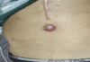
what is this and what is the disease?
abdominal straie
cushing’s syndrome

Sister Mary Joseph Nodule
metastatic disease

Caput Medusae
portal htn

cullen’s sign
periumbilical ecchymosis: bluish discoloration around umbilicus
cullen’s sign usually occurs due to
hemoperitoneum
hemorr panc
uterine tube rupture: ectopic pregnancy
grey turner’s sign: flank ecchymosis
- need to “turn” them over to see
grey turner’s sign usually occurs during
retro-peritoneal bleeding
hemorr panc
why would you perform auscultation of abdomen before palpation anad precussion?
bowel motility
auscul bowel sounds with the _________ of your stethoscope
diaphragm
bowel sounds: normal
5-34/min
gurgling, clicking
borborygmi
loud, audible sounds
prolongeded gurgles of hyperperistalsis
NORMAL
high pitched bowel sounds
tinkling (raindrops on barrel)
signs of early intestinal obstruction
increased bowel sounds are indicative of
diarrhea
early intes obstruction
decreased bowel sounds are indicative of
what is the parameter?
none for a minute - decreased gut activity
ab sx
ab infection/peritonitis/injury
absent bowel sounds are indicative of
parameters?
no sounds ofr 2 minutes
caused by:
- longer-lasting intestinal obstruction
- intestinal perforation
- intestinal ischemia/infarction
vascular abdominal sounds
pathology!
bruit: AAA, renal arteries, fem arteries
friction rubs: spleen, liver
venous hum: b/w xiphistenum & umbilicus
percussion of abdomen helps to assess (5)
amount & distribution of ab
solid or fluid masses
percussion tenderness
size of spleen and liver
ascites
percussion technique
light, then harder
ab percussion: tympany
gastric air bubble
gas filled portions of intestines
excessive tympany with abdominal percussion indicates
excess gas like in an obstruction
ab percussion: dullness
solid organs
unexpected dullness in percussion of ab =
megaly
full stomach
mass
how to proceed with palpation of abdomen?
ask pt if they have pain then palp that place last
palpation technique
encourage relaxtion & palp during exhalation - mouth breath
light: pads
deep: with both hands
what decreases with ab palpation while pt is breathing with mouth open and jaw dropped?
voluntary guarding
You ask your patient to tell you “ which hurts more when I press or let go” and then you proceed to firmly press your fingers down into her abdomen and then withdraw your hand quickly. By doing this technique you have assessed for the presence of:
a) voluntary guarding
b) Blumberg’s sign
c) rigidity
d) Cullen’s sign
Blumberg’s sign
direct assessment of liver is difficult due to…
liver!
percussion - liver span
measure vertical span of liver, mid-clav
- umb –> lower border
- nipple line –> upper border
liver span
mid sternal: 4-8cm
mid clav: 6-12 cm
larger is men & taller people
liver palpation technique
- L (posterior hand) - move liver anterior
- ask pt to take a deep breath
* inhale = liver moves “down” - 3 cm below costal margin - palp “down and up”
- liver should feel: soft, sharply defined, regular (mild tenderness = normal)
what is an alternative way for palp the liver?
hooking technique
spleen percussion
two techniques
- L lower naterior border from cardiac dullness @ 6th rib –> anterior ax line –> costal margin –> Traube’s space
- L anterior ax line = tympanic: ask pt to take a deep breath = still tympanic
spleen palpation
pt supine
- L (posterior hand) to bring spleen anterior
- “take a deep breath”
- spleen will move “down”
- repeat with R lateral fetal (knees partially flexed)
spleen can be palpated in what percent of adults?
5%
kidney palpation
pt supine
- L (posterior) hand to push kidney “fwd”
- “take a deep breath” - will move kidney “down”
- press down with R hand
- R kid = sometimes palp
- L kid = rarely palp
CVA tenderness
- fingertip palpation first
- fist percussion
* use fish to pound on hand - painful jar/thud
bladder can be palpated if…
distended above pubic symphysis: round/smooth dome
usually cannot be palp
palpation of aorta
- press firm and deep on upper ab slightly L of midline, one hand on each side of aorta
- feel for pulsations
- assess width: >3cm = AAA
ascites
protuberant ab with bulging flanks
flud = sinks with gravity while bowel (filled with air) will rise

ascites occurs with (8)
MOCCHIN’
malnutrition
ovarian ca
cirrhosis
constrictive percarditis
heart failure
IVC/hepatic vein obstruction
nephrotic syndrome
how to test for ascites?
shifting dullness when pt turns onto one side
fluid wave
- tap one side –> should feel on other side
- false positive: sometimes not until it is too late or in people without ascite

the presence of what makes the dx of ascites highly likely
positive fluid wave
shifting dullness
peripheral edema
appendicitis symptoms
fever, nausea, vomiting
periumb (vague) –> migration of pain to RLQ
McBurney’s point
2in from ASIS

rovsing’s sign
RLQ pain during palp of LLQ –> referred rebound tenderness

psoas sign
pain with resisted flexion on R side

obturator sign
appendicities
pain on passive internal rotation of flexed thigh

tests for appendicities
- Mcburney’s
- rovsings
- psoas
- obturator
You are concerned that your patient has acute appendicitis. You press on her left lower quadrant and she states she feels pain in her right lower quadrant. You have just determined that the patient has a positive:
a) Obturator sign
b) Rovsing’s sign
c) Turner’s sign
d) Murphy’s Point Tenderness
e) Psoas Sign
Rovsing’s sign
cholecystitis
inflammed gallbladder

murphey’s sign
cholescystitis
- place hand firmly @ RUQ
- “inhale deeply”
- pain/catch breathe –> inflammed!
ventral hernia
hernias of ab wall exclusive of groin hernias
- protrustion intestines thru ab wall
can be seen better if pt raises head and shoulders off table
how tell a hernia from an intra-ab mass?
ask pt to raise head and shoulders off table
“strain down”
hernia = palp
mass = obscured by ab M
- but can feel ab mass in ab wall!!
peritonitis assessment
ask pt to cough and ID the pain
palp with 1 finger, then hand
check for rigid, rebound, percussion, tenderness
peritonitis usually signals
acute abdomen
modifiable breast ca risk (6)
postmenopause
obesity
breast feeding
contraceptives
HRT
ETOH
physical inactivity
breast ca risks: non-modifiable (4)
age:
- > 50
- 1st full-term preg
- menarche: <12
- menopause: >55
breast
- previous ca
- atyp hyperplasia
- density
previous chest wall radiation
probable: hxn no
t breastfeeding
selective risk factors for breast ca
fam hx breast/ovarian ca on both sides
order of breast exam
- inspection
- arms @ sides
- arms over head
- hands on hips
- leading fwd
- palpation
- ax: seated
- breast: supine
- nipple
- if mass felt –> recheck nodes
axilla exam
seated
- “relax with L arm down”
- support pt’s wrist/hand
- cup fingers of R hand –> reach apex
- feel for nodes

nodes of the breast
location and final drainage
pec: anterior ax fold (pec major)
subscapular: post ax fold (lateral border of scapula)
lateral: upper humerus
—-> central —-> supra/infra-clav
pec nodes of breast will drain
anterior chest, most of breast
subscapular nodes of breast
posterior chest, part of arm
lateral breast nodes
most of arm
fibroadenoma
FIBROADENOMA: firm, round/rubbery, nontender, mobile
15-25 y/o
no retraction
breast self exam timing:
5-7 days after onset of menses
BSE for R breast
- lie down, pillow under R shoulder with R arm behind head
- palp with L 3 middle fingers: vertical stripe pattern
- L breast
- soapy shower, 1 arm behind head: repeat steps 2 & 3
- inspect in mirror: arms @ sides, hands on hips
must of a chaperone present for what exams?
breast
rectal
pelvic
auchincloss maneuver
hands on hips, shoulder roll lateral and medial
inspection of breast should happen in what order
pt sitting
- arms at sides
- arms over head
- hands on hips, shoulder roll
- leaning fwd
draping for breast exams
full exposure for exam
drape 1 while palp other
clinical breast exam
pt supine - vertical strip
- circ motion with pads of 3 middle fingers: light –> medium –> deep
- from tail of breast: axilla –> medial: clav to boob fold
pads of 3 fingers
change positions for lateral and medial breast
clinical breast exam: lateral breast
- pt roll to opposite hip
- “put hand you’re not laying on on head”
- palp ax –> nipple
clinical breast exam: medial breast
- “lay supine with shoulders flat”
- “put hand on neck”
- lift elbow to lvl of shoulder
- palp nipple –> midsternum: under boob –> clavicle
assessment of nipple discharge
compress circumfrentially on areola
note # ducts discharge is from
lithotomy position
stirrups
obtain urine specimen _________ GYN exam
before
GYN exam order
- external inspection, palp: genitalia, pubis, bartholin glands
- speculum: cervix - obtain specimen: pap smear then cultures
- internal spection: vag walls, M tone
- bimanual: cervix, uterus, adnexa
- rectovag
- rectal: guaiac/hemoccult
ectocervix
visible portion of cervix
red columnar epith around os, pinik shiny squamous continuous with vag lining
round or slit-like
endocervical canal
lined with columnar
squamocolumnar jxn
@ pub: columnar encircling ox replaced by squamous
later risk of dysplasia
PAP SMEAR
lymphatics of gyn
ingunal nodes: vulva, lower vagina
- only ones accessible to exam
pelvic/ab nodes: upper vag, internal organs
for the best results with cervical ca screening
not on period
nothing in vag for 48 hours: sex, douches, tampons, contraceptive foams/creams, vag suppository
most important risk factor for cervical ca
HPV 16, 18
basic risk factors for cerv ca (8)
early sex
multiple partners
STIs
age
no
PAP
nutrition
smoking
immune status
drape in lithotomy pos
mid-ab –> knees, depress in middle for eye contact
first contact for max comfort in litho pos
inner thigh
lithotomy pos: pos of pt anatomically
thighs flexed & abducted
hips ext rot
examiner ____ for speculum exam and _____ for bimanual exam
sits
stands
gloves for gyn exam
dbl glove dominant hand
bartholin glands palpation
only if hx/inspection suggest problem
“pinch vag @ 4 and 8 o’clock”
speculum insertion steps
- lub speculum
- enter @ 45 angle, closed, over other index finger (in vag)
- direct down and posterior
- remove finger
- rotate speculum to horizontal pos
- open blades: warn of click!
- look into vag
- pos ends of speculum cupping cervix and lock
if cervical discharge is mucopurulent, culture for…
chlamydia
gonorrhea
pap smear
plastic:
- place longer end into os
- press, turn, scrape in full clockwise circle
- smear on glass slide
endocervical brush:
- cone shaped brush into os
- roll in circle b/w thunb and index
- smear onto glass with rolling motion
preg = cottom tip & saline
spray slide with fixative
liquid base test
pap smear
benefits
- less false negatives
- no slides
- no fixatives
use broom –> obtain specimen –> put into container
withdrawl of speculum steps
- warn pt of click with closign the speculum
- release speculum and slowly close while pulling out: inspect vag walls
- rotate speculum @ same 45 degrees with insertion
bimanual exam intro steps
lub index and middle finger of dom hand (has 2 gloves)
insert fingers: palm up, thumb abducted
pain with mvmt of cervix =
chandelier’s sign
CMT: cerv mvmt tenderness
what signs are suggestive of PID?
positive chandellier’s
CMT
adnexal tenderness
what is a blueish hue to cervix or vag walls
Chadwick’s sign of early preg
bimanual exam of uterus
palp hand pressing down & inward b/w pubic symphysis and umbilicus
slide into anterior cervix and sandwich cervix:
- pelvic hand = anterior uterus
- ab hand = posterior uterus

bimanual exam: ovaries
- put ab hand on RLQ
- put pelvic hand in R lateral fornix
- sandwich ovaries: press in and down with outside hand and up with inside hand
- repeat for L side
ovaries should not be palpable in….
postmenopausal women
assessing pelvic M strength
- withdraw fingers so not touching cervix
- spread fingers in vag wall
- “please squeeze my fingers”
- compress snugly and move inwards/upward
- last over 3 sec
rectovag exam
- inform pt
- reglove! remove glove from dominant hand
- lub fingers 2-3
- place index in vag and 3rd in anus
- “strain down” (will relax sphincter during insertion)
urethral exam
perform if urethritis or inflammation of paraurethral glands suspected
insert index into vag and milk outwards
observe for discharge
benign breast mass characteristics
skin changes
smooth, soft
firm, mobile
well-defined
malignant breast mass characteristics
hard, immobile
fixed to skin/soft tissue
irreg margins
skin changes
pre-preg, when to test for rubella?
3 months prior
folic acid
0.4-0.8mg
horm changes in preg
increase estrogen, progesterone, placental hormones (HCG)
estrogen and preg
endometrial growth –> supports early embryo
progesterone and preg
lowers esop sphincter tone –> results in gastroesophageal reflux
relax ureter and bladder tone –> hydronephrosis, incr risk of bacteriuria
coag and preg
hypercoag state
Cv changes in preg
increase:
- RBC mass
- plasma vol
- CO
decrease:
- vasc resistance
- BP
musc-skel changes in preg occur due to…
wt gain
relaxin (horm)
musc-skel changes in preg
lumbar lordosis
lig laxity in SI joints & pub symp
breast changes in preg
stim by horm: increases:
- vasc
- glandular tissue (hyperplasia)
- sens
more nodular by 3rd month
breast changes mid-to-late preg
colostrum expressed
areolae darken
more pronounced montgomery glands
increasingly vis venous pattern
uteral changes during preg
rotates R to accom rectosigmoid struct on L
enlarge –> results in:
- freq voiding
- round lig pain
- R side hydronephrosis
the uterus is most easily palpable above pubic bone @
12-13 weeks of preg
vag secretions during preg
thick, white, more profuse
vag walls during preg
thickens, deeply rugated
cervical changes during preg
chadwicks: increased vasc and edema
increased secr
hegar’s sign
mucous plug
hegar’s sign
palp softening of cer isthmus (portions of uterus that narrows into cervix)
mucous plug fx
protects uterine environ from outside pathogens
scehduled screenings during pregnancy include: (3)
- aneuploidy testing: 1st and 2nd trimester
- oral glucose tol test: 24028 weeks
- rectovag swab for group B strep: 35-37 weeks
preg and constipation
slow GI transit
- horm changes
- dehydration
- iron (prenatal vitamins)
hemorrhoids during preg
constipation
decreased venous return
compression by fetus
changes in activity lvl
when does the center of gravity shift during preg?
3rd trimester
exercise during pregnancy assists: (7)
preeclampsia
preterm birth
decrease length of labor and complications during delivery
DM-G
DVT
varicose veings
wt gain
tobacco and pregnancy
low birth wt
placenta previa/abruption
preterm labor
fetal digit anomalies
spont abortion/fetal death
alcohol and preg
fetal alcohol syndrome
what is the leading cause of preventable mental retardation in the US?
fetal alcohol syndrome
foods to avoid during preg
unpasteurized, raw, undercooked
preg recommends 2 servings of _________ per week
selected fish and shellfish
Gravida
total # times preg
para
babies delivered during viable period
TPAL
term deliveries - preterm deliveries - abortions - living children
G7P5 (4-1-1-5)
7 preg, 5 living children (4 term preg - 1 preterm deliver - 1 abortion - 5 live children)
establishing the expected date of delivery (EDD)
Naegele’s rule:
- first date LMP
- subtract 3 months
- add 7 days
how to establish EDD when actual date of conception is known
conception age which is 2 weeks less than menstrual age can be used
how to verify EDD
- doppler fetal HR: positive @ 10-12 weeks
- ultrasound: 1st trimester
- fetoscope: heard at 18 weeks
- fetal mvmt: quickening @ 18-24 weeks
vital sign changes during preg
BP: falls in middle months –> return to normal in 3rd trimester
HR: increased resting
RR: gen unchanged
chronic versus gestational htn
SBP > 140, DBP > 90 @ 20 wks gestation
chronic: BEFORE
gestational: AFTER
preeclampsia
after 20wks gestation:
- elev BP
- SBP > 140, DBP > 90
- proteinuria
recommended wt gain during preg: low BMI
< 18.5
28-40
Recommended Weight Gain in Pregnancy: normal BMI
18,5-24.9
25-35
Recommended Weight Gain during pregancy: high BMI
25-29.9
15-25
recommended wt gain during pregnancy: obese GMI
> 30
11-20
what is the mask of pregnancy?
chloasma-hypermelanosis of sun-exposed areas DURING preg: 50-70% affected
hair and pregnancy
dry and thinning
mouth and preg
periodontal disease common
nose and preg
congestion and nose bleeds more common
eyes and preg
can be pallor –> anemia
examine retina is BP elevated
lungs/thorax and preg
may complain of SOB but no change in RR
heart and pregnancy
venous murmur common in adv preg
apical pulse be rotated up and L
striae gravidarum
stretch marks
stretch of skin and tear of collagen in dermis
fundal height
pub symp –> top of fundus
auscultate fetal HR with _____ @ 10 weeks and _______ @ 18 weeks
doptone
fetoscope
change in fetal HR from 1st weeks to term
150-160: 1st weeks
120-160: term
during pregnancy, the uterus is in the pelvis until…
12-14 weeks
leopolds maneuver
detms: fetal pos beginning 2nd trimester
greatest accuracy after 36 wks
helps detm readiness for vag delivery
leopolds maneuver helps detm readiness for vag delivery by assessing: (5)
which side fetus back is facing
what part is @ pelvic inlet
upper and lower fetal poles
fetal desc into maternal pelvis
est size and wt
cervical dilation
2, 4, 6, 8, 10 cm
2 = penny
4 = oreo
6 = soda can
8 = donut
10 = roll of cheap TP
freq of prenatal visits
usually individualized but typ
- 0-28 wks: 1/month
- 28-36: every 2 weeks
- 36-deliver: weekly
anorectal jxn
pectinate/dentate line - serrated: sep anal canal from rectum
boundary b/w somatic and visceral N supplies
columns of morgagni
anal columns
- each contain an A and a V –> hemorrhoid!
folds of mucosa from rectum to anorectal jxn
prostate gland location
in front of anterior wall of rectum
surround bladder neck and urethra - 15 to 30 ducts into urethra
fx of prostate
thin, milky, alkaline fluid –> helps sperm viability
prostate structure
bilobed: round/heart shape
2. 5cm long
average length of examining finger of uterus
6-10cm
peritoneum and rectum
covers superior 2/3s
–> rectovesical pouch: males
–> rectouterine pouch: females
valves of houston
3 semilunar txverse valves
lowest one is palpable –> do not mistake for intrarectal mass

technique for rectal exam
left lateral decubitus
pilonidal cyst/sinus
congenital - sinus tract opening with slight drainage
generally symptomatic

anal fistula
inflam tract/tube
openings @ skin, anus, rectum

anal hemorrhoids
chronic increased venous pressure
difference b/w internal and external hemorrhoids?
internal - above dentate line with painless bleeding
external - below dentate line with painful swelling

Pruritus Ani
usually due to pinworms: esp younger pts


Enterobius vermicularis: pinworm
egg deposit on perianal folds
most common symptom = perianal itching
common reinfections -> can affect entire household
anal fissure
oval ulveration - sentinel tag
risk due to anal sex –> easy to transmit HIV, STI’s
anal fissure is usually associated with
prior abscess
proctitis
crohn’s disease

Condyloma Acuminata: HPV - warts
Condyloma lata - secondary syphillis
flat and velvety“moist”
rectal prolapse is a…
projection of pink mucosa seen when pt bears down
what happens to the anal sphincter with pressure?
first reflex tighten and then relax with con’t pressure
anal angle
towards the umbilicus
palpation of anus with severe tenderness
do not force –> ask pt to bear down
maybe use lidocaine jelly
rectal polyps
common
may be pedunculated (on stalk) or sessile (flat)
rectal ca
irregular border - firm, nodular, rolled edge
central ulceration
rectal shelf
peritoneal metastasis to peritoneal reflection anterior to rectum
can be felt with tip of examining finger: firm or hard
occult blood in stool can indicate:
bleed in GI
colon ca/polyps
single negative stool sample does not rule out ca
how can you get a false-positive occult blood test?
ingestion of red meat w/in 3 days of test
grade for colorectal ca screening guidelines
A: 50-75 y/o: sigmoioscopy/colonoscopy
C: 76-85
D: > 85: no screening
I: insuff evid for CT colonography and DNA testing as screening modality
colorectal screening test intervals
annual high-sens fecal occult blood test
or
sigmoidoscopy every 5 yrs with occult every 3
or
colonoscopy every 10 years
DRE and colorectal ca screening
not recommended as stand-alone test
reach is limited
checking stool from DRE will miss >90% color abnorm
DIPSS
discharge from penis
infection
pain in scrotum
swelling in scrotum
sores/growths on penis
phimosis
inability to retract foreskin over glans
tx for phimosis
circular or dorsal slit
paraphimosis
retracted foreskin cannot be returned to regular position
how does one usually get paraphimosis
usu by healthcare personal
hypospadias
displace urethral meatus to underside
balanitis
inflammed glans
balanosposthitis
inflammed glans and foreskin
how to inspect penis for discharge?
compress glans with pinching technique
if no discharge, ask pt to milk
what do you ask the pt to replace before the scrotal exam?
replace foreskin
what is the most ocmmon viral STI in the US?
it can occasionally form…
HPV: warts
large exophytic masses: interfere with poo and sex
tx for anogenital warts
immune therapy
sx
difference in HPV locations in men
circumcised: shaft
uncircumcised: glans
what do anogenital warts look like?
pink or colored lesions
smooth, flat papules –> verrucous papilliform
genital herpes
HSV1/2 - small vesicles –> painful ulcers on red base

uniqueness about herpes
latent state
average incubation after exposure = 4 days (2-12 days range)
syphilis incubation period
2-3 weeks: papule –> ulcer (chancre)
chancre
syphilis ulcer: 1-2cm with raised, indurated margin
syphilis tx
long acting pcn
chancre healing
heal spont w/in 3-6 weeks even in absence of tx
chancre base
usually non-exudative

Syphilis Chancre
peyronie’s disease
fibrotic tunical albuginea –> crooked erection
palp non-tender palques beneath skin of dorsum of penis

penile fx
rupture of tunical albugnea of corpus cavernosum
penile ca
undurated, nontender nodule/ulcer

increased risk of penile ca with
HPV
HIV
smoking
PUVA exposure (tanning beds)
AA (african american)

pearly penile papules: normal varient seen in Af-Am. and circumcised men
asymptomatic acral angiofibromas: corona and sulcus of glans
most freq after puberty
transillumination of the scrotum
if scrotum is swollen, light it up
red glow = serous, hydrocele
dark = blood, testis, tumor, most hernias
hydrocele
non-tender: fl-filled in tunica vaginalis
fingers can get above mass within scrotum
scrotal edema
pitting, taut skin
scrotal edema may be seen in
CHF
nephrotic syndrome
epidermoid cysts
firm, yellowish, nontender skin nodules on scrotum

cryptorchidism
hidden testicle: absent or undescended
- usually desc spont by 6 months: if not –> sx!
most common location: just outside external inguinal ring
not corrected cryptorchidism is an increased risk for
testicular ca
infert
torsion
inguinal hernia
acute orchitis
inflamed painful, tender, swollen testicle: hard to distinguish from epididymis
possible RED scrotum, usually unilateral
due to: mumps (viral)
testicular length is usualy…
less than/equal to 3.5cm
Klinefelter’s syndrome
less than/equal to 2cm small firm testes
small, soft testes can suggest: (5)
- atrophy in cirrhosis
- myotonic dystrophy
- estrogen use
- hypo-pituitary
- follows orchitis
risk factors for testicular tumors
cryptorchidism
ca of contrallat testicle
mumps orchitis
childhood hydrocele
testicular tumor markers
AFP
bHC
what is the most common neoplasm in men ages _________
15-35: test ca
dev of testicular ca
early = painless nodule –> late = replaces testicle (feels heavier than usual)
epididymitis
inflammed vas deferens
usually alongside acute prostatitis
may have red scotum, inflammed vas deferens
varicocele
“bag of worms”
varicose veins of spermatic cord: usually L
why is standing the best pos for locating varicocele?
varicocele collapses when scrotum is elevated
TB of epididymis
chromic inflammation –> firm enlargement of epididymis
thickening/beading of vas deferens
spermatocele v epididymal cysts
s = > 2cm
e = < 2 cm
epididymal cyst
painless, movable mass above testis
transilluminates
testicular torsion
twist testicle on spermatic cord –> necrosis
painful, tender, swollen and retracted UP
cannot elicite cremasteric reflex
inguinal hernia palpation
invag scrotal skin –> travel up inguianl canal –> ask pt to cough/strain down –> hernia will touch fingertip
what is the most common groin hernia in men and women?
indirect inguinal
indirect inguinal hernia
goes through inguinal canal to touch fingertip
defective obliteration of fetal processus vaginalis –> mostly congenital
indirect inguinal hernia are more freq on _______ b/c…..
right, descends last
direct inguinal hernia
usually in men > 40: hesselbach’s triangle (weak inguinal canal floor)
bulge near external inguinal ring - RARELY enters scrotum
femoral hernia
below inguinal lig: women > men
usually more lateral than inguinal hernias
most common methods for screening for prostate ca is….
PSA
DRE
PSA
glycoprotein released by prostate epith cells: biomarker for prostate ca
shortcomings of DRE for prostate ca screening
DRE only reaches posterior and lateral surf of prostate
many false positives
when does prostatic hyperplasia usually begin?
5th decade
during what exam will the pt feel the urge to urinate?
prostate exam: rotation of finger anterior to palp prostate
BPH
symm enlarged, smooth, firm, elastic
protrudes into rectal lumen
median sulcus may be obliterated
prostate ca
area of hardness (nodule) –> usually distinct
can be irregular if prostate enlarged
symptoms of BPH
irritative: urgency, freq, nocturia
obstructive: decreased stream, incomplete emptying with straining
what in men can cause urinary obstruction and chronic UTI?
BPH
what should always raise suspicion for prostate gland pathology
new onset ED
only ____ cases of prostate ca palp on rectal exam
50%
prostatitis
80% = gram negative bacteria
suspect STI in men under 35 y/o
elevated PSA
PSA for ca screening sould be done _________ after prostatitis episode
1 month
both due to elev PSA
prostatitis may cause:
urethral discharge
lower urinary tract obstruction