Erhand Hohenester - Cell Signalling Flashcards

You may prefer our related Brainscape-certified flashcards:
1
Q

What is one way of conceptualize/understanding cell signalling?

A

One way to conceptualize cell signalling is to visualize a cell as device that converts inputs signals into output signal (in response to the input)

Focus of lecture serious - Input signals from other cells

Output – very varied

Not a unidirectional process – feedback/communication between output & inputs

2
Q

What type of signalling does this lecture series focus on?

A

Main focus – inter (between) cellular signalling

Involves a cell releasing an extracellular signalling molecule (peptide/protein/hormone) that cannot pass through membrane unaided.

Hence, it binds to a receptor molecule - yields a change in the receptor which leads to a sequence of intracellular signalling events, ultimately influencing an effector protein

3
Q

Outline the difference between receptor activation and Signal transduction/downstream signalling?

A

Receptor activation - refers to information being transferred across the P.M

Signal transduction/downstream signalling - refers to transfer of information within the cytosol and nucleus

4
Q

What are the two main families of receptors which will be discussed?

A
  1. G protein-coupled receptors
  2. Receptor tyrosine kinases

What’s not included: Notch, Wnt, Hedgehog, NFκB, cGas-STING

What’s also not included: Signalling pathways in bacteria and plants

5
Q

What are the different types of intercellular signalling?

A
  1. Contact dependent – signalling cell physically targets the target cell via two transmembrane cell membrane proteins  widespread but common in the Immune system
  2. Paracrine - local mediator meaning that it interacts with neighboring cells
  3. Synaptic - neuron releases neurotransmitter – released into synapse
  4. Endocrine - can interact with cells over large distances as the hormone/signalling molecule is released into the blood stream

This lecture series will focus on Paracrine and endocrine intercellular signalling

6
Q

What is the difference between paracrine and endocrine?

A

Paracrine & endocrine – Both involve the release of soluble mediators by signalling cell which interact with a target cell, but the difference is distance that the signalling molecule can travel and effect

Paracrine - Short distances effecting neighbouring cells

Endocrine - Longer distances

7
Q

What is autocrine signalling?

A

Autocrine - form of paracrine signalling –> cell is both a signaling cell and target cell

8
Q

What type of signalling are growth factors, cytokines and hormones involved in?

A
  1. Growth factors - Paracrine & Endocrine
  2. Cytokines - Paracrine & Endocrine
  3. Hormones - Endocrine
9
Q

Outline what the following types of signlling molecules are/where they are produced/what is their role?

  1. Cytokines
  2. Chemokines
  3. Hormones
  4. Growth Factors
A

Types of signal molecules

  1. Cytokines are secreted mainly by immune cells and modulate the immune response.
  2. Chemokines are a subset of cytokines that function as chemoattractants.
  3. Hormones are produced by endocrine glands and distributed by the bloodstream. They can be small organic molecules (adrenaline), peptides or proteins, and have a wide variety of effects.
  4. Growth factors stimulate cell growth, proliferation and differentiation. Some cytokines and hormones act like growth factors.
10
Q

Do most signalling molecules pass straight through the membrane or interact with PM receptors?

A

Most molecules are hydrophilic meaning they need a cellular receptor

Example – Beta-adrenergic receptor –> epinephrine binds resulting in conformation change – change intracellular domain which initiates as signal transduction cascade

11
Q

Do hydrophobic molecules also bind to receptors?

A

Most small hydrophobic molecules can diffuse through the P.M., allowing them to bind to intracellular receptors instead

e.g. Steroid & sex hormones

12
Q

Do cells normally respond to singular or a multitude of singalling inputs?

A

Animal cells respond to multiple signal inputs – never just receive one signal –> coordination between a multitude of signals

Act like a control panel receiving a mutlitude of signals, from which it decides it’s next step/action

Signal integration - The process of responding correctly to a number of different signals –> receive multitude of signals and respond with the appropriate output

Example attached is a simple representation of this concept

13
Q

Can the sam signalling molecule elicit different responses?

A

YEAHHHH BOI, context is important as it can influence/alter the effect of signalling molecules, this case cell type influencing the ACh effects

14
Q

What ‘changes/modifications’ are commonly used to alternate between active and inactive states in a signalling cascade?

A

Changes in state link signal inputs to outputs

Signalling information is transduced through the cell and goes through a signalling nodes which switch from an inactive to active state. This switch from off to on can be as a result of…

  1. Conformational changes
  2. Addition or removal of post-translational modifications
  3. Formation or dissociation of complexes
  4. Changes in subcellular localization
15
Q

What effects do PTMs have on the states of signalling molcules/nodes?

A

Effects of post-translational modifications

Changes in state are often associated with PTM as it can alter…

  1. Conformation
  2. Promote partner binding
  3. Prevent protein binding
  4. PTM can influence localization – nuclear vs. cytoplasmic
  5. Proteolytic stability
16
Q

What writers, readers and erasers system when discussing PTM?

A

Most common PTM is phosphorylation –> takes place on Tyrosine, serine or threonine

  1. Kinase adds phosphate (Writer)
  2. Phosphatase removes phosphate (Eraser)
  3. In on the on state, molecules bind (reader) to the activated form of the protein and mediate the output/response

System known as the writer, reader and eraser system

This system is not limited to phosphorylation but also applies to other PTM – Acetylation, ubiquitination

17
Q

How many different kinases & phosphatases are known to exist in the human genome and what is their conserved structure (Kinase)?

A
18
Q

How is kinase activity regulated?

A

Kinase activity is regulated by conformational changes and phosphorylation of the activation loop

Kinase is regulated by phosphorylation itself – autophosphorylation

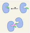
  1. Inactive state – activation loop (orange) blocks the binding site for ATP + C-helix (purple) is in up conformation inhibiting the formation of Lys-Glu salt bridge
  2. Active state – Loop undergoes conformational change that opens up the active site for ATP and substrate binding + C-helix (purple barrel) switches from the ‘up’ to ‘down’ conformation which forms a salt bridge (Lys-Glu) which is required for catalytic activity

As long as the Loop is phosphorylated the kinase remains in its active state until a phosphatase comes in to remove them

19
Q

Why are many anti-cancer drugs protein kinase inhibitors? Provide one example.

A

Many kinases become mutated and overexpressed in cancer cells - driving forward cell growth and progression. Thus, making it an attractive drug target

Imatinib (GleevecTM, GlivecTM) is an ATP-competitive inhibitor targeting the Bcr-Abl fusion protein (fusion protein caused by chromosomal translocation - Abl is a tyrosine kinase) encoded by the Philadelphia chromosome (abnormal Chromosome 22) – constitutively active (constantly)

Imatinib binds to between the Lobes and blocks action - was developed for the treatment of chronic myelogenous leukaemia and has more than doubled the 5-year survival rate of this disease.

20
Q

Outline how GTPases are used as molecular switched.

A
21
Q

What is one very large obstacle that the cell needs to overcome in order to acheive effective signalling?

A

Signal transduction takes place in a crowded environment

Cell’s are extremely busy environment - difficult to coordinate these changes of activation and inactivation –> Still one of the big areas of research

22
Q

How can the formation of signalling complexes increase signalling specificity/overcome cell crowded environment?

A

Signalling complexes increase signalling specificity

23
Q

How does multivalency and membrane association help increase signalling specificity and overcome the busy cellular environment?

A
24
Q

Is there a high degree of response time variability when it comes to cellular signalling?

A
25
Q

What factors are associated with a transient response and a more long lasting response?

A

Signalling systems – persistence of response can be different

Transient response - rapid response and decay

  1. Fast turnover of signal mediators
  2. Negative feedback loops
  3. Rapid Adaptation

Long-lasting (permanent) response - often the case in developmental signals

  1. Switch-like behavior
  2. Positive feedback loops
26
Q

What should you remember when it comes to signalling notation?

A
27
Q

What are the general different scenarios when it comes to positive and negative feedback loops?

A
28
Q

Why is having a positive feedback loop important for a fast response? Graphically what does it do?

A
29
Q

What are the general negative feedback loops that shut down receptor activation?

A
30
Q

What is the effect of having a negative feedback loop with a delay?

A

Negative feedback may lead to dampening of the signal  reduces the amplitude

But…

Negative feedback tend to have a delays resulting in oscillating outputs

Why?

Takes time for the cell to dampen the signal due to noise in biological systems leading to weak negative feedback loop response which diminishes quickly

Solution to this?

If a robust biological oscillations are required, we need positive feedback loops to counteract the effect of the negative feedback - amplify the entire system

31
Q

Outline the example of the robust natural oscillator in Xenopus oocyte cell cycle.

A

CDK oscillates with a period of 1 hour – Robust oscillations

Background - Cell cycle activated by cyclin which activates a CDK – drives cell cycle progression

Core negative feedback loop:

  1. The CDK-cyclin complex phosphorylates the anaphase-promoting complex (APC)
  2. Promotes Cdc20 binding
  3. The APC-Cdc20 complex polyubiquitylates the cyclin, targeting it for destruction.

Core negative feedback loop is sharpened/counteracted by two positive feedback loops

Positive feedback loop 1:

  1. The CDK-cyclin complex phosphorylates the kinase Wee1
  2. Inactivating Wee1
  3. Prevents phosphorylation of CDK-cyclin complex - preventing CDK-cyclin complex inhibition –> Promoting phosphorylation of APC

Positive feedback loop 2:

  1. The CDK-cyclin complex phosphorylates the phosphatase Cdc25
  2. Active Cdc25 dephosphorylates the CDK-cyclin complex,
  3. Activating it (double positive loop) –> Promoting phosphorylation of APC
32
Q

Apart from +ive and -ive feedback loops what other system do cells adopt to control signalling?

A

Logic gates – apart from feedback loops we also have logic gates

  1. A and B need to be present to lead to an output
  2. Neither A and B can be present in order to achieve the output
  3. Either A or B OR Both can lead to an output
  4. XOR – either A or B but not Both leads to output
33
Q

Outline how logic gates and feedback loops can be combined to create sustained input detector?

A

Sustained input detector – used so that only a sustained input yields an output but not a transient input

The system consists of…

  1. Fast branch - happens quickly straight to the gate molecule - alteration in protein activity
  2. Slow branch - happens slowly - doesn’t interact quickly with gate molecule –> change in gene expression

AND Gate –> Means that both fast and slow branch products need to be present to lead to an output

Hence….

  • Only during sustained input do we get elevated levels from the fast and slow branch
  • During transient input the fast branch increases but decays quickly which is followed by slow branch expression - both of which are not aligned in time yielding no response
34
Q

Outline how the sustained input detector is used in mitogen signalling

A

Mitogen signalling – signal molecule leading to cell division (correct output)

For cell division - we need the signalling molecule to induce an sustained input

  1. Signalling by a mitogen leads to activation of MAP Kinase (Mitogen activated protein)
  2. MAPK phosphorylates TFs directly and indirectly – leads to Fos1 transcription and translation
    - Fos1 is TF that is associated with transcription of genes involved in cell cycle progression
  3. Fos1 itself is unstable, so it needs to be phosphorylated itself by MAPK
  4. Hence, upon Fos1 transaltion, the input signal needs to be present and active to stimulate MAPK activity so that Fos1 can be phosphorylated and stabilize - allowing it to act as an effector.

Slow Pathway - Fos1 transcription/translation

Fast pathway - Fos1 stabilization via MAPK

Both of the processed need to be present/aligned in time in order to obtain the desired output of cell division.

Note - Feedback loops in oscillating signals require the mediator to have an intrinsic half-life/stability. Otherwise it remains active preventing the oscillations

35
Q

Outline the general structure of a G-protein coupled receptor and the general activation mechanism.

A
  1. GPCR – Receptor protein has 7 T.M helices  Change’s conformation when binding ligand
  2. Conformational change allows it to associate with inactive G protein, triggering subsequent exchange of GDP to GTP, creating an active complex
  3. The active complex then dissociates from the receptor to form an alpha and Beta subunit both of which are membrane anchored
  4. Either the alpha subunit or the Beta/gamma dimer can activate effector proteins
36
Q

How many GPCRs are there in the human genome and what type of signals are capable of activating GPCRs?

A

There are ~800 human GPCRs, which makes them the largest family of cell surface receptors - 1/3 of all drugs target GPCRs

The signals that activate GPCRs are very varied:

1, Photons (vision)

  1. Exogenous small molecules (smell, taste)
  2. Neurotransmitters (e.g. serotonin)
  3. Small molecule hormones (e.g. adrenaline)
  4. Peptide hormones (e.g. glucagon)
  5. Proteins (e.g. chemokines)
37
Q

Define the following terms in the context of GPCRs:

  1. Full agonist
  2. Partial Agonist
  3. Neutral antagonist
  4. Inverse agonist
A

Most GPCRs have a certain level of basal/constitutive activity – absence of ligand there is still a couple % activity

Full agonist (native ligand) will activate the receptor to 100%

Partial agonist – less than 100%

Neutral antagonist suppresses activity to the basal level

Inverse agonist – reduce activity below basal level

38
Q

What is a popular example of a GPCR?

A

Popular example of GPCR - The β2-adrenergic receptor

Class A GPCR/rhodopsin-like

  • First GPCR to be cloned (Lefkowitz laboratory, 1986).
  • Target of beta blockers (used to treat angina pectoris since 1964) - antagonist that resemble adrenaline and inhibit activation of the adrenergic receptor

Image - 2 structures superimposed – Blue - Inactive / Green - active

39
Q

What is the following protein?

A

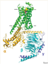
β2-adrenergic receptor in complex with heterotrimeric G proteins

Represents the entire Heterotrimeric G protein receptor complex

40
Q

What are the different families of GPCRs?

A

Variety of different classes/families of GPCRs

  1. Class A – Rhodopsin like receptors
  2. Class B – Larger Extracellular domain – receptor for peptide hormones (glucagon)
  3. Class C – Different ligand binding domain which is coupled to a normal GPCR –> form dimers – Metabotropic glutamate receptor
  4. Class F – Even larger of Extracellular domain

Note - The coupling mechanism with the heterotrimeric G protein is the same for Class A and B

41
Q

What structural determination technique has allowed for the structural detemrination of several GPCRs?

A

Due to advances in Cryo-EM we were able to obtain structures for a variety of Class A and B GPCRs as well as GPCR + arrestin

42
Q

Outline the step by step process by which GPCR is activated.

A

Activation of heterotrimeric G protein by ligand bound GPCR

(1) Agonist binding leads to a conformational change in the GPCR, which stimulates G protein coupling and GDP/GTP exchange in the G protein (alpha-subunit)
(2) Active, GTP-bound, G protein dissociates into α subunit and βγ dimer (both are membrane-bound due to lipid modifications).
(3) The α subunit and βγ dimer regulate effector proteins (e.g. AC, adenylyl cyclase – ATP to cAMP).
(4) GTPase activity of G protein will lead to GTP hydrolysis deactivates the α subunit, which eventually reassembles back with a βγ dimer.

43
Q

What are the different families of G-proteins?

A
44
Q

What are the three different models of GPCR activation?

A
45
Q

What is Adenylyl cyclase (Gα protein effector)?

A

Adenylyl cyclase is an effector protein - regulated by Gα protein effector

Converts ATP in cyclic nucleotide (cAMP) – removes diphosphates

Adenylyl cyclase – membrane spanning protein with two intra-cellular globular domains C1a and C2a (homologous to each other) which possess enzymatic activity

1997 the globular domains coupled with alpha G protein domain were crystallized

46
Q

What does the complete adenylyl cyclase structure resolved in 2019 tell us?

A

2019 – full length structure was determined using Cryo-EM

12 TM helices form bundle in membrane

C1A and C2A associate with each other

47
Q

What are some examples of hormones that rely on AC for signalling?

A
48
Q

Outline the effects that cAMP have on Cyclic AMP-dependent protein kinase A.

A
49
Q

Give an example of a slow response mediated by protein kinase A.

A

Example - Somatostatin signalling –> activates a slow cell signalling response via PKA

Somatostatin is an inhibitory hormone.

  • In the digestive system, it decreases gastric emptying and the release of pancreatic hormones.
  • In the nervous system, it decreases the release of pituitary hormones (growth hormone, thyroid- stimulating hormone, prolactin)
50
Q

Outline a mechanism by which somatostatin induces a slow response.

A

Mechanism:

  1. cAMP created and binds to inactive PKA to release activated PKA
  2. Active PKA enters the nucleus and phosphorylates the TF CREB
  3. Activated CREB can bind to the CRE promoter element – leading to gene transcription
  4. Products of gene transcription create desired effect

CRE = cAMP response element

CREB = CRE-binding protein (a transcription factor)

51
Q

Provide an example and brief mechanism of a fast response mediated by PKA

A

Example of a fast response mediated by PKA: fight-or-flight response

Fight or flight

  1. Adrenaline binds to receptor - B-adrenrgic receptor –> activation of Heterotrimeric G-protein
  2. Leads to an increase in cAMP via adenylyl kinase
  3. Leads to activated PKA
  4. Leads to phosphorylation of the inhibitory subunit that normally binds & inhibits the calcium channel –> when phosphorylated it is released
  5. Calcium channel open –> allows for the subsequent influx of calcium ions
52
Q

Provide a summary of the downstream signalling that occurs from Adenlyl cyclase.

A
53
Q

Apart from AC, what other effector is important downstream of GPCRs - G-Alpha?

A

Phospholipase C-β is another important Gα protein effector

Phospholipase C-β is a enzyme that is associated with the inner leaflet of the P.M. via its CT domain

Upon activation of GPCR –> the G-alpha subunit bound to GTP allosterically activates PLC-β which cleaves the phospholipid PIP-2 (located on the inner leaflet of the membrane) –> releasing a soluble hydrophilic head group (IP3) and leaving behind DAG (diacylglycerol)

54
Q

Outline the cleavage reaction performed by PLC-β.

A
55
Q

What are some examples of cell responses mediated via PLC-β?

A
56
Q

What effects do the PLC-β cleavage products (IP3 and DAG) have on a cell?

A

Schematic showing/elaborating on the PLC-β action

  1. Diacylglycerol - remains in the membrane and associates with PKC (protein kinase C)
  2. After cleavage, IP3 acts as a secondary messenger and diffuses to the ER and associates and opens Ca2+ channels resulting in the movement of Ca2+ from the lumen [high] to the cytosol [lower]
57
Q

Outline how IP3 and ryanodine receptors produce Ca2+ waves?

A

Two types of receptors in the ER which are called

  1. IPs receptors
  2. Ryanodine Receptors

We get positive and negative feedback loops which result in waves of calcium being released

  1. Positive feedback loop - IP3 binding to IPs receptors leads to movement of Ca2+ into the cytosol which induces/activates other channels (Ryanodine Receptors) to activate/open – amplification of signal at lower Ca2+ concentrations
  2. Negative feedback loop - once high calcium concentrations are reached they inhibit the activity of the channels preventing the movement of Ca2+ into the cytosol
    - Ca2+ pumps in the ER restore low Ca2+ cytosol concentration until IP3 binds again to induce a release of another wave
58
Q

What does the following graph depict?

A

Vasopressin-induced [Ca2+] oscillations in liver cells

Illustration of the wave phenomenon produced IP3 and ryanodine receptors in ER

Graph illustrates relationship between Vasopressin concentration applied to cells and the concentration of Ca2+

  • High Vasopressin concentration, result in increased IP3 release resulting in quicker oscillations of calcium within the cells (waves move closer together)
  • In response to stress, vasopressin induces liver cells to release glucose from glycogen.
  • The liver vasopressin receptor is a class A GPCR, and downstream signalling occurs via the PLC-β-IP3 pathway.
59
Q

What is the role of Calmodulin in Ca2+ signalling in the cell?

A

Calmodulin relays the increase in [Ca2+] to other proteins

Concentration of calcium is sensed by calmodulin

Presence of Ca2+ –> Calmodulin binds to 4 Ca2+ via positive cooperativity –> induces a conformational change whereby the linker forms a stable helix which can interact with effector molecules (e.g. CaM kinase peptide)

60
Q

What is the one of the main proteins that active calmodulin interacts with?

A

Ca2+/calmodulin-dependent kinases (CaMKs)

CaMK II is a dodecamer, consisting of two rings of six subunits each.

The subunits consist of a kinase domain and a non-catalytic hub domain, which are connected by a linker containing the CaM-binding site and an autophosphorylation site.

61
Q

Outline the mechanism by which Calmodulin activates CaMKs

A

CaMKs is a large oligomeric enzyme consisting of 2 rings of 6 subunits – Hub domain, kinase domain and linker

  1. Inactive kinase domain can undergo conformational change (pops out) into the tighlty associated active state - known as molecular flickering
  2. Calmodulin-Ca2+ comes in and binds to the linker region –> permanently moving the kinase domain to the disassociated active state – Stabilizing the active conformation
  3. In the active state the kinase domain can phosphorylate targets and autophosphorylate itself –> by autophosphorylating it is stabilizing the disassociated active state
  4. Ca2+ may decrease and the calmodulin will dissociate from the linker but since the linker was auto phosphorylated it remains active until the phosphates are removed by a phosphatase –> example of a form of molecular memory as the kinase remains active even in when Ca2+ drops
62
Q

What consequence does CaMK II molecular memory on its activity?

A
  1. When calcium spikes are low (e.g. when hormone concentration is low) – there is sufficient time between the Ca2+ waves for the phosphorylation on the linker to be removed by a phosphatase - reduced CaMK II activity
  2. When calcium spokes are high/frequent (e.g. when hormone concentration is high) – there is not sufficient time for the phosphatase to act to inactive the CaMK II, so we get a rapid increase in its activity (memory from the previous wave) until the system saturates
63
Q

Provide a summary of the different downstream effects of phospholipase C-β signalling?

A
  1. GPCR activated - activates G-alpha protein
  2. Activates PC-β which results in PIP cleavage
  3. DAG remains in the membrane and activates protein kinase-C
  4. IP3 is released in the cytosol which interacts with Ca2+ channels in the ER, causing a wave of Calcium release
  5. Ca2+ can then bind and activate Calmodulin which in its active state can interact with various effector molecules (CaMKs)
64
Q

Are different GPCR states associated with paritcular disease states?

A

GPCR variants have also been associated with particular disease states

Example of a monogenic disease - congenital stationary night blindness

Linked with to a constitutively activating mutation which increases GPCR activity

65
Q

What are Orthosteric and allosteric sites in context of GPCRs? What role do allosteric modulators play?

A

Orthosteric site - Binding pocket accommodating endogenous receptor ligand

Allosteric site - binding site that is distinct from the orthosteric site

Allosteric modulators are classified as negative allosteric modulators or positive allosteric modulators (NAMs and PAMs, respectively) –> They modulate the function of the ligand in a positive or negative manner – can be endogenous (Sodium – NAM / Cholesterol – PAM or NAM) or synthetic

66
Q

What are bitopic ligands?

A

Bitopic ligand - Molecules that interact with both the orthosteric and allosteric sites, may be either agonists or antagonists.

It is clear that there are multiple intermediate conformational states that exist for GPCRs (alone and in complex with their effectors) - as shown by the ternary model many different activation states and protein/ligand bound are possible – some of which have been corroborated using structural data

67
Q

Provide an overview of the olfactory system anatomy.

A

Brain structure called the olfactory bulb which sits above our nasal cavity

There are gaps in the ethmoid bone that allow for the passage of Olfactory sensory neurons into the olfactory epithelium

Specialized neurons - Olfactory sensory neurons (OSN) connect the olfactory bulb to the nasal cavity – goes through openings in the ethmoid bone –> These neurons have cilia that responsible for detection odorant molecules

Olfactory epithelium - most composed of OSN and support cells called sustentacular cells. This layer also contain olfactory stem cells which regenerate OSN neurons which are one of the few neurons that can be regenerated in the human body

68
Q

In general terms how does the olfactory system work?

A

Olfactory system

Each olfactory sensory neuron expresses only one type of odorant receptor (OR) – each receptor is encoded by different gene product

ORs constitute the largest GPCR family in mammals

~700 human OR genes (~350 are functional) – the other 350 are pseudogenes which are most likely being lost during evolution

~1,200 mouse OR genes (~800 are functional) – number of receptor genes is larger as they rely on smell much more

Mice express 5-10 million OSNs – all the neurons that expresses the same kind of receptor connect to same glomeruli (region on the olfactory bulb) – 1000-fold convergence onto this brain region

69
Q

Explain what is meant by the combinatorial coding of olfactory information

A

We don’t recognize 350 different smells, instead we recognize much more than that

This is possible due to the different combinations of receptors activated by a particular odorant –> each combination generating a unique odour

Odorants can interact with multiple receptors

70
Q

How does the signalling take place in odorant receptors?

A
  1. Odorant molecule binds to OR
  2. Activates heterotrimeric G-protein - in particular Gαolf activates adenylyl cyclase.
  3. Adenylyl cyclase increase cAMP concentration
  4. The increase in [cAMP] opens cAMP-gated Na+/Ca2+ channels – directly opens channel – leading to the influx of ions, depolarization and an action potential
  5. The initial depolarization is amplified by Cl- efflux through Ca2+-dependent Cl- channels – calcium enters and binds to the Ca2+-dependent Cl- channels stimulating Cl- efflux.

Grey zone – less relevant to cell signalling - explaining how only a single OR is expressed in a neuron

71
Q

What is the one receptor, one neuron rule and how is it enforced?

Olfactory system

A

One receptor, one neuron –> refers to the fact that OSN only express a single type of neuron

How this rule is enforced is not clear but…

One study in zebrafish, OR gene silencing was dependent on OR activity: signalling through G protein βγ subunits was both necessary and sufficient to suppress the expression of other OR genes - βγ subunits responsible for suppression of OR genes?

(Ferreira et al. (2014), Neuron 81:847-859)

72
Q

Outline the anatomy of the visual system/retina.

A

Photoreception – anatomy of the retina - back of the eye

  1. Sclera – connective tissue
  2. Choroid – vasculature – nutrients and oxygen
  3. Retinal pigment epithelium
  4. Photoreceptors - Cone and Rod Cells

The human eye contains ~120 million rod cells - more sensitive, localized to the periphery of the retina, little color distinction.

~6 million cone cells - less sensitive, localized to the center of the retina, three different types: red, green, blue.

  1. Two further layers of cells - Bipolar cells – connect light sensitive cells to ganglion cells (cells connect to the brain)

Note - Strange setup of cells - Two layers of cells (Ganglion and Bipolar cells) between light source and photoreceptors

73
Q

Outline how GPCR receptors in rod cells help us detect light

A

Rod (most sensitive) photosensor cells

All photoreceptors hyperpolarise in response to light -interior becomes more negative

Dark – GPCR called rhodopsin is inactive - Cation channels are open in the outer segment – Cell depolarization (more positive) and high release of neurotransmitter to Bipolar cells.

Light – GPCR rhodopsin is activated, closing the cation channels - resulting in hyperpolarization of the cell –> the net result is that no neurotransmitters are released which is interpreted by the Bipolar cell, ganglion cell and brain as light being perceived

74
Q

What are the two different types of bipolar cells for cone cells?

A

There are two types of bipolar cells for cone cells:

ON bipolar cells depolarize in response to light – pass on a signal

OFF bipolar cells hyperpolarize in response to light – passes on a different signal

A similar ON/OFF system exists for rod cells, but it involves a different type of neuron (amacrine cells).

The ON/OFF pathways are important for the detection of light increments and decrements.

75
Q

How does rhodopsin changes conformation in response to light?

A
76
Q

Outline what happens to rhodopsin in rod cells in the dark?

A
77
Q

Outline what happens to rhodopsin in rod cells in the light?

A
78
Q

Outline the signal amplification that takes place in rod cells.

A

Signal Amplification in Rod Cells

  • This signalling pathway is characterized by a significant amount of signal amplification
  • As we can see from the cascade 1 photon absorbed by rhodopsin leads to a change in membrane potential of 1 mV (106-107 Na+ ions prevented from entering in one second)
  • Human retina has a sensitivity of around 5-10 photons
79
Q

What are the four different mechanisms by which we reduce Rhodopsin/GPCR activity?

A

Return to resting state and adaptation – Move from Light to dark

Four different mechanisms…

  1. GAP binds to the transducin Gα subunit and stimulates GTP hydrolysis – leads to a decrease conversion of cGMP to GMP – increase cGMP
  2. Closing of cGMP-gated channels leads to a decrease of cytosolic [Ca2+], which in turn stimulates the guanylyl cyclase to make more cGMP.
  3. The falling Ca2+ levels activate rhodopsin kinase (GRK1) by causing the dissociation of the inhibitory EF-hand protein recoverin. GRK1 phosphorylates the cytosolic tail of rhodopsin, partially inhibiting transducin (G-protein) activation.
  4. Arrestin-1 binds to phosphorylated rhodopsin, further inhibiting transducin activation.
80
Q

Outline how GAPs are used to help reduce Rhodopsin/GPCR activity.

A

GAPs are tethered to the cGMP phoshodiesterase - help to convert active G-alpha (GTP) to inactive form (GDP bound), preventing it from stimulating cGMP phoshodiesterase

Transducin GTPase is the rate-limiting reaction in rod photoresponse deactivation (completed in <0.5 seconds) - GTPase converts active G-Alpha to inactive form by change it from the GTP bound state to the GDP state –> the presence of the GAP increase the rate of the rate-limiting reaction

  1. RGS9-Gbeta5 –> GTPase complex
  2. Membrane anchor - R9AP (TM-protein) anchors complex to photoreceptor disc membranes
81
Q

Outline how GRK1 and arrestin help reduce rhodopsin activity - help return to resting state

A

Return to resting state and adaptation – GRK1 and arrestin (Slower response)

  1. GRK1 –> Falling Ca2+ levels activate rhodopsin kinase (GRK1) by causing the dissociation of the inhibitory EF-hand protein recoverin.

Active GRK1 phosphorylates the cytosolic tail of rhodopsin, partially inhibiting transducin (G-protein) activation

  1. Arresting binds to the phosphorylated tail – inhibiting its activity – completely inhibits any activation of transducin
82
Q

Outline the different stages within the rhodopsin life cycle.

A
83
Q

Outline the steps within the retinoid cycle (Reformation of 11-Cis-retinal from the All-trans-retinal)

A

The entire cycle takes several minutes to complete (recovery time after photo-bleaching).

Retinoid cycle doesn’t take place in the photoreceptor cell but instead in the retinal pigment epithelium – adjacent to photoreceptor cells

84
Q

Apart from shutting down Rhodopsin activity, what other roles do Rhodopsin-arrestin play?

A

Arrestin is the terminating step for rhodopsin but…

β-arrestins appear to be involved in signalling - desensitization, internalization, MAPK signalling and transcriptional activation

This means that when the Rhodopsin in bound to β-arrestin it can itself become involved in signal transduction - different pathway

85
Q

Using Rhodopsin in bound to β-arrestin as an example, explain what is meant by Bias of the system

A

G-protein bias vs. Arrestin Bias

Bias of the system - Refers to the fact that ligands can bind to either the G-protein bound state or the arrestin bound state depending on its bias - Bias depends on the nature of interaction with the receptor

86
Q

What is meant by the following terms…

  1. Biased Ligand
  2. Biased receptor
  3. Biased system
A
  1. Biased ligand – ligand induces a unique receptor conformation that results in differential coupling to the signal transduction cascade and a biased response relative to a reference agonist - different agonists or even the same agonist with different PTMs binding to different sites - e.g. Differential glycosylation of FSH
  2. Biased receptor - modifications of the receptor influence its ability to bind ligands or transducers - ultimately influencing the response

E.g. Number of phosphorylation sites on the rhodopsin tail – the receptor will be biased towards or against arrestin vs G-protein binding

E.g. mutation of glutamic acid, serine or threonine residues in the c-terminus of neuropeptide Y4 receptor disrupts agonist-induced recruitment of β-arrestin 2 and receptor endocytosis

  1. Biased systemdifferential expression of transducer elements proximal to the receptor, such as the receptor itself, β-arrestins or GRKs, as well as expression of amplification cascades distal to the receptors -
    e. g. cell increases GRK expression leading to more phosphorylation and β-arrestins binding - β-arrestin bias

E.g. Certain agonists targeting the dopamine 2 receptor (D2R) have different effects in the striatum and prefrontal cortex, which could be related to the differential expression of β-arrestins and GRKs across brain regions

Note - GPCR is very conformationally labile – very mobile molecule. Hence, depending on what molecules bind we get different conformations

87
Q

What are the different classes of Enzyme-coupled receptors/

A

Class of Enzyme-coupled receptors - Ligand binding to the receptor activates an intrinsic catalytic activity of the receptor

  1. Receptor tyrosine kinases
  2. Tyrosine kinase-associated receptors - similar but the tyrosine kinase is a separate polypeptide (different gene) that is permanently associated with the receptor
  3. Receptor serine/threonine kinases
  4. Receptor-type tyrosine phosphatases (not discussed)
  5. Receptor guanylyl cyclases (not discussed)
88
Q

How do enzyme-coupled receptors differ to GPCRs?

A

Unlike in GPCRs (associates with heterotrimeric G protein), the catalytic activity of enzyme-coupled receptors (e.g. tyrosine kinase) is associated permanently with the receptor, either as part of the same polypeptide chain or non-covalently.

89
Q

Outline the basic structure and acitivity of receptor tyrosine kinases (RTKs)

A

Receptor tyrosine kinases (RTKs)

  • Receptor tyrosine kinases are single-span transmembrane proteins (single alpha-helix traversing the membrane) with an extracellular ligand binding domain and an intracellular tyrosine kinase domain.
  • Ligand binding results dimerization allowing for trans-autophosphorylation of the receptor –> generating binding sites for signalling proteins
90
Q

How many RTKs are encoded by the human genome?

A

The human genome encodes 58 RTKs

  • Extracellular ligand binding domains is varied
  • But the intracellular is always a intracellular tyrosine kinase
91
Q

What are some examples of signalling molecules that signal through RTKs?

A

Examples of signalling molecules that signal through RTKs.

  1. Epidermal growth factor (EGF) - important
  2. Insulin
  3. Nerve growth factor
  4. Fibroblast growth factor
92
Q

Why is it important to understand RTKs in the context of cancer?

A

RTKs are frequently over-expressed or mutated in cancer

93
Q

What is a common experimental technique used to detect auto-phosphorylation?

A

Receptor activation is commonly studied by Western blotting using antibodies against phosphotyrosine (anti-pTyr).

Procedure –> Cell treated with ligand, cell lysed, immunoprecipitated, run on SDS-page and blotted with antibody against phosphotyrosine.

Increasing ligand conc. results in increased tyrosine phosphorylation

But!

To ensure that the total level of RTKs is not changing we…

Control for the total level of receptor – you strip the blot and re-probed with an antibody for the receptor itself. Hence, we can safely deduce that the increase in phosphotyrosine is due to receptor trans-autophosphorylation rather than an increase in receptors

94
Q

Are a large proportion of RTKs activated by ligand-induced proximity of kinase domains?

A

Yes, many RTKs are dimerised by their ligands.

The proximity of the kinase domains in the RTK dimer favours trans-autophosphorylation.

95
Q

What are some examples of RTK ligand dimers?

A
96
Q

What is an example of a monomeric ligand that can induce RTK activity?

A

Epidermal growth factor (EGF) is a monomer. When it binds to monomeric EGF receptor, a dimerization arm is in receptors is exposed, which promotes EGFR dimerization.

Binding of EGF to monomeric EGF induces a conformational change which exposes the dimerization arm which allows EGFR to dimerize

97
Q

How does the insulin receptor go against the classical norm of a RTK?

A

Insulin receptor is a covalent, disulphide-linked dimer –> receptor is already dimerized!

Insulin receptor is composed of the polypeptide chains (alpha and beta). The dimers are held together by disulphide bridges

Activity

When insulin binds, the extracellular domain changes conformation which ultimately brings the two intracellular kinase domains together allow for phosphorylation

Takeway - Highly variable

  1. Some RTKs are non-covalently associated in the absence of ligand (e.g. DDRs)
  2. Some RTKs require higher-order clustering for full activation (e.g. DDRs)
  3. Some RTKs require co-receptors (e.g. FGFRs)
98
Q

Explain the importance of Heparan sulphate acting as a co-receptor for fibroblast growth factor

A

Heparan sulphate is an essential co-receptor for fibroblast growth factor

99
Q

Outline the changes that occur in the kinase domain between the active and inactive state using Insulin receptor

A

Kinase Domain – Active v.s. Inactive – example Insulin Receptor Kinase

Inactive state – Activation loop (green) blocks the activation site – Tyr1162 adopts a pseudosubstrate conformation, meaning it occupies the site where the actual substrate tyrosine would be located

Active state - the activation loop undergoes a conformational change so that it no longer blocks the active site + activation loops three tyrosine residues are phosphorylated preventing returning to the inactive state - Substrate tyrosine (other kinase activation loop) occupies the active site

100
Q

Outline the activation loop dyamics that take place in RTK?

A

The unphosphorylated activation loop is predominantly in the inactive conformation – blocking the active site.

But there are transient excursions out of the active state

When dimerization occurs…

For transphosphorylation to occur, two kinase domains with the activation loop in the active conformation have to meet; this is much more likely if the kinase domains are in close proximity –> One loop acts as the substrate the other free’s the active site

Two requirements are needed for transphosphorylation

  1. Transient excursion of the loop so that it can be phosphorylated – but once one is phosphorylated it is locked (untill phosphatase removes it) so it can easily phosphorylate the other
  2. Two Kinase are in close proximity to each other – dimerization

Note - Auto-phosphorylation of the activation loop is the first step in the activation of most RTKs –> this followed by phosphorylation of other sites in the cytoplasmic domain

101
Q

Outline the process by which EGF receptor tyrosine kinase is activated by an allosteric mechanism (does not follow classical example)

A

Best studied receptor EGF - does not follow the classical mechanism just described

  1. Dimerization of the inactive monomers in presence of EGF
  2. When dimerization occurs, they form a asymmetric dimer - one kinase (activator) allosterically activates the receiver

3, Activation results in the autophosphorylation of the C-terminal tail not in the activation loop

102
Q

Why is the phosphorylation of RTKs intracellular domain so important?

A

Once the Receptor is phosphorylated, the phosphorylation sites become docking sites for downstream mediators and effectors

103
Q

What are the three makin classes of intracellular signalling proteins for RTKs?

A
104
Q

What protein domain is responsible for recognizing phosphotyrosine docking sites?

A

SH2 (and PTB) domains recognize phosphotyrosines

The human genome encodes ~100 SH2 (Src homology region 2) domains, which mediate pTyr binding.

Structure

Small globular domain with a conserved pocket that recognizes and bind phosphotyrosine as well as a specificity pocket which accommodates an apolar residue (located 3 residues upstream)

105
Q

What does the modular architecture of signalling refer to and why is it important?

A

Modular architecture –> refers to the fact the signalling protein are composed of different domains (i.e. lego blocks), each providing it’s own specificity

E.g. SH2 domains are found together with other domains, for example PH and C2 which allow these mediators/signalling molecules to recognize the plasma membrane

Why is it useful?

The modular architecture enhances specificity (e.g. membrane targetting/binding domains) and allows the formation of multiprotein signalling complexes – target multiple proteins at the same time

106
Q

Outline the mechanism for Src Kinases.

A

Mammals have several Src family kinases - Rous sarcoma virus has captured and integrated the tyrosine kinase into its genome but a truncated version - viral oncogene (promotes tumour formation)

Inactive form - Inactive form is autoinhibited allosterically by a SH2 domain which recognizes a phosphotyrosine at the C-terminal - this binding forcrs Src tyrosine kinase in a conformation where the C-helix is in its Up position + the activation loop is blocking the active site, rendering the active site inactive

Active form - Phosphatase removes the phosphate of the C-terminal Tyr 527, preventing SH2 binding and thus resulting in a conformation in the active site (C-helix down + active loop swings out) –> rendering the receptor active

Note - V-src lacks the regulatory C-terminal region so the SH2 domain does not recognize phosphotyrosine and thus the Src Kinase remains in the active state

107
Q

Does EGF stimulate a whole range of intracellular signalling networks?

A

Yes, a whole host of signalling pathways are activated but there will be a focus on three in the lecture course

108
Q

Outline the cascade of reactions in the Ras-MAP kinase pathway.

A
109
Q

What is the key region in RAS that changes depending on GDP/GTP bound states?

A

Ras is a small GTPase – Central to Ras-MPK pathway

Structural difference - GTP and GDP bound states

The switch helix is one of the regions that changes most between the GDP- and GTP-bound states.

In the GTP-bound state of Ras, the switch helix is able to interact with Ras effectors

110
Q

What are the two possible states of RAS?

A

Ras – Molecular Switch

Two forms

  1. Off state – GDP bound - created by intrinsic GTPase activity of the GTPase but process is accelerated by GAPs
  2. On-state – GTP bound - created by an exchange of nucleotides – GDP leaves and GTP binds which is catalyzed by GEF
111
Q

Where in the cell is Ras located?

A
112
Q

How many members are ther in the Ras family?

A

Ras is a member of a superfamily of small GTPases

Three members of the Ras family which is part of a larger family of GTPases

  1. H/K/N-Ras
  2. Rheb
  3. Rap1

Rho family GTPase - change the cytoskeleton in response to the cell surface

113
Q

What function does the Rho family of GTPases perform?

A

Digression: Rho family GTPases regulate actin dynamics

114
Q

Outline the cascade of reactions that result in Ras activation in response to RTK activation via Grb2 and Sos

A
115
Q

Outline how activated Ras-GTP activates the MAP kinase pathway

A
116
Q

What is the benefit of having a kinase cascade within cells e.g. Raf –> Mek –> Erk?

A

Signal amplification by kinase cascades

Benefit - allows for significant amplification as each kinase can activate/act on multiple kinases

117
Q

In the MAP kinase pathway are there many different kinases?

A

Yes, There are a multitude of MAPKKKs, MAPKKs and MAPKs

In order to create some order scaffold proteins are used.

118
Q

What are the scaffold proteins responsible for organising Raf, MEK, Erk and MLK, MKK and JNK?

A

Scaffold help organise the different MAP kinase pathways ensuring that correct output is produced by organising all the relevant players

119
Q

How does Erk (the last kinase in a MAP-Kinase pathway) regulate gene transcription/cell cycle?

A
120
Q

Outline how a sustained input from a mitogen signal is required to drive cell division via the MAPk pathway

Note - Sustained input detector (already discussed)

A

c-fos is involved in the sustained input detector system

Sustained mitogen signals through Ras-MAP kinase pathway activating ERK leads….

  1. Slow response - Fos1 transcription via TCF and SRE
  2. Fast response - Phosphorylation of Fos1 via an activated ERK

Only if Fos1 is stabilized by phosphorylated and is able associate with c-Jun to form the AP-1 TF, do we get cell division

Note - the signalling pathway is constructed in this way so that a brief mitogen input doesn’t lead to cell division but sustained mitogen signalling does drive cell division as the Fos1 can get transcribed and phosphorylated.

121
Q

How is the MAPK Erk pathway regulated/switch off?

A

This pathway is controlled by… Multiple negative feedback loops

Two examples of -ive Feedback loops

  1. Fast response - Erk phosphorylates Sos breaking Grb2-Sos interaction
  2. Slow response - One of the gene’ transcribed as a result of Erk action is MKP (phosphatase) which directly dephosphorylates Erk

MKP = MAPK phosphatase - (family of dual-specificity phosphatases, i.e. act on pSer/Thr and pTyr)

122
Q

Summary of key points Ras-MAP kinase signalling – Central to cell division

A
  1. Ras activation by RTKs is short-lived (intrinsic activity + GAPs) + RTKs are inactivated by specific phosphatases
  2. The MAP (mitogen-activated protein) kinase pathway amplifies the signal and conveys it from the cell surface to the nucleus.
  3. Only a sustained input drives cell division
  4. Various positive and negative feedback loops exist to modulate the strength and duration of the signal.
  5. Scaffold proteins help prevent crosstalk between parallel MAP kinase modules.
  6. Due to the importance of Ras-MAP in cell division - Ras and B-RAF are proto-oncogenes. Ras mutations are found in ~15% of human cancers. B-RAF mutations are found in ~60% of melanomas
123
Q

What type of receptor is normally recognizes cytokines?

A

Signalling by cytokines (JAK-STAT pathway)

Cytokines are recognized by receptors that are similar to RTKs – Tyrosine Kinase associated receptors (Tyrosine kinase is a separate protein bonded to the receptor – e.g. JAK)

124
Q

Briefly outline the JAK-STAT pathway - cytokine signalling

A

Mechanism

  1. Ligand binds – dimerization
  2. JAKs phosphorylate each other as well as receptor
  3. Creates docking sites for mediators (STAT)
  4. STAT become phosphprylated and dissociates
  5. After dissociation, STATs form a dimer which enters the nucleus in order to influence transcription

Note - Phosphorylation is required for dimerization and thus activity

125
Q

Briefly outline the signalling cascade intiated by transforming growth factor-β (TGFβ)

A

TGFβ – important for development and wound healing

Mechanism

  1. TGFβ dimer binds to receptor - forming a heterodimer between type 1 and 2 TGFβ receptors
  2. Type 2 receptor phosphorylates type 1 receptor (serine/threonine kinase)
  3. Active Type 1 kinase phosphorylates a Smad2/3 (TF) which can form a dimer with Smad4
  4. Moves into the nucleus where it binds to TGFβ cis-regulatory sequence act as a transcription regulatory complex
126
Q

What is one of the main advanatages of STAT and SMAD signalling relative to regular RTK pathways?

A
  • RTK pathways are longer & involves more players
  • STAT and SMADs pathways are much more direct and quicker – involve 1 TF which is phosphorylated by the receptor which can then translocate to the nucleus in order to influence transcription –> faster response
127
Q

Apart from the central Ras-MAP kinase pathway, what other two mediators in the EGF pathway will also be examined?

A

The two mediators present in EGF signalling – PI3K and PLC-γ

128
Q

Outline how RTK activation (EGFR) by EGF results in PIP2 cleavage as well as the consequences of cleavage reaction

A

PIP2 cleavage by PLC-γ generates the second messenger IP3

Pathway

  1. Activated EGFR/RTK is bound by PLC-γ which has a 2xSH2 domain
  2. Activated PLC-γ causes the cleavage of PIP2 - releasing IP3 and diacylglycerol
  3. IP3 interacts with Ca2+ channels in the ER, releasing Ca2+, which is sensed by Calmodulin and thus Calmodulin dependent kinase
  4. Diacylglycerol interacts with protein kinase C activating it (binds Ca2+ as well)
    - Identical to what has been described for GPCR
129
Q

How is PIP3 produced in the plasma membrane?

A

Phosphoinositide 3-kinase produces PIP3

PIP2 is composed of 2 fatty acids, glycerol and Inositol sugar (each hydroxyl group in the sugar ring can be phosphorylated) – PIP2 – biphosphate (4,5)

To generate PIP3, PIP2 is phosphorylated by PI 3-kinase at position 3.

130
Q

What domain is capable of recognizing PIP3?

A

PIP3 is recognised by pleckstrin homology (PH) domains

131
Q

Outline the PI3K/Akt pathway responsible for regulating cell survival and growth

A
  1. Activated RTK bound by survival signal
  2. Activated PI-3 kinase binds to activated RTK (allosteric activation) leading to conversion of PIP2 to PIP3
  3. PIP3 acts as a docking site for 2 different kinases (PDK1 and Akt)
  4. Akt become phosphorylated by PDK1 and mTor
  5. Active Akt dissociates and phosphorylates a protein called bad
  6. Inhibitory action of Bad terminates releasing an active apoptosis inhibitory molecule (Bcl-2/Bcl-xl) –> P-Bad binds to 14-3-3 protein
  7. Bcl-2/Bcl-xl inhibit apoptosis - If signal is not present the cell would undergo apoptosis
132
Q

How is PIP3 converted back to PIP2?

A
133
Q

Summarize the different roles of PIP2, PIP3, IP3 and DAG.

A
134
Q

What are the two main ways to terminate RTK signalling?

A
  1. Dephosphorylation - Carried out by protein tyrosine phosphatases (PTPs)
  2. Endocytosis – remove the receptor from the cell surface –> Receptor recycling or degradation

Note - Does not always terminate the signal as signalling can continue in endosomal compartments  same topology

135
Q

Explain how ubiquitination of the EGF results in its internalization.

A

Ubiquitination of activated EGF receptor by the E3 ligase Cbl

When EGF becomes activated and phosphorylated it can recruit both Grb2 and Cbl

Cbl has a E3 ligase that can ubiquitinate the receptor signalling for internalization – terminating the signalling

136
Q

Is there overlap between GPCR and RTK pathways?

A

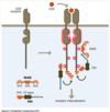
Yes there is Overlap of GPCR and RTK signalling pathways (“cross-talk”)

Example….

  1. Phospholipase C/IP3/diacylglycerol pathway are stimulated by both receptors
  2. Arrestin can itself be a signalling mediator and can turn on MAP kinase pathway
  3. Recent publication – activated RTK can influence G-proteins as well
137
Q

If NGF and EGF both the same Ras-MAP kinase pathway, how come NGF binding to NGFR causing cell differentiation and EGF binding to EGFR causes cell proliferation?

A

Different feedback loops determine signalling outcomes

Famous experiment - PC12 cells expressing both EGF and NGF were exposed to either EGF or NGF resulted in different outcomes

a) EGF – resulted in proliferation
b) NGF – resulted in differentiation

EGF and NGF both activate the Raf-MEK-Erk pathway in PC12 cells but different feedback loops at play

EGF - A negative feedback loop downstream of EGFR produces transient activation of the MAPK pathway, resulting in cell proliferation - Erk phosphorylates SOS which inhibits Raf via Ras

NGF - A positive feedback loop downstream of TrkA (NGF receptor) creates a bistable system, resulting in cell differentiation and neurite outgrowth –> Erk phosphorylates protein kinase C which phosphorylates Raf stabilizing the system meaning that the signalling remains activated

138
Q

But how exactly does negative and positive feedback loops determine proliferation (EGF) vs. differentiation (NGF)?

A

Erk is responsible for both c-Fos transcription as well as phosphorylation

Hence, in EGF as we only get a brief stimulation of ERK only small quantities of c-Fos will be stimulated resulting in proliferation.

Whereas for NGF the sustained signalling results in increasing quantities of stable pc-Fos created stimulating differentiation

Basically different quantities of pc-Fos produced as shown by graphs in figure

139
Q

How does Erk know what to do in the NGF and EGF scenarios - how does it know to phosphorylate SOS or PKC?

A

Possible ideas

  1. Another player involved – e.g. another player that activates with NGFR which allows PKC activity – effect on PKC that is unique to NGFR
  2. Possibly internalization?
140
Q

What experiment was performed to validate the two-responses generated by ERK and NGF?

A

Different feedback loops determine signalling outcomes

Introducing players to modulate feedback loops in order to examine the outcome

  1. Add PMA to cells – stimulates PKC  even EGF will lead to differentiation as we have artificially stimulated PKC  resulting in a NGF like profile
  2. Add Gö7874 inhibits PKC action - converts the normal NGF profile (differentiation) into the EGF profile of proliferation as it inhibits the +ive feedback loop that sustains the signal
141
Q

Is everything that bossman Hohenester told us a over simplifcation?

A

‘YESSSSS’ - Borat accent

142
Q

What receptor does bossman Hohenester focus his research on?

A

Hohenester’s own research of a specific RTK – DDR1/DDR2

DDR1 and DDR2 are 50% homologous and are thought to function in a similar way

143
Q

How was the discoidin receptor discovered?

A

Authors were searching for proteins related to the insulin receptor and screened a human placental cDNA library with a 32P-labelled antisense oligonucleotide against a region that is conserved in many tyrosine kinases

Basically, probing a cDNA library for new kinases

They discovered a new RTK with a discoidin-like domain in the extracellular region –> at the time it was known as an orphan receptor - unknown ligand

144
Q

What ligand activated discoidin domain receptor tyrosine kinase?

A

Collagen –> Surprise that this structural protein also had a functional role as a ligand

145
Q

What was unusual about the activation kinetics of DDR by collagen?

A

Activation kinetics was very unusual

Experiment

Cells expressing the receptor are exposed to the activating ligand for different time periods and the lysed cell is probed for antibodies against phosphotyrosine (activation) and followed by probing for receptor using antibodies (control)

Results

It takes 2-4 hours of ligand exposure to obtain saturation of autophosphorylation of the discoidin domain receptor

Plus the levels don’t of phosphotyrosine don’t drop quickly (sustained for several days)

146
Q

What are the physiological and pathological roles of DDRs?

A

Physiological and pathological roles of DDRs

Discovered from gene knock-out studies and studies in human patients

DDRs are involved in Tissue and organ development

  1. Mammary gland (DDR1)
  2. Inner ear, kidney (DDR1)
  3. Long bones (DDR2)

Range of Pathologies where DDRs are associated

  1. Atherosclerosis (DDR1)
  2. Kidney disease (DDR1)
  3. Arthritis (DDR2)
  4. Fibrosis (DDR1 and DDR2)
  5. Cancer (DDR1 and DDR2)
147
Q

What were the questions addressed in Erhard Hohenester’s research?

A

How does collagen bind to the DDR receptor?

How does collagen activate the receptor?

How is it regulated?

148
Q

What was the ligand-binding assay performed in DDR research by Erhard Hohenester?

A

First thing required before performing further experiments is a ligand binding assay

  1. Collagen (ligand) is immobilized to wells in the 96 well plate and the receptor is added with increasing amounts
  2. Add an antibody against the receptor –> so if receptor binds the antibody will also bind – antibody is coupled to an enzyme that creates a visual color when substrate is added (coupling) –> colour change is an indicator of receptor binding
  3. Plot O.D. (color) against receptor construct added (His-DDR2) - we get an asymptotic curve

Results

Collagen I – high affinity but when heated/denatured affinity is lost.

Furthermore, other extracellular proteins (Laminin and fibronection) are also tested - low affinity

149
Q

What technique allowed Erhard Hohenester to study the collagen binding site with DDR?

A

Collagen is a highly repetitive structure – glycine at every 3rd position to allow for the superhelix to be created + other common A.A. is proline

Not easy to recombinantly form collagen + break into small peptides. Thus, making it hard to run experiments to determine the binding site

BUT!!!

Collagen toolkit allows us to create small collagen fragments that retain triple helical structure

Native guest sequence (actual sequence) is flanked by host sequences (synthetic sequences Gly + Pro – high propensity to form triple helix)

Able to create a library of different collagen fragments that harbour a region of the native sequence

150
Q

Using the collagen toolkit library, how were they able to determine the collagen binding sequence for DDR?

A

Identification of the DDR2 binding site in collagen

Collagen toolkit library containing 56 peptides that span the entire native collagen sequence

Used the ligand binding assay to scan the entire library

Results?

Recognition sequences overlapped between two peptides, hence producing the two central peaks – sequence given in attached image (key residues highlight - O = hydroxyproline)

151
Q

What did the crystal structure DDR2 DS domain bound to a GVMGFO peptide reveal?

A

Having identified the key collagen sequence – the peptide was co-crystallized with DDR2 DS domain (extracellular region)

M, F and O play an important role - substitution of these residues eliminate binding

Important residues come from 2 different chains in collagen. Hence, explaining why the denaturation of the triple helical structure results in a loss of binding - residues need to be present and positioned in the specific conformation relative to eachother

152
Q

What disorder are DDR2 mutations linked to?

A

Bone disorder related to DDR2 – SMED

Defect in the growth of long bones and is accompanied by abnormal calcification

153
Q

What impact do SMED mutations have on DDR2 trafficking?

A

Examining cells expressing DDR2 – either WT or mutants

Cells were stained for DDR2 and Ras - Ras is used simply as a marker for the Plasma membrane

Merge – combination of DDR2 and Ras stain  allows for identification of any differences

Results

  1. WT and E113K are normally trafficked
  2. Kinase domain mutants of DDR2 are not trafficked to the surface - these mutants remain interior (likely in ER) explaining why they are disease causing
154
Q

Why does E113K mutant in DDR cause disease if it is trafficked correctly?

A

The E113K mutation abolishes collagen binding

E113 –> Glutamic acid is one of the key residues involved in hydroxyproline binding

155
Q

What is one experimental method that can be used to study where ligand induced RTK dimerization? i.e. where do the receptors dimerize?

A

One way to study RTK dimerization can be studied is using disulphide cross-linking

Idea – make number of mutants where you replace residues with Cys in the linker and transmembrane region

Subsequently, you expose mutants to the ligand, extract them and run them on non-reducing SDS-page gel to detect which species dimerize (only be present as dimer if disulphide bridge formed) allows us to identify mutants that readily cross-link

Example using EGF

156
Q

What were the results from the DDR1 cross-linking experiment?

A

Performed the cross-linking experiment in the presence and absence of ligand

Results

  1. Strong dimerization signal for all the mutants created in presence and absence of collagen

Meaning that even in the absence of a ligand DDR is a dimer + there is efficient cross-linking –> different to EGF mutants on previous card

  1. Cross-linking had no effect on DDR1 activation – all were able to bind to phosphotyrosine antibody - normal behaviour
157
Q

What did fluorescent DDR1 visualization reveal about DDR1 signalling behaviour?

A

Collagen induces the formation of large DDR1 signalling clusters

Absence of Ligand (unstimulated) - DDR1 forms clusters

Presence of ligand (10 min) - DDR1 redistribute to form peculiar worm like tubular structures – unusual for RTKs

Clustering is not yet fully understood

158
Q

What section of the cytosolic juxtamembrane region is required for DDR1 activation?

A

DDRs have a very long juxtamembrane region – unstructured

BUT!

Only the last quarter of the JM region is required for DDR1 activation

Experiment - Deleted different regions of the JM region and examine impact on DDR1 activation (Phosphotyrosine antibody)

JM4 was identified to be required for DDR1 activation as its deletion abolishes activation

JM4 must carry out some functional role

159
Q

What did the crystal structure of DDR1 JM4-kinase region reveal about JM4’s role?

A

Crystal structure illuminated importance of JM4 - this region inserts itself into a cleft in the kinase domain above the activation loop.

JM4 region reinforces autoinhibition by the activation loop - basically also sterically blocks the kinase in the inactive state – preventing ATP binding + positioning of C-helix for kinase activity

160
Q

Outline the experiments performed on JM4-kinase domain to show that JM4 was phosphorylated before the activation loop.

A

Experiment A) –> Expose JM4Kinase-WT and mutants to ligand for different time periods and run on native-PAGE

Results

  1. JM4K-WT showed increase in movement in the first 5 minutes and after 3 hours –> increase in movement due to phosphorylation as it increases the charge of the molecule

BUT! –> JM4K tyrosine mutants (residues in JM4 loop) –> did not exhibit this increase in movement –> Not getting phosphorylated

Experiment B) –> Run a similar experiment but stain the gel with site-specifc tyrosine antibodies –> showing us that JM4 tyrosine residues (orange) are phosphorylated before activation loop (green)

What the fuck does this all mean???

Tyrosine residues in the JM4 region become phosphorylated quickly, followed by the slow phosphorylation of the activation loop –> unusual as normally the activation loop become phosphorylated first in RTKs

JM4 phosphorylation is required first before activation loop phosphorylation –> removal autoinhibtory effect

161
Q

What type of kinetics does DDR1 kinase exhibit?

A

Graph shows us that only normal Michaelis-Menten are obtained when both JM4 and A-loop are phosphorylated

There is some reduced activity when JM4 is only phosphorylated

This holds true for both substrates – Peptide and ATP

162
Q

Main takeaway from the research performed on JM4 DDR?

A

JM4 phosphorylation is required for DDR1 activation

  1. JM4 Y569F mutation didn’t have a significant effect on receptor activation
  2. JM4 Y586F - reduction in activity
  3. JM4 Y569F/ Y586F - abolished activity – receptor is basically inactive
163
Q

Summary of the findings from Erhard Hohenester DDR research?

A
  1. A triple-helical peptide library was used to identify the DDR binding site in collagen - normally recombinant dissection is used
  2. X-ray crystallography has revealed the details of the DDR-collagen interaction
  3. Disulphide cross-linking experiments ruled out ligand-induced dimerization or conformational changes as mechanisms of DDR activation
  4. Using Super-resolution microscopy has revealed large-scale DDR redistribution in response to collagen binding - tubular structurs
  5. Structural and biochemical experiments have shown that JM4 plays an important role in DDR autoinhibition and activation –> act as a second level of control of DDR above simply the activation loop.
164
Q

How do OR in Olfactory sensory neurons switch off their activity when a odorant is no longer present?

A

Decrease in odorant - GPCR is no longer activated

  1. The cAMP concentration lowers as it hydrolyses to AMP
  2. Alpha subunit of G-Protein effectively terminates its own activity, hydrolysing GTP to GDP, re-joins to the beta and gamma subunits and retains its resting (OFF) state
  3. Cell regains its electrical neutrality and maintains Ca2+ homeostasis by pumping Ca2+ out of OSNs by sodium-calcium (Na+/Ca2+) exchangers, voltage-gated chloride channel (ClCn) and transport proteins Ca2+ ATPase
165
Q

How do OSN adpat to a odorant in the short term?

A

Adaptation / Short-term adaptation (STA) refers to a decrease in the response to the odors by OSNs - reduction in excitation even if odorant is still present

CNG - cyclic nucleotide-gated channel

OSN excited –> Influx of Calcium

  1. Calcium bound calmodulin (Calm) binds to CNG channel reduces channels sensitivity to cAMP, thus CNG channel closes
  2. Ca2+/Calm activates enzyme Phosphodiesterase (PDE) which transforms cAMP to 5’AMP
    - cGMP is also produced during odorant stimulation - [cGMP] decreases CNGs close
  3. cGMP-activated PDE2 hydrolyses both cAMP and cGMP, hence terminating their action
  4. Regulators of G-protein Signaling (RGS) also suppress GPCR mediated signals by accelerating the hydrolysis of GTP bound to the Gα subunit
166
Q

How does OSN desensitization (longer term) occur in response to an odorant?

A

Desensitization involves loss of responsiveness by ORs in the repetitive presence of odorants or stimulus

Mechanism includes phosphorylation of the ORs resulting in uncoupling from its heterodimeric Gα-olfprotein –> phosphorylation also signals for their internalization to the cytosol and down-regulation of the cellular components of OR

PKA, PKC and GPCR specific kinases (GRKs), phosphorylate the serine and threonine residues present in the intercellular loops and C-terminal regions of ORs.

Note - GRKs promote binding of arrestins, cytosolic co-factor proteins, causing steric uncoupling of the receptor and G-protein

167
Q
A