Bill Wisden - Neuroscience Flashcards
Why do we want to study the brain?

What are the different levels one can study the brain?

From an evolutionary standpoint why would we need neurons?
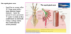
Hypothetical multicellular organism with sensory cells (S) that control motor cells (M) (e.g., cilia) by releasing a chemical transmitter or hormone into the common fluid space – diffusion like this takes time…
Direct connections between sensory and motor cells by means of nerve axons speed up this communication
Communication is quicker and more specific – Wired in speed and specificity
Note - the ultimate action on the motor cells is still chemical
Hence, a sensible speculation could be that the nervous systems evolved to hardwire and improve the specificity of communication (Sensor direct connects to motor cell)

Why can’t we rely on diffusion?
Simple organism could rely on this but this would hinder communication in larger/more complex organisms…
On what timescale does diffusion normally occur?

What is Stokes-Einstein law?

How long would it take for a molecule to diffusion along 1 cm of axon (rough calculation)?
Why 6D?

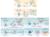
How was axonal movement studied?

What were the three types of movement identified from pulse-chase radiolabeling?
Discovered three types of movement – faster than passive diffusion but not fast (hours – days) –> facilitate movement along these long axonal regions
Fast - 50-400 mm/d –> 3 hours for movement from cell body to terminal
Slow b - 2-8 mm/day
Slow a - 0.2-1 mm/day
How is material moved inbetween the cell body and axon terminal?

What are the two main cytoskeletal elements?

What are the different motor proteins and what do they transport?

Outline the general process by which signals are transferred from one dendrite of one nueron to the dendrite of another

Why are membrane channels needed in neurons?
Movement of ion through the lipid bilayer would require a lot of energy – thermodynamically not favourable
Membrane - Negatively charged phosphate on head + hydrophobic environment

What is the basic idea that allows for electrical signal creation in neurons?
Basic idea of electrical signals – Driven by differences of ion conc. on opposite sides of membrane
What is the role of pumps in neurons?
Active pumping –> The action of the pumps is crucial for the maintenance of ionic concentration differences within membranes (essential for the creation of electrical signals)
There are many different kinds of pumps - Most use ATP as an energy source to build up a gradient of ions.
A large proportion of the energy intake of a human is devoted to the operation of ion pumps - explains the increase of mitochondria in neurons
How does a sodium pump work?

What are the distribution of ions across neuronal membranes at a resting state?
Distribution of ions across neuronal membranes
Different animals, varied numbers for the exact concentrations.
But the relative concentrations, high K in, low K out, high Na out, low Na in, are always the same - High K+inside and High Na+ outside
Other things to note…
A- –> anions of other ions e.g. Carboxylate groups (COO-) of amino acids, sugars, etc.
Chloride is high on the outside and low on the inside
Other important ion for signalling is Ca2+ - tends to be higher outside
Balancing out the charges of all ions…
we get an electrochemical gradient where the inside of the cell is more negatively charged relative to the outside – between -60 to -75 mV

Using the example attache below (K+ permeable membrane), outline what is meant by electrochemical equilibrium

All systems move towards equilibrium – this case electrical & chemical equilibrium
Setup - membrane permeable only to K+ ions + high concentration of K+ and A- in one cell
- K ions start to diffuse down their concentration gradient from one side to the other – left to right down chemical gradient
- This results in a build up of charge (Negative - left + positive - right) - electrical potential difference builds up
- Chemical forces causing a net diffusion of K from left to right are now countered by a growing electrical force which opposes the flow of K+
- Eventually an electrochemical equilibrium potential is reached where the electrical force equals the chemical (or diffusional) force, and no exchange occurs

How can we calculate membrane potential at equilibrium for a given ion species?

What kinds of stimuli can open channels?

In relation to neurons, what are changes in membrane potential/voltage used for?

Definition of depolarization and hyperpolarization?

What happens to voltage as an electrical signal passes through an axon?

How can the concept of the space constant, λ, be used to explain the necessity for action potentials in larger organisms?

What is meant by passive spread?

How do neurons use action potentials to overcome the loss of voltage described by the space constant?
Action potential - Cyclical process
- Depolarization further upstream in the axon
- Electric field spreads along axon - passive spread
- Change in voltage changes membrane permeability via the action of voltage gated channels
- Trigger another round of depolarization
Cycle continues - like a positive feedback loop

Outline what is meant by the all or nothing concept for action potentials?
All or nothing –> refers to the fact that a threshold change in membrane voltage has to be reached in order to drive another membrane potential
If the threshold is not reached - signal is NOT propagated

Is the threshold for action potential fixed?
The threshold is not fixed.
For a short period of time after the firing of the action potential, the threshold is much greater than normal.

How does the rate of action potentials firing act as information?
Since the change in membrane potential only fluctuate within a given rang (can’t be used)…
The only way to convey intensity is by changing the frequency/rate of action potential firing – runs on a rate code
Increased firing - increase intensity of signal being conveyed
There are some exceptions where information is coded in graded sub-threshold potentials

Why was the squid axon particularly useful to study?

How were the first recording of an action potential performed?
The squid giant axon can be up to 1 mm in diameter – a thousand times thicker than in humans.
The first recording of an action potential using a microelectrode inserted into an axon was done by Alan Lloyd Hodgkin and Andrew Fielding Huxley in Plymouth UK in 1939 –> allowed them to see changes in voltage/membrane potential
What are the different phases of an action potential and their corresponding changes in mVs?

During the course of an action potential, how does sodium and potassium conductance change?
After reaching threshold gNa (sodium conductance) increases quickly, but inactivation then reduces gNa to zero.
gK (potassium conductance) increases more slowly, and only decreases once the voltage has hyperpolarized.

What is the difference between absolute and relative refractory period?
The absolute refractory period is when the sodium channels are inactivated
The relative refractory period is when gK dominates following the action potential

How are the kinetics of Na and K currents governed?
Current (movement of charge)
- K potassium currents activate following depolarisation but show little inactivation
- Na currents are governed by two kinetic processes activation and inactivation following depolarisation
Both Na and K channels deactivate (i.e. close) when the membrane potential is hyperpolarised
Definitions of Activate, Inactivate and deactivate?
Activation – opening following depolarisation
Inactivation – closing independent of voltage
Deactivation – closing following hyperpolarization
What does the following illustration depict?


During the resting potential, what is going on in terms of channel and the ion currents?

During the depolarization stage, what is going on in terms of channel and the ion currents?

During the repolarisation stage, what is going on in terms of channel and the ion currents?

During the refractory period, what is going on in terms of channel and the ion currents?
Channel inactivation & deactivation?

After an action potential, how are ion gradients re-established?

Do we need a lot of ions to change the membrane potential?
Very few ions are needed to change the membrane potential
To change the membrane potentially from -65 to 0 by sodium ions – we only need to change the sodium ion concentration by 10-5% - e.g. 9x10⁻¹⁰ moles
Outline how action potential propagate through a neuron?

What is myelination and what advantages does it provide?
Myelin is formed by glial cells and creates a high-resistance, low-capacitance, sheath. This, effectively, greatly increases the space constant (λ) and the action potential jumps from node to node (Nodes of Ranvier), thus increasing the velocity of the action potential by 20 times or more.
Consequently, voltage gated channels are concentrated in at the nodes of Ranvier as action potentials only arise there
In the central nervous system, the glial cells are oligodendrocytes, in the peripheral nervous system they are Schwann cells –> myelinating cells of the N.S.

What is saltatory conduction?
Saltatory Conduction – action potentials jumping between the Nodes of Ranvier
Clustering of the Voltage gated channel at these Nodes - resulting in Saltatory conduction between these nodes

Give an example when myelination of neurons goes wrong?
Multiple sclerosis
Involves - Episodic autoimmune destruction of myelin (immune system attacks myelin) surrounding the nerves of the central nervous system leads to a progressive burden of neurological deficits (conditions) from monocular blindness to total paralysis.
Outline the structure of sodium voltage-gated channels?
Sodium voltage-gated channel - A single protein threads through the membrane four times to form the voltage-gated sodium channel.
Consists of 4 domains – D1-4 each containing 6 TM domain helices – these domains extend into the central pore only allowing the passage of dehydrated sodium ions
Voltage sensor - sensitive to changes in voltage in combination with Gate mechanism that ensure channel remain shut

How can we study properties of Sodium voltage channel and what have we learnt from these studies?
Study single channel properties of a Sodium voltage channel using patch clamp recordings
- Opening and closing of the channel are random events, but the frequency with which they occur is influenced by transmembrane voltage.
- The transition rate between open and closed states is <10 μs.
- The flux rate through the pore when it is open is of the order of 107 ions per second
- Following opening, voltage-gated Na+ channels enter an inactivated (non-conducting) state in which they are refractory to subsequent depolarization – conformation where the sensor unable to detect voltage changes

Outline the sturcture of Voltage-gated potassium channels (Kv1 series)

What are the two ways in which information can pass from one neuron to the next?

Outline the process by which axon terminal depolarization results in neurotransmitter release

Why is Ca2+ important for neurotransmission?
Depolarization causes voltage gated calcium channel to open –> allowing Ca2+ to diffuse down its electrochemical gradient into the cell
Ca2+ is excitatory as it causes membrane depolarization and acts as a secondary messenger that binds to various accessory proteins which activate effector proteins –> results in vesicle release

Describe the life-cycle of vesicles within the axon terminall?

What are the five steps in which chemical neurotransmission can be divided into?
- Synthesis of the neurotransmitter in the presynaptic neuron
- Storage of the neurotransmitter and/or its precursor in the presynaptic nerve terminal
- Release of the neurotransmitter into the synaptic cleft
- Binding and recognition of the neurotransmitter by target receptors
- Termination of the action of the released transmitter - e.g. degradation or uptake mechanism
Outline the generic life of a nuerotransmitter from synthesis to degradation/recycling

Why would the vesicle fusion in theory be thermodynamically unfavourable?

What is the role and structure of the SNARE complex?

What is the process of SNARE complex formation?
Ca2+-dependent vesicular release
- Vesicle docks - SNARE complex forms pulling the membrane together (Syntaxin, Synaptobrevin and SNAP-25) – Priming
- Ca2+ caused by depolarisation
- Binds to Synaptotagmin - cause a conformational change which drives the fusion – exact process is not 100% known
- Heavily dependent on ATP – not shown

Outline the thermodynamic of SNARE fusion - how does it provide enough energy to overcome membrane fusion?

What is the effect of clostridial toxins on SNARE complex formation?
Clostridial toxins - Cleava SNARE proteins

Just take a second to look at the beauty of cell biology

What is our current view/understanding of synaptic vesicles?
Surface of the vesicle coated in proteins – many of which the function is not known
Examples:
V-ATPase - set up proton gradient –> as many transporters utilize a H+ gradient to counter transport the transmitter
VGLUT – Vesicular glutamate transporter – uptake of glutamate
What are the two major degenerative neurological diseases?
Two major degenerative neurological diseases
a) Alzheimer’s Disease
b) Parkinson’s Disease
What is ageing? What are some common change we observe?
No clear explanation for aging - 2nd law of thermodynamics - everything tends to disorder?
Interestingly for the brain - Neurons don’t divide and except for 2 exceptions we don’t have stem cells to replenish/replace neurons – we have our neurons from birth

What do we consider healthy aging?
In “healthy ageing”, there is little or no neuronal loss, but synaptic function changes.
Dementia is not “healthy ageing” – pathology
Remember to think about ageing beyond the neurons and brain: e.g. hypertension & diabetes increase the risk of dementia
Whole range of systems interacting with the brain – basically remember the environmental factors not only think about the hardcore molecular changes
General definition of dementia?
Stedman’s Medical Dictionary
The loss, usually progressive, of cognitive and intellectual functions, without impairment of perception or consciousness; caused by a variety of disorders including severe infections and toxins, but most commonly associated with structural brain disease
Characterized by disorientation, impaired memory, judgment, and intellect.
What is Alzheimer’s disease?
A type of dementia
Alzheimer’s is a dementia caused by progressive neurodegeneration. The symptoms get worse over time, and progress over several years
Symptoms of dementia?
- Thinking, memory & decision-making all fail (all patients).
- Communication and language fail.
- Depression.
- Anxieties or phobias appear.
- Sleep problems. Disrupted circadian rhythms.
- Anger or agitation.
- Poor motor coordination. Frequent falls.
Basically no proper neuronal function - Disease spreads from the cortex & hippocampus (Thinking & memory) to other areas impacting other homeostatic functions
How does a healthy brain compare to a AD brain?
Healthy brain cross section - Important things to note…
- Sulcus - Fold in brain
- Gyrus - Substructure of cortex
- Highlights region associated with language and memory
- Ventricle – Cavity in the middle of brain that supplies cerebral spinal fluid
Observed changes to an AD Brain:
- Substantial loss of brain volume – neuronal loss
- White matter (Myelin sheath) attacked
- Enlarged ventricles
- Widened sulci
- Thinned Gyri

With AD, can we not simply replace all the lost/damaged neurons with new cells dervied from neural stem cells?
Neurons are permanently postmitotic – no new neurons are created to replace those lost + no nueral stem cells
There are 2 exceptions (Anita lecture) but these regions don’t correspond to language and memory
What does this brain scan from an AD patient show?

Images were each acquired 1 yr apart and show progressive hippocampal (H) atrophy as the individual progressed from memory complaints (left column, t ¼ 0) to mild congnitive impariment (center, t ¼ 1y) and on to fulfil criteria for AD.
Hippocampus region degenerates – neuronal/mass loss – memory loss
What characterisitcs of AD pathology can be observed on the cellular level?

What are amyloid plaques and Neurofibrillary tangles?

What genes are responsible for Autosomal-dominant AD – early-onset AD?
Mutations that cause autosomal-dominant, early-onset AD occur in the following 3 genes:
- APP (amyloid precursor protein),
- PS1 (presenilin 1) – protease – cleave & process APP
- PS2 (presenilin 2) – protease - cleave & process APP
These are dominant mutations - only need 1 mutant copy needed. Hence if you have one these mutations, you’ll most likely get AD
But this familial form of AD accounts for about only 1% of AD cases.
What are the genes typically associated with late-onset AD?
Late-onset AD - these genes only influence the risk
- The APOE4 allele (or e4 allele) is the strongest genetic risk factor for late-onset AD.
- TREM2 (triggering receptor expressed on myeloid cells 2) allele
Variants in the TREM2 gene and APOE ε4 increase late onset AD-risk by 2-4 fold
Outline how APP is processed by Beta & gamma secretase to form Amyloid Peptides

What order of events does APP cleavage by β -secretase and γ- secretase occur in?

Do gene duplication and missense mutations cause inherited forms of Alzheimer’s disease?

Do you require gene duplication or missense mutation to acquire AD?
Nope, this is the confusing part
Some patients with AD can sometimes not have any of these mutations
Outline the role of presenilin enzyme located within the membrane

Do we know how Amyloid β is toxic towards cells?
No consensus on how β amyloid is toxic
There is evidence for higher levels of amyloid being associated with increased dementia risk but is it just a consequence of the disease or is it actively contributing?
What is amyloid plaque and is it associated with other diseases apart from AD?

Summary on the difference between familial AD (early-onset) and spordic (late-onset)?

What is tau protein?

How does tau pathology in the brain look like?

How does tau differ between healthy neurons and in tauopathies?

What are tauopathy diseases?
Tau and amyloid proteins misfolding and aggregation occur concurrently in AD
But tauopathies are diseases categorized by tau precipitation but lack amyloid – e.g. Frontal temporal dementia – neurodegeneration of neurons in the frontal cortex
Do mutations in the Tau gene - FTLD17 result in pathology?
YEAHHHHH BUDDYYYYY

What has recent cryo-EM research revealed about tau filament structures?

Outline the structure of the tau filaments obtained from cryo-EM research?
Illustrating a cross section from the Tau filament
Individual Tau polypeptides folds over on itself to form a C-like conformation – held tightly together by anti-parallel β sheets interactions (H-bonding?)
Two Tau conformations were identified in the filaments – PHF & SF
Diagram illustrates which regions of the tau protein specifically interact in the filament Core region (C-shaped) is composed of regions R3 and R4 and the rest of the protein forms a ‘fuzzy coat’ – IDP

What are the two main hypothesis behind non-familial AD?
Non-familial Alzheimer’s disease - Is it due to amyloid and tau or inflammation?
Amyloid & tau commonly present in people with AD but not all people with Amyloid and Tau deposits exhibit AD symptoms
Some researchers believe that either…
- Inflammation leads to tau hyperphosphorylation and APP hyper cleavage
- Tau hyperphosphorylation and APP hyper cleavage leads to inflammation
Underlying theme - inflammation is driving neurodegeneration
But interestingly in early-onset dementia APP and tau mutations are the cause of the disease –> so they must play some role?
What mutation is most commonly associated with increased risk of late on-set AD?
ApoE4 (Cholesterol binding protein) which has 4 different alleles
ApoE4 homozygous - higher risk of AD in later age

What effect do APOE alleles have on the propensity to develop AD with age?
The age-of-onset is scored as a function of the individual’s APOE genotype.
Graph depicts the proportion of people unaffected (lower means higher incidence) at a given age with different allele combinations
Homozygous for APOE 4 carries a higher risk of disease onset as we age relative to the other allele combinations – nonetheless even though there is a higher risk there is no certainty that one develops AD

What is APOE’s normal function?
What is it doing? Bottom line we don’t know bruhhhh
Protein expressed in many cell types including neurons
Extracellular protein – binds to cholesterol
Amyloid plaques not only contain Aβ peptides but ApoE as well - diagram illustrating possible scenarios

What did a nature paper reveal about a possible relationship between TREM2, seeding and APOE?

What are microglia and what role do they play in the brain?

What roles can microglia be potentially playing AD?

What interesting findings did Shi Y et al., 2019 discover about Microglia?
Microglia drive APOE-dependent neurodegeneration in a tauopathy mouse model
- Brain’s innate immune system, specifically microglia, are a driving force behind neurodegeneration and tau pathogenesis
Microglial depletion in TE4 mice decreased neurodegeneration and pTau pathology progression
Postulated that microglial depletion either directly prevented microglia-mediated inflammation or acted more indirectly by decreasing ptau neurotoxicity
- ApoE’s effects on neurodegeneration and ptau pathogenesis are mediated by microglia
ApoE’s effects on neurodegeneration, ptau stage progression and ptau immunoreactivity in TE4 mice were abolished as soon as microglia were ablated
What link has been made between TREM2 and AD on a molcular level?
TREM2 is expressed on the surface of microglia –> receptor for cholesterol or amyloid beta - essential for plaque recognition
If you lack TREM2 or have mutations - Microglia less likely to associated with Amyloid plaques
Futhermore, TREM2 is associated with a signalling cascade linked to phagocytosis

Outline the process that might be taking in nuerons during the inflammation hypothesis?
Step 1 - microglia are maintaining homeostasis and repairing any damage –> released by neurons and endocytosed + broken down by microglia
Step 2 - But we can get increased inflammatory stress (cause yet unknown), we get…
Increased axonal varicosities
APP synthesis
Tau phosphorylation
Step 3 - Results in Microglia priming overactivation
Step 4 - Neuronal death

Is there a consensus on whether the plaques form first or there is an increased inflammatory response?
No consensus on order of events, either…
- Plaques and tangles form first - leading to increased immune response
- Increased immune response first - leading to increased plaque and tangle formation
But remember that mutations in APP, P1 and P2 cause AD
Potential role of TREM2 in the inflammation hypothesis?
TREM2 - allow microglia to recognize and mount an immune response
What other environmental factors may be influencing the onset of AD?
Evident that Aβ and pTau play a central role are they a primal cause? Consequence?
We know that Aβ and pTau are is influenced by the interplay between vasculature, innate immunity and neurons
On top of that there are a host of different variables to consider

How does Aβ fluctuate during wakefulness and shlompage? Effect of sleep restriction?
Aβ is increased in wake and decreased in sleep.
Aβ increases during waking hours and decrease during sleep
Same trend was seen in mice
Sleep restriction in humans causes Aβ elevations - what happens to long term sleep deprivation?
We also know that APP gene is transcribed at higher levels when neural activity is high - promoter of gene is partially driven by Ca2+ signalling

What was Maiken Nedergaard’s lab hypothesis for why Aβ decreases at night?

What treatments are currently used to tackle AD?
Currently, there are no effective treatments
Acetylcholinesterase inhibitors (increase Ach levels) and NMDA receptor (glutamate receptor) antagonists increase cognitive performance transiently – counteract symptoms to increase memory but only has a temporary effect
Why is it important that we figure out ways to diagnose AD prior to symptom appearance?

What are the potential different phases of AD?
Possible Hypothesis
- Biochemical Phase - Aβ/pTau build up
- Cellular phase - inflammation
- Clinical Phase - AD diagnosis

What are some examples of disease modifying agents in development for treating AD?

Symptoms of parkinsons disease?
Motor Symptoms: Tremor (4-6 Hz): happens at rest but disappears during voluntary movement
Rigidity: posture
Akinesia: slowness in movement execution (bradykinesia) or lack of spontaneous movements (hypokinesia)
Non-motor features: autonomic defects (Gut), cognitive defects, sleep disorders, depression, and in about 30% patients, dementia
What are the different possible origins of PD?

Relation between PD and dopamine neurons?
Dopamine: a neuromodulator (tyrosine based) - loss of this neurotransmitter is common – dopamine neuron death
PD cause death of dopamine producing neurons.
Dopaminergic neurons are particularly vulnerable to degeneration in Parkinson’s disease, and it is the loss of dopamine signalling in the brain which causes severe motor symptoms
Note - AD which causes more generalized cell death

Outline the mechanism for dopamine production and where dopamine neurons are located/organised in the brain

Produced in the brain stem
- Starts with L-tyrosine
- Tyrosine hydroxylase - adds hydroxyl to form L-dopa
- DOPA decarboxylase, decarboxylates L-DOPA to form Dopamine
- Vesicular monoamine transporter used to transport dopamine into vesicles
- Dopaminergic neurons are found in the middle of the brain at the top of the brain stem - Substantia Nigra & Ventral tegmental area – limited number of these neurons
The cell body of the Dopaminergic neurons are located in these 2 regions, but the axons spread and innervate across the whole brain
In PD Dopaminergic neurons only die in the Substantia Nigra
Volume transmission = neuromodulatory transmission

Upon binding to dopamine receptors, what effects does dopamine have on the cells?
Dopamine is a nueromodulator
Typically doesn’t have immediate actions - triggers second messenger cascades – take minutes/hours
5 Dopamine receptors (D1-5) – all G-protein coupled receptors that increase AC activity – ultimately influencing Short- and long-term gene responses – many of the immediate-early gene proteins act as TFs – leads to long gene responses
Short term responses - phosphorylation of ion channels (leak K+channels) –> result in membrane potential change which allow the neurons to enter a more excited state
D1-D5 receptors tend to be on different cells - consequence? Different effects of dopamine on different cells

What effect does dopamine have on the brain?
Dopamine pathways do many things: Hard to pinpoint at every process seems to involve dopamine in some way, for example…
- Control flow of blood through the brain
- Motor control (nigrostriatal) system
Behavioural control
Dopamine is the brain’s motivational chemical. It works on glutamate synapses to modulate their excitability.
A shortage of brain dopamine causes an indecisive personality, unable to initiate even the body’s own movement. Parkinson’s disease. Time stops. L-DOPA therapy. ‘Awakenings’ film. (Oliver Sachs)
Excess dopamine, more arousal. Attention defecit disorder. May cause schizophrenia. Dopamine’s action is essential for habbit formation and drug addiction.
Did L-dopa therpay prove succesful in rabbits? Is still used as a therapy today?

What is the theory behind neural esembles?
Neural ensembles
Ongoing theory - when you are think/paying attention to something –> those thoughts are represented by an ensemble of neurons – basically you have an ensemble of neurons that become active in response to certain stimuli

What does dopamine do to all the neural ensembles?

Outline what is going on in the attached image?


When thinking about neural ensembles + dopamine, what happens during PD?

What are Lewy bodies?

Do mutations or duplication in α-synuclein increase the chance of aggregation?

In-vivo is it easy to convert α-synuclein into it’s aggregate/toxic structure?
YEAHHHH BBBYYYYY Conversion of α-synuclein to toxic structure containing β sheets/cross β structure
Convert soluble α-synuclein into toxic structure - gently shake α-synuclein in salt solution for several days to form fibrils
Easy synthetic interconversion

What structure do α-synuclein fibrils form?

What are the stages of PD pathology and do they always follow the same order?

What does the following image show?


What is PDs real origin? In what region of the body is it thought to start?
PD could actually start in the gut
- α-synuclein deposits could form first in the enteric nervous system, and then move into the central nervous system
- Presence of a-synuclein inclusions in the large intestine may be a biomarker for PD
What are prion proteins?

How is Prpsc formed?
Prions: defined as alternatively folded, self-propagating protein conformers
Unknown reason – we get interconversion from Prpc to PrPsc
PrPsc is soluble a can convert healthy PrPC to PrPsc

Could the prion hypothesis be applied to PD and AD?

How does Aβ/Tau and α-synuclein in AD and PD spread throughout the brain?

Is there any proof of the prion hypothesis in AD?

Is there any proof of the prion hypothesis in PD (C. Luk et al., 2012)?
Paper showing α-synuclein inoculation causes Parkinson-like neurodegeneration
What did they do?
α-Syn preformed fibrils were generated by incubating purified α-syn (5 mg/mL in phosphate buffered saline) at 37°C with constant agitation for 5 days - shaking + salt solution results in α-Syn fibrils/precipitation
Sonication of the fibrils created Lewy bodies which were then injected into the striatum (essential for motor movement
Using an antibody they examined the spread of the Lewy bodies after 30d, 90d and 180d

How do prions spread from cell to cell?

Do different conformations of Tau and alpha-synuclein cause different diseases?
Different diseases caused by the same protein with different conformations (prion strains)
Alpha-synuclein – can cause PD, dementia with lewy bodies and Multiple system atrophy
Referred to as Tau/synuclein prion strains

Give an example of a different tau prion strain causing a different neurological condition?
Tau protein disease – Picks Disease - disease specific fold of the same protein
Different form of pTau that spreads from the cortex
Diagram illustrates the difference between the Pick fold and the AD fold
Different conforms of filamentous Tau

What are some therapeutic ways to treat the prion-like aspect of these diseases?

What is one hypothesis that describes ways in which mis-folded protein deposits kill neurons?
Hint - Protein misfolding mechanism

Have attempts been made to block the PERK/eIF2a pathway?

Summary of PD main points

Is there any reason why neurons are post-mitotic?
Neurons have permanently exited the growth cycle – reason not exactly known
Consequence - cannot regenerate neurons if there is neurodegeneration
Exceptions - Granule Neurons in the Olfactory bulb can regenerate neurons – cell bodies can divide + Dentate granule cells in the hippocampus divide at a slow rate in adults but reason for their division is not known
How are neurons kept alive without replacement?
Exact reason not really known, but…
No replication - low DNA damage via replication - possible contributing factor
Given that there are a large number of vesicles primed at the synaptic terminal, how does Ca2+ confer any specificity with regards to the vesicle/neurotransmitter released?
No specificity just depends on the vesicle/neurotransmitter present – Stochastic process
but there may be some specificity that we don’t know about as there are a large array of proteins present on the vesicles surface
Is Knocking-out APP viable therapy?
In Mice APP knockouts can survive - thus making it hard to ascertain its function
But it is a very conserved gene across species so from an evolutionary standpoint it should have a function
Why do Glial cells uptake neurotransmitters?
One reason is to maintain the precision of synaptic transmission by aiding in signal termination (removal from synaptic cleft) – e.g. Glutamate neurotransmitter are taken up by Astrocytes.
Another reason is to provide metabolic support to the neurons - Glutamate converted to glutamine which is supplied to neurons which can convert it back to glutamate –> replenish neurotransmitter pool
Relation between Presenilin and Gamma-secretase?
Presenilin is subunit of the Gamma-secretase
Gamma-secretase - appears to be a general protease in the membrane as it has a number of targets with include APP.
Do voltage gated Ca and Na channels have the same threshold?
Generally speaking, they should have the same threshold to induce an action potential
But you can get…
Voltage Gated Ca2+ with different thresholds e.g. T-Channel induces an action potential when there is hyperpolarisation – used in the generation of rhythm (e.g. pattern of breathing and heart beating)
How does stroke lead to neuronal loss?
Blood vessel bursting or damage - leads to reduced oxygen delivery to neurons which are very metabolically active cells
Lack oxygen –> No ATP –> inability to maintain the membrane potential - mass release of neurotransmitters
For example, glutamate binds to glutamate receptor resulting in excessive Na+ influx which is toxic + triggers apoptosis
Can new neuronal synapses be established?
Yes, synaptic plasticity - theory behind how memories are formed –> cytoskeleton regulates growth of synapses
Synapses can regenerate but Axons and Cell bodies can’t
Can you have two types of neurotransmitter in the same vesicle?
Yes, vesicle may have two different co-transporters – e.g. GABA and glutamate - which would be both excitatory and inhibitory signal – not fully understood
What are the normal models used to study AD?
- Using mouse or monkey models with specific Human Knock-in mutant genes for APP, Tau, etc - interestingly mice themselves don’t exhibit significant cognitive decline with high levels of plaque
- Using pluripotent stem cells to grow neurons in-vitro - organoids
What is the language of communication in the nervous system?
Action potentials are essentially the language of communication – frequency of which can change (Quantitative)
Do all neurotransmitters rely on graded depolarization?
All neurotransmitters basically cause graded polarisation as this allows for the integration of multiple signals – balance of Excitatory and inhibitory postsynaptic potentials that dictate the outcome
Inhibitory Signal - GABA binds to the GABAA receptor – results in Cl-influx
Excitatory signal - Glutamate/Nicotinic acetylcholine signal – Results in Na+influx
Action potential initiates at the Axon hillock – High Conc. of Na Voltage gated channels

If you stimulate an axon in the middle, action potentials are conducted in both directions. Yet when an action potential is generated at the axon hillock, it only goes toward the axon terminals and does not backtrack. Explain why action potentials are bidirectional in the first example and unidirectional in the second.
Due to the refractory period of Na channels become desensitized resulting in the forward movement of the action potential
- Middle of axon - If we artificially stimulate the axon in the middle, we get bidirectional movement as the refractory period won’t be blocking the movement of the action potential
- Axon hillock - Action potential moves down the axon
a) Presence of Voltage gated Ca+ channels in dendrites move depolarization down axon - unsure whether this form of signalling is used in-vivo
b) Lower conc. of Na gated channels in the cell body
What are dense-core vesicles? How are they formed?
Dense core vesicles – vesicles which release peptide (e.g. somatostatin, opioid peptides)
Many neurons may release this alongside the neurotransmitters increasing information diversity
How are they created?
Long peptide is cleaved into smaller peptides and commonly circularized (occurs in ER) - Packaged in vesicle which is transported to synaptic terminal + Peptides normally gate G-protein coupled receptors
Contrast with other small molecules are made in the cytoplasm of the cell and pumped into vesicles
Outline the role of Orexin (peptides neuromodulators) in the brain
Orexin - Created by small group of neurons in the lateral hypothalamus – only transcribed in this region + packaged into dense core vesicles –> the orexin neuron axons innervate the whole brain
Orexin role - Keeps you awake – neuromodulator (released outside of synapses) –> cause excitation via cAMP and Ca/IP3
How does the gaseous neurotransmitter e.g. nitric oxide work?
Permeable to the membrane - Nitric oxide synthase is triggered by Ca2+ (depolarization) to produce Nitric oxide - diffuses to the next cell and stimulates cGMP synthase
How does it not spread everywhere?
Nitric oxide decomposes quite quickly