Type of EPITHELIA
SIMPLE
STRATIFIED
PSEUDOSTRATIFIED
Types of SIMPLE EPITHELIA
Squamous
Cuboidal
Columnar
Types of STRATIFIED EPITHELIA
Squamous
Cuboidal
Columnar
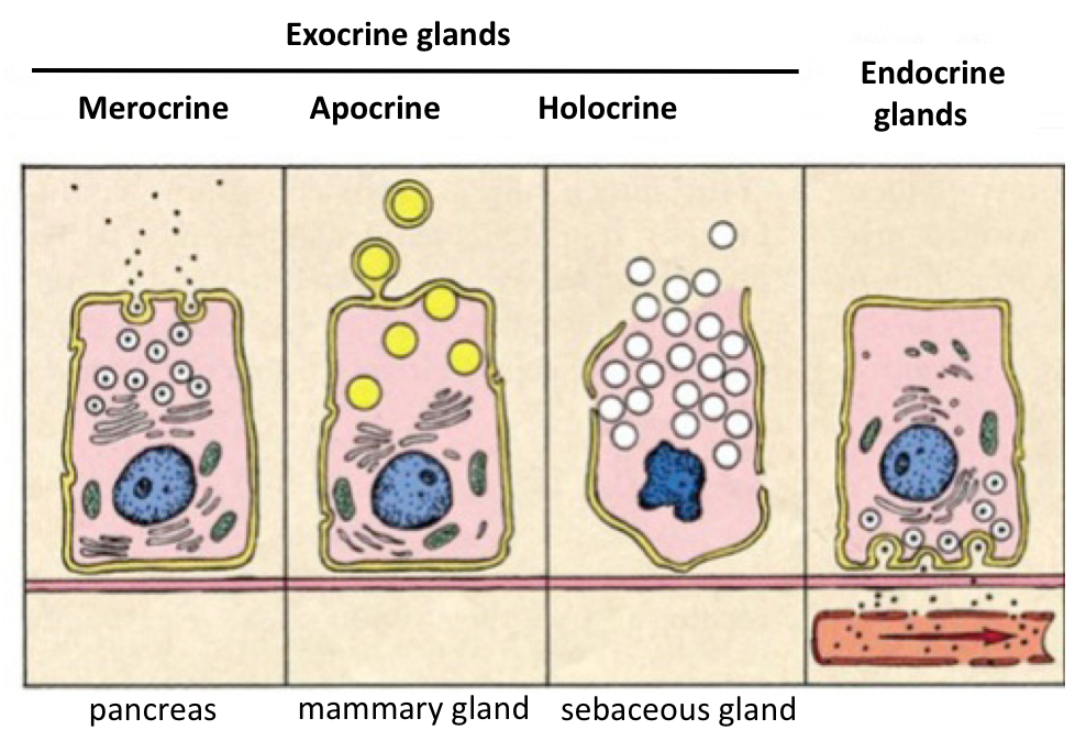
Types of GLANDS
Unicellular
Multicellular (Simple, unbranched, and Compound, branched)
Exocrine
Endocrine
Types of secretion
Serous
Mucous
Mechanisms of secretion
Merocrine
Apocrine
Holocrine
Name the cell type
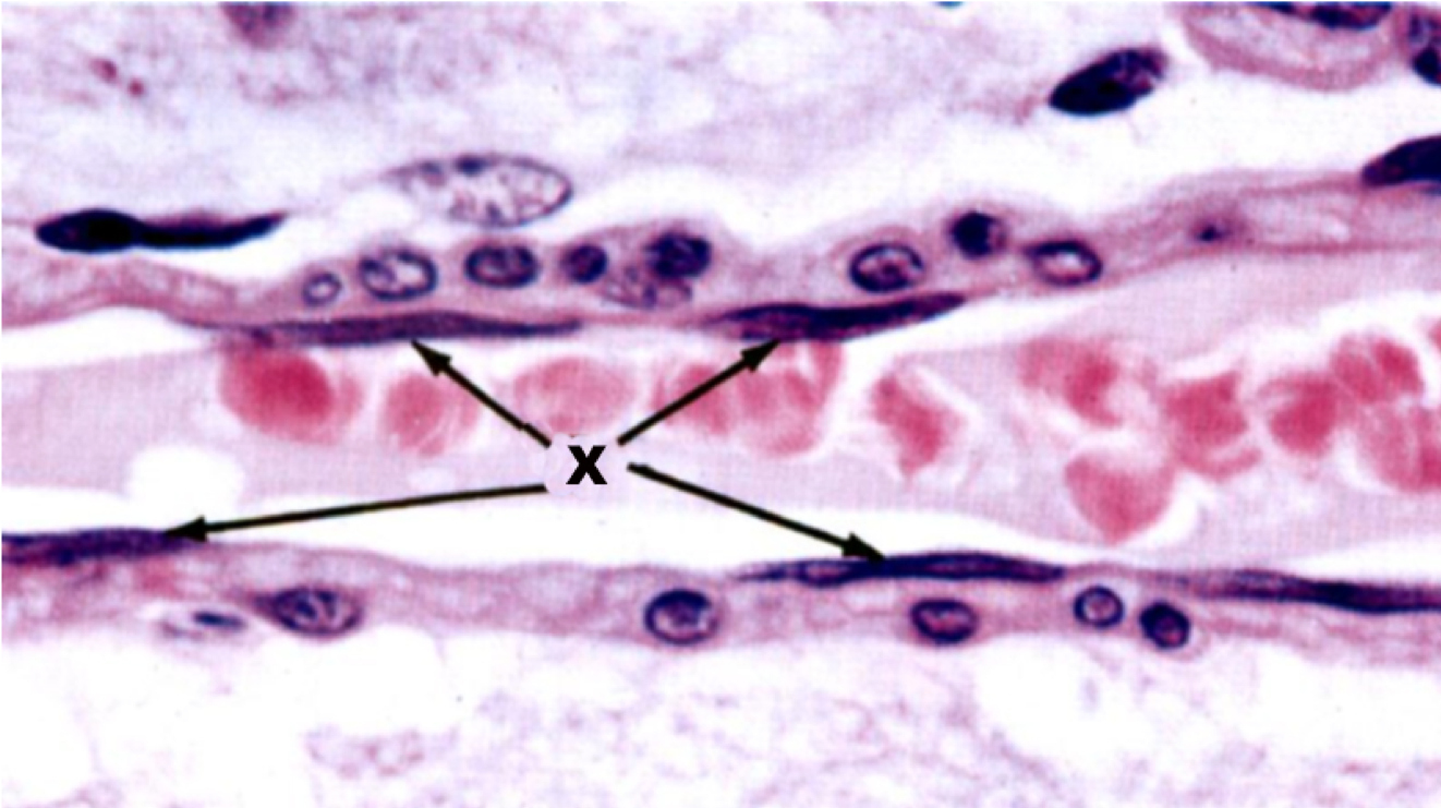
Endothelium cells
Simple Squamous
Endothelial nuclei are indicated by arrows
Important characteristics & functions of EPITHELIUM
1) covering/lining of all body surfaces – it is derived from all three germ layers
2) have specific apical, laterial, and basal domains
3) Basement membrane: Extracellular membrane attachment of cells to underlying connective tissue
4) Avascular
5) Majority of glands derived from epithelia
5) High regenerative capacity
6) Diversity of function (e.g. protections, secretion, absorbtion)
Name the cell type
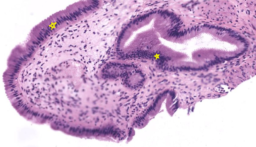
Epithelium (gall bladder)
Simple columnar
Always look for the simplest form of an epithelium as it will usually be representative (plane of section artifacts can lead to mis-diagnosis)
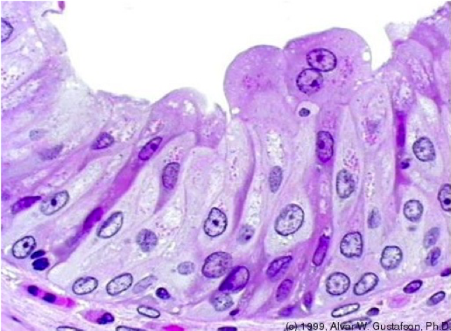
Name the cell type
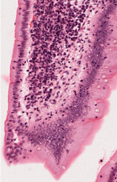
Epithelium (small intestine)
Simple Columnar
Always look for the simplest form of an epithelium as it will usually be representative (plane of section artifacts can lead to mis-diagnosis)
Transitional cells
Large surface cells [umbrella, dome cells], often binucleate. Unique to urinary system – thus also called UROTHELIUM
Cytokeratins
Intermediate filaments
All epithelia contain keratin intermediate filaments
Therefore, cytokeratins are diagnostic of epithelia
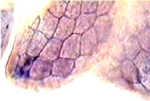
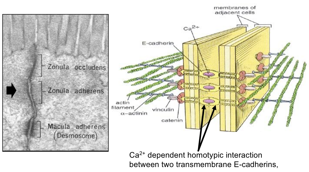
Terminal Bar: Junction Complex
Can be either Zonula Adherens or Zonula Occludens
(can’t distinguish between the two in that picture)
Zonula Occludens: Principal Functions
Selective permeability barrier
- seal intracellular space
- regulate paracellular transport
- facilitate/promote transcellular transport
Cell polarity influence
- separate apical from basolateral domains
- restrict movement within cell membrane
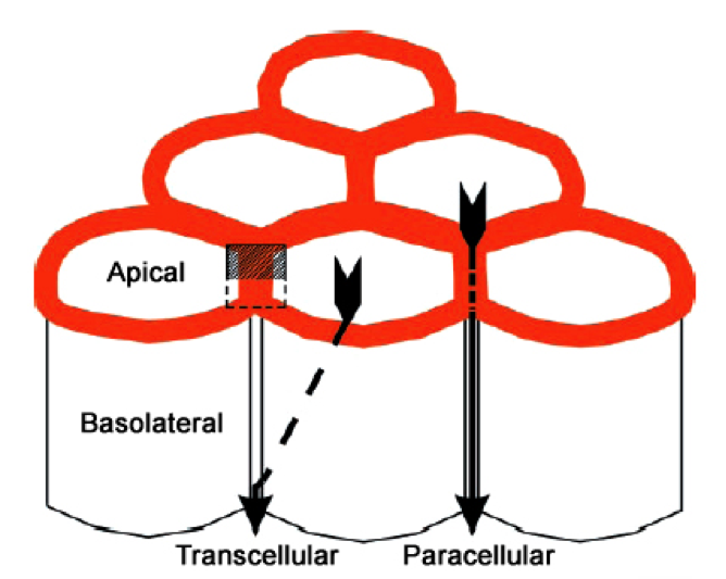
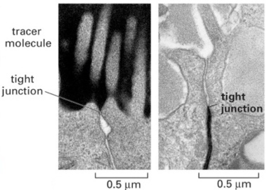
Tight junctions AKA zonula occludens
Zonula Occludens: Principal Components
Major integral membrane proteins
- Claudins
- Occludin
- Tricellulin
Peripheral membrane proteina (intracellular)
- Zonula occludens (ZO) ZO-1, ZO-2, ZO-3
- Others (eg. cingulin, spectrin)
Cytoskeleton proteins
- Actin
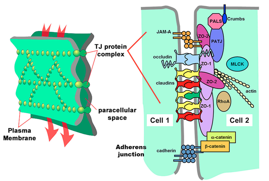
Tricellulin
regulation of paracellular flow
Zonula Adherens: Principal Functions
Important belt-like cell-cell adhesion units
(circumferential = zonary)
Functions:
- Thought to mediate folding and other 3D shapes of epithelia via actin and myosin
- Tranduce signals from adjacent cells
- resist mechanical stress
Zonula Adherens: Principal Components
Integral membrane proteins (E-cadherins: Ca++ binding, homotypic interactions)
•Peripheral membrane proteins (intracellular)
- Catenins (α-, β-, γ-)
- α-Actinin
- Vinculin
•Cytoskeleton proteins
- Actin (filamentous) – they are continuous with actin cytoskeleton
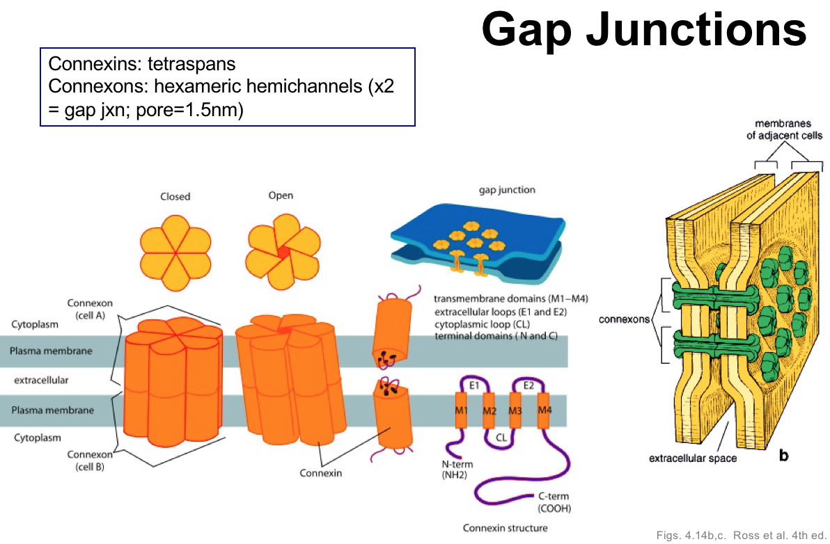
Gap junctions
A type of lateral surface specialization - Communicating junction
Important spot-like (punctate) cell-cell communication and resource-sharing unites
Channels formed as adjacent connexons align between cell membranes - 6 connexins make up one connexon
found in many tissues and function in electrical synapses
Appoximation to syncytium (multiple nuclei sharing cytoplasm)
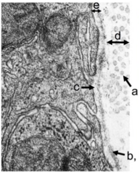
(Basal Lamina cell)
a. Collagen III
b. Collagen IV, Lamina Densa
c. Lamina Lucida
d. Reticular Lamina
e. Basal Lamina
Basement Membrane
Made up of the basal lamina, retucular fibers, and anchoring fibrils (Collagen VII)
Provides physical support; trasmits forces to adjacent CT
Has selective permeability; filter for macromolecules and cell migration barrier

Basal Lamina
One component of basement membrane (along with reticular lamina)
Made up of Type IV collagen, laminin (both of which form chicken wire networks), and nidogen

Reticular Lamina
One component of the basement membrane (along with basal lamina)
Made up of reticular fibers; mostly collagen III


