Elbow Flashcards
Static Palpation - Elbow

MCL:
–Anterior bundle
–Posterior bundle
–Oblique bundle
LCL:
–Annular ligament
–Radial collateral ligament
–Lateral ulnar collateral ligament

Active Range of Motion - Elbow
Flexion of the Elbow:
140°to 150°
Extension of the Elbow:
0°to 10°hyperextension
Supination of the Forearm:
90°
Pronation of the Forearm:
80°to 90°

Passive Range of Motion - Elbow
End Feel:
- Elbow flexion –Tissue approximation
- Elbow extension –Bone to bone
- Forearm supination –Tissue stretch
- Forearm pronation –Tissue stretch or bony end feel
- Capsular Patterns
Consider muscle length and tone:
- Shoulder extension (Extended elbow)
- Shoulder flexion (Flexed elbow)
- Wrist flexion
- Wrist extension
Resisted Assessment - Elbow
- Elbow Extension
- Elbow Flexion
- Forearm Pronation
- Forearm Supination
- Wrist Flexion
- Wrist Extension

Cozen’s Test
(lateral epicondyle)
Purpose:
- assess the integrity of wrist extensors/common extensor tendon at the lateral epicondyle
Procedure:
- pt is seated and practitioner stands adjacent to elbow
- stablize the elbow and palpate the lateral epicondyle
- pronate the forearm and extend the wrist
- pt then resists force against the practitioners resistance
Positive:
Pain over the lateral epicondyle
Indicates:
- Lateral epicondylopathy

Mill’s Test
(lateral epicondyle)
Purpose:
- assess wrist extensors/common extensor tendon at the lateral epicondyle
Procedure:
- pt is sitting and elbow is fully extended
- practitioner pronates the forearm and flexes the wrist
Positive:
- Pain along the lateral epicondyle region of the humerus
Indication:
- Lateral epicondylopathy

Middle finger sign
(wrist extensors and radial nerve)

Purpose:
- assess integrity of the wrist extensors at the elbow insertion
- integrity o the radial nerve at the arcade of frohse and radial tunnel
Procedure:
- pt is sitting with forearm pronated and fingers extended, practitioner is facing the patient
- examiner resistes the middle finger in extension (extensor digitorum)
Positive:
- pain over the lateral epicondyle
- pain can be distal to lateral epicondyle in case of radial tunnel syndrome
Positive:
- lateral epicondylopathy if pain at lateral epicondyle
- radial neuropathy if there is neuropathic pain or paraesthesia
Test for Medial Epicondylopathy
(medial epicondylopathy)

Purpose:
assess the wrsit flexors/common flexor tenson atr the medial epicondyle insertion
Procedure:
- pt is sitting or standing and makes a fist. practitioner faces the patient
- palpate along the medial epidcondyle with one hand and grasp the wrist with the other
- passive supination of the forearm and extend the elbow, wrist and fingers
Postive:
- complaints of discomfirt along the medial aspect of the elbow
indication:
- medial epicondylopathy
Tinel’s Sign: Elbow
(ulnar nerve)
Purpose:
- assess the integrity of the ulnar nerve about the elbow
Procedure:
- pt is sitting/standing/supine and practitioner is beside the arm
- tap and percuss over the cubital tunnelfor for 10 seconds
Positive:
- tingling sensation in the ulnar distribution of the forearm and hand distal to the point of compression of the ulnar nerve
Indication:
ulnar neuropathy abou the elbow

Elbow Flexion Test
(ulnar nerve)
Purpose:
- assess the integrity of the ulnar nerve at the cubital tunnel
Procedure:
- pt sitting or standing with elbows max flexed and wrists extended
- pt holds position for 3-5 minutes
- practitioner can apply shoulder depression and contralateral cervical lateral flexion to senzitize
Positive:
- Radiating pain into the ulanr nerve distribution in the pateint arm or hand
Indication:
- cubital tunnel syndrome

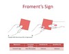
Froment’s Sign
(ulnar nerve)
Purpose
- to assess the integrity of the ulnar nerve
Procedure
- pt grasp paper between thumb and index finger
- examiner tries to pull away the paper and observes the thumb
Positive:
- IP joint flexion of the thumb is a positive Froments sign
Indicates:
- ulnar nerve palsy
Note:
- normal nerve supply (ulnar nerve) to adductor policis is required to perform test
- pt will cheat using flexor policis (median) if there is a ulnar palsy
Test for Pronator Teres Syndrome
(median nerve)
Purpose:
- To assess the integrity of the median nerve at the level of the pronator teres muscle
Procedure:
- part a - pt sits with elbow flexed and resists attempt to supinate forearm, while supinating the forearm passively extend the patients arm
- position is held for 30 seconds
- part b - pt is asked to relax and then pressure is applied to the belly of the pronator teres for one minute
Positive -
- tingling or paresthesia in the median nerve distribution
Indication:
- pronator teres syndrome
Note:
- Part a - used to enhance blood flow to deep and superficial muscle bellies to minimise space for emdian nerve
- part b used to compress the median nerve

Pinch Grip Test (OK Sign)
(anterior interosseous nerve) AIN

Purpose:
- assess the integrity of the anterior interosseous nerve
Procedure:
- pt is sitting or standing and makes an ok sign with the thumb and index finger
Positive:
- inability to touch the tips of the thumb and index finger together or touching the pads of the thumb and index finger together
Indication:
- neuropathy of the anterior interoeous nerve (AIN)
- common due to neuritis not entrapment
Note:
the AIN is the primary motor branch off the median nerve which innervates flexor pollis longus, flexor digitorum profundus of the index/middle and pronator quadratus
Valgus Stress Test
(medial ulnar collateral ligament)
Purpose:
- assess the integrity of the structures which stabalise the elbow against valgus stress
Procedure:
- pt is sitting or standing
- elbow is flexed 20-30 degrees with forearm pronated
- pt’s arm is stablised with the practitioner’s primary contact over the lateral elbow, secondary holds the wrist
- valgus force through elbow and secondary
- assess the end feell of the ulnar collateral ligament and degress of opening at the medial elbow joint
Positive:
- pain
- laxity
indicates:
- mcl ligament sprain with or without joint instability

Moving Valgus Stress Test
(medial collateral ligaments)

Purpose:
- assess the integrity of the structures which stablise the elbow against valgus stress during movement
Procedure:
- pt is standing or sitting and practitioner stands adjacent to affected elbow
- abduct arm to 90 and externally rotated
- pts elbow is then flexed to approx 130 degrees
- stablize the arm at the lateral distal humerus (elbow), primary is at the pt’s wrist
- abduction and external rotation are maintained while valgus force is applied at the elbow by the examiner’s secondary hand
- elbow is moved into extension
Positive:
- pain between 70 to 130 of flexion
- laxity
- crepitus
- replication of ulnar nerve symptoms
Indicates:
- MCL ligament sprain with or without joint instability
- ulnar nerve neuropathy
Valgus Extension Overload Test
(elbow impingement)
Purpose:
- to detect the presence of posteromedial olecranon osteoppyte or olecranon fossa overgrowth
Procedure:
- pt sitting or standing and practitioner is adjacent to affected elbow
- arm is abducted to 50 and flexed to 30
- stablise humerus with primary and pronate the forearm
- create vlagus force at elbow using primary and secondary
- maintain position and move into extension
Positive:
- pain posteromedially as olecranon tip osteophytes enegae the posteromedial olecranon fossa
Indication:
- posteromedial elbow impingement
- stress fracture of the olecranon

Radiocapitellar Compression Test
(radiocapetallar joint)
Purpose
- assess the integrity off the radiocapitellar joint
Procedure:
- pt is sitting or standing and practitioner is facing the pt
- secondary cups the posterior elbow with either a thumb or index finger palpating the radial head
- practitioner holds the pt wrist in slight extension and radial deviation
- axial load through the forearm to the radiocapitellat joint via pateints hand and wrist
- Active or passive pronation and supination o the forearm are preformed during midrnage elbow extension/flexion motion
Positive:
- Pain and crepitus in the radiocapitellar joint
Indicates:
- radiocapitellat joint degeneration
- OCD
- panner’s disease
- radial head/neck fracture

Varus Stress Test
(lateral radial collateral ligaments)

Purpose
- Test the integrity of the structures which stablise the elbow against varus stress
Procedure:
- sitting or standing and prac adjacent to elbow
- elbow flexedto 20-30 wirh supinated forearm
- stabilise medial elbow and wrist and apply varus force
- assess and feel the lateral ulnar collateral ligament and degree of opening
Positive:
- Pain
- Laxity
Indication:
- LUCL ligament sprain with or without joint instability
Posterolateral Pivot-Shift Apprehension Test
(posterolateral rotary instability)
Purpose:
- assess for elbow posterolateral rotary instability
- LUCL laxity
Procedure:
- pt is supinewith test arm overhead and externally rotated
- grasp wrist and extend elbow
- supinated force applied to forearm at the wrist
- pts elbow is then flexed while valgus stress and compression are applied to the elbow
- must contact the humerus to create valgus
Positive:
- apprehension as the elbow is flexed 20-30 degrees
- reduction (clunk) of the radial head around 40-70 degrees of flexion
Indication:
- posterolateral rotary instability
Notes:
- set up used to luxate the humeroulnar/radiohumeral joint
