ear infections, allergic rhinitis Flashcards
heat and moisture lead to swelling and maceration of the skin in the EAC; breakdown of skin allows bacteria to enter
Clinical presentation:
- ear pain, worse with movement of the external ear; pruritis; discharge
- EAC may be erythematous and edematous
- may have decreased hearing
- TM may or may not be intact

otitis externa : “swimmer’s ear”
etiology of otitis externa
- psueomonas (38%)
- step epidermidis (9%)
- staph aureus (8%)
- fungal: asperigillus or candida albicans (2-10%)
treatment of fungal otitis externa

- clotrimazole 1% solution BID x 14 days
- acidifying solution (acetic acid)
treatment of bacteria otitis externa
- cortisporin otic suspension (polymixin B; Neomycin; Hydrocortisone) ** avoid is suspected TM perforation
- Floxin otic: indicated for perforated TM
- Use ear wick: if marked swelling of EAC; remove wick after 2-3 days and continue meds as directed

management of otitis externa (things to do other than medication)
- pain control
- keep canal dry (no swimming 7-10 days)
- most cases resolve within 5-7 days: if not, consider fungal infection
- prevention: 2% acetic acid to acidify the EAC
what population is susceptible to malignant otitis externa? what organism causes it?
- seen in diabetics and immunocompromised
- pseudomonas in 95% of cases
* complications: osteomyelitis; meningitis; mortality
clinical presentation
- intense ear pain and otorrhea (discharge)
- red, granulation tissue in EAC
- possible periauricular lymphadenopathy, edema and trismus (spasm of jaw muscle)
- elevated ESR or CRP

malignant otitis externa
treatment for malignant otitis externa
- admit
- IV Ciprofloxacin
peak incidence of acute otitis media. Why?
- 6-18 months old
- usually precipitated by a viral URI; eustachian tube becomes obstructed with fluid and mucus; accumulated fluid become sedentary
- horizontal eustachian tube allows for migration of organisms from nasopharynx
- enlarged adenoids prevent adequate drainage
most common pathogens of acute otitis media
- streptococcus pneumoniae (40-50%)
- haemophilus influenzae (45%)
- moraxella catarrhalis (10%)
predisposing risk factors of acute otitis media
- age and immature anatomy in children
- secondhand smoke
- day care
- use of pacifier
- season (fall/winter)
clinical presentation for pediatric patient
- irritability
- decreased appetite
- +/- fever
- tugging on ear
- hearing loss
- may also see conjunctivitis, rhinorrhea, ear dx, vomiting, and diarrhea

acute otitis media
clinical presentation in adult
- otalgia
- rare to have fever
- opaque or reddened TM
- bulging TM
- decreased TM mobility

acute otitis media
otitis media: criteria for diagnosis of children 6 mo -12 years
- moderate to severe bulging of the TM OR
- new onset of otorrhea not due to acute OE OR
- mild bulging of TM and recent ear pain ( <48 hr) or intense erythema of the TM
tympanometry
quantitative measure of acoustic impedance
- compliance or resistance of the middle ear in response to changes in air pressure
- probe inserted into ear canal to deliver positive and negative pressure
type of tympanogram. what conditions will cause this

- type B: little or no mobility
- fluid or TM perf
type of tympanogram. what conditions will cause this

- type C: retracted
- eustachian tuve dysfunction
you should trea of Acute otitis media with Abx under what conditions
- < 6 months
- 6-23 months: severe signs/symptoms or bilat AOM
- >24 months: if diagnosis is certain and illness is severe
** severe: moderate or severe otalgia, otalgia for > 48 hrs; temperature above 102.2F
you can treat acute otitis media with abx or observation with close follow up (48-72 hrs) if?
- 6-23 months: non severe unilateral AOM
- > 24 months: non-severe unilateral or bilat AOM
treatment of acute otitis media
amoxicillin 80-90 mg/kg/day (q 12 hrs) x 7-10 days
**should not be used in kids who are at high risk of AOM caused by resistant organism
what puts a kid at high risk of AOM caused by resistant organism
- abx in last 30 days
- concurrent purulent conjunctivitis
- hx of recurrent AOM resistant to amoxicillin
treatment for high risk of AOM caused by resistant organism
- amoxicillin/clavulanate = Augmentin
- 90mg/kf amoxicillin and 6.4 mg/kg clavulanate
- do not use if PCN allergy
- omnicef
should OTC cold preparations be used in children under 4 yo
no
how should you treat a recurrent acute otitis media
- IM rocephin (ceftriaxone)
- augmentin
- consider need for tympanostomy tubes if 3 or more episodes in 6 months
when does otitis media become Chronic
- drainage from middle ear > 2 weeks
- associated with TM perforation that is usually painless (conductive hearing loss)
clinical presentation
- +/- pain, otorrhea
- associated with acute or chronic OM
- conductive hearing loss
- no movement with pneumatic otoscopy
- vertigo
TM perforation
** most heal spontaneously
clinical presentation
- extension of OE or AOM into mastoid air cells
- postauricular pain, edema and erythema
- protrusion of pinna
- fever
- deep temporal pain

mastoiditis
tx: IV abx; ENT consult
etiolofy of otitis media with effusion (OME)
- middle ear effusion secondary to inflammation or eustachian tube dysfunction
- following a viral URI; AOM; or in association with allergic rhinitis
clinical presentation
- afebrile
- amber-colored (straw) fluid behind TM
- may see air fluid level and bubbles
- neutral or retracted TM
- conductive hearing loss
- tympanogram type B

otitis media with effusion (OME
if you have an adult patient with persistent unilateral otitis media with effusion, what must you do
refer to ENT to r/o nasopharyngeal carcinoma
management of otitis media with effusion (OME)
- “watchful waiting”; reevaluate 4-6 weeks
- intranasal steroids in underlying allergic rhinitis
when would you present a patient with otitis media with effusion (OME) to ENT for T-tubes
- persistent fluid and/or hearing loss > 3 months duration
clinical presentation
- inflammation or blockage resulting in negative middle ear pressure
- ear fullness, hearing loss
- retracted TM; prominent bony landmarks
- type C tympanogram
eustachian tube dysfunction
treatment of eustachian tube dysfunction
- steroid nasal spray; decongestant
- topical nasal decongestant
- phenylephrine (neo-synephrine) or oxymetazoline (Afrin)
- limit therapy to 3 days to avoid rebound congestion
clinical presentation
- discomfort or damage to ear due to pressure differences (associated with altitude changes)
- ear fullness; pain; tinnitus
- bloody otorrhea if TM perforation
ear barotrauma
treatment for barotrauma
supportive
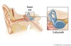
what is labyrinthitis
- benign, acute infection or inflammation of vestibular system
- most commonly associated with viral infection
- seen commonly in 30-60 yo
clinical presentation
- acute onset of severe vertigo, 1-2 days duration
- N/V
- tinnitus and/or unilateral hearing loss. No CNS deficits
- (+) head thrust: cannot maintain visual fixation
- horizontal nystagmus
labyrinthitis
treatment of labyrinthitis
symptomatic
- bed rest; hydration
- treatment of N/V: meclizine (antivert) 25 mg TID
- benzodiazepines
perennial rhinitis
occurs year round: usually due to dust mites, mold, animal dander
clinical presentation: exam finding
- nasal speculum exam: pale, blue mucosa, boggy; clear discharge
- eyes and periorbital: palpebral conjunctiva may be pale, swollen
- allergic shiners
- denie morgan lines: skin folds under eyes
allergic rhinitis
treatment for allergic rhinitis
topical intranasal corticosteroids
- beclomethasone (beconase)
- Triamcinolone (Nasacort): OTC
treatment for allergic rhinitis: specific symptoms of sneezing, rhinorrhea, and itching
antihistamines: first generation
- chlorpheniramine 4mg q4-6 hr or 8-12 mg BID for sustained release
- Diphenhydramine (Benadryl) 25-50 mg BID-TID
**side effects: dry mouth, constipation, sedation
benefit of second generation antihistamines for treatment of allergic rhinitis
less sedating
- claratin 10 mg daily
- Allegra 60 mg BID
- Zyrtex 5-10 mg daily
treatment for allergic rhinitis: specifically nasal congestion
decongestant: causes vasoconstriction
pseudoephredrine (sudafed) 30-60 q 6-8 hr or 120 mg BID for SR
*** caution flag: HTN, cardiac disease
last resort treatment for allergic rhinitis
immunotherapy: hyposensitization
- stimulates production of IgG against allergens
- success rate 85%
- abnormal autonomic responsiveness triggered by stress, sexual arousal, ciggs, temp changes or anti-HTN medication
- nasal congestion and rhinorrhea
- no itching or sneezing
- nasal mucosa is normal; IgE normal
perennial non-allergic (vasomotor) rhinitis
treatment of perennial non-allergic (vasomotor) rhinitis
- avoid triggers
- topical steroids
- topical antihistamines (azelastine)
- topical anticholinergics
treatment of nasal polyps
** commonly seen with allergic rhinitis, vasomotor rhinitis
intranasal glucocorticoids
refer to ENT for obstruction
- see tachyphylaxis (diminished response of drug) with overuse of topical decongestants
- what is the condition when you see severe rebound congestion after stopping topical decongestant?
- nasal mucosa is erythematous
rhinitis medicamentosa
**treatment: stop medication and treat with intranasal glucocorticoid
treatment of otitis media after treatment failure
IM rocephin (ceftriaxone) 50 mg IM or IV once daily for 3 days
