Dr. Mhawi 5 Connective Tissue Flashcards

1
Q

Explain this image

8.5.7

A
  • Primitive CT (undifferentiated)
  • Found in the embryo
  • Mesodermal origin
  • Gives rise to almost all tissue in adult
  • Contains elongate MESENCHYME* cells

nLoosely organized and loosely attached

nLarge nuclei

nProminent nucleoli

nThin cytoplasmic processes

•Abundant, viscous extracellular substance with few collagen and reticular fibers

2
Q

Explain this image

A

At the upper left corner is a low magnification of a cross section of the umbilical cord in which major blood vessels are visible. The mucoid tissue consists of fibroblasts and mesenchyme cells embedded within a gelatin-like extracellular matrix rich in collagen fibers. Note the simple cuboidal layer of the amniotic cells that cover the umbilical cord.

3
Q

Explain this image

A

Mucoid connective tissue. Collagen fibers, appeared light blue, are visible among the fibroblasts. Simple cuboidal amniotic cells are visible at the upper left corner. Trichrome stain.

4
Q

Explain this image

A

Mucous tissue. A section of an umbilical cord showing fibroblasts surrounded by a large amount of loose extracellular matrix composed of ground substance and collagen fibers. H&E stain.

5
Q

Explain this image

A

Loose (areolar) connective tissue. Whole mount preparation. This tissue is taken from a mesenteric spread. You are looking down on a mesentery that was stretched on the slide, fixed, then stained. The slide shows a random sampling of the cells and fibers that make up loose connective tissue. The thicker fibers which have a reddish color, are collagen fibers. The very thin fibers, which tend to appear dark

6
Q

Explain this image

A

Loose (areolar) connective tissue. Whole mount preparation. Collagen fibers appear brown. Elastic fibers dark blue. The stars are placed beside mast cells (appear red).

  • Plays important role in:
  • immunity
  • inflammation
  • diffusion of nutrients,

oxygen, CO2, and waste

7
Q

Diagram of loose connective tissue

A
8
Q

_________

Consists mostly of bundles of Type I collagen fibers and fibroblasts

•Collagen fibers and fibroblasts aligned in parallel

nProvide resistance to prolonged or repeated stresses

poorly vascularized

nRepair of injury is slow

A

Regular dense connecitve tissue

•Found in:

nTendons

nLigaments

nAponeuroses (flat tendons)

9
Q

Explain this image

A

Longitudinal section of regular dense connective tissue from a tendon. A: Thick bundles of parallel collagen fibers fill the intercellular spaces between fibroblasts. Low magnification. B: Higher magnification view of a tendon of a young animal. Note active fibroblasts with prominent Golgi regions (G), nucleus (N) and dark cytoplasm rich in RNA. PT stain.

10
Q

Explain this image

A

Longitudinal section of tendon. BV, blood vessel; TC, tendon cell nuclei. dashed line, arbitrary cross-sectional cut of tendon (see next slide).

11
Q

Explain this image

A

Dense regular connective tissue of tendon (cross section). BV, blood vessel; Ent, endotendineum; TC, tendon cell nuclei.

12
Q

Explain this image

A

Electron micrograph of a fibrocyte (cross-sectioned) in regular dense connective tissue. The sparse cytoplasm of the fibrocytes is divided into numerous thin cytoplasmic processes that interdigitate among the collagen fibers. x25,000.

13
Q

Explain this image

A

Section of dense irregular connective tissue. This figure shows fibroblasts (arrow) with many thin cytoplasmic extensions (arrowheads). As these cells are pressed by collagen fibers, the appearance of their cytoplasm depends on the section orientation; when the section is parallel to the cell surface, parts of the cytoplasm are visible. PT stain. Medium magnification.

14
Q

Explain this image

A

Irregular dense connective tissue. Note the irregular arrangement of the collagen fibers (green). N, nucleus of fibroblast responsible for synthesizing the fibers; BV, blood vessel.

15
Q

Explain this image

A

None lactating mammary gland showing loose connective tissue (arrows around and between the glandular tissue) and irregular dense connective tissue (upper and lower right parts of the micrograph). Masson trichrome. X 200. Human breast glandular epithelium.

16
Q

What are the 3 fibers that surrounds and support the connective tissue and the 2 ground substances

8.5.7

A

•Fibers: collagen fibers

elastic fibers

reticular fibers

•Ground substance: proteoglycans

multiadhesive glycoproteins

  • fibronectin
  • laminin
17
Q

Explain this image

A

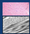
Upper panel: light micrograph of bundles of collagen fibers (blue arrows). Note the wavy appearance. Fibroblasts, responsible for their synthesis, are indicated by the green arrows. Lower panel: At the TEM level collagen fibers are composed of fibrils. The latter are cut in cross (upper left) and longitudinal (lower right) sections. When cut longitudinally each fibril appears to consist of regular alternating dark and light bands. Ground substance completely surrounds the fibrils. x100,000.

18
Q

Synthesis of collagen molecules is _______

A

vitamin C-dependent

8.5.7

19
Q

Explain. this image

A

TEM of a fibroblast which is responsible for the synthesis of collagen molecules in the rER and for the assembly of these molecules EXTRACELLULARLY into collagen fibers.

20
Q

______ collagen is most common

•occurs in:

nloose & dense connective tissue

nbone

______ collagen is present in

  • hyaline & elastic cartilage
  • Vitreous body of the eye
A

Type I

Type II

21
Q

______– (reticular fibers) reticular connective tissue

•Synthesized by fibroblasts

______ – meshwork sheets – basal/external laminae

•Synthesized by epithelial cells, muscle cells, Schwann cells

______ – fibers – dense irregular connective tissue, placenta, blood vessel walls

•Synthesized by fibroblasts, smooth muscle cells

______ – anchoring fibrils – epidermal-dermal interface

•Synthesized by fibroblasts

A

Type III

Type IV

Type V

Type VII

22
Q

______ forms the Stroma of certain organs (support

A

Reticular fibers

Reticular fibres in the spleen. The spleen is covered by dense connective tissue capsule (left) from which a trabecula is extended into the parenchyma of the organ.

23
Q

Explain this image

A

Reticular fibres form a delicate supporting framework (network) for many highly cellular organs such as liver (this picture).

24
Q

In loose CT, reticular fibers they are formed by _____, in hematopoietic and lymphatic tissues they are formed by______

A

fibroblast

reticular cells

25
Q

True or False: Cannot be identified by H&E

8.5.7

A

True

Demonstrated with:

•silver staining

ni.e., they are argyrophilic

•PAS reaction

nDue to higher sugar content than regular collagen

Reticular fibers (red arrows) in the lymph node. Silver stain.

26
Q

_______ are

Selectively stained with (Van Gieson’s) (resorcinol)

Abundant in

elastic arteries (aorta)

ligaments (ligamentum nuchae and ligamentum flava)

vocal folds of the larynx

elastic cartilage (external ear)

A

Elastic Fibres

Made up of 2 components:

  • Central core of elastin molecules
  • Fibrillin microfibrils

found within and around elastin core

27
Q

Explain the images

A

Upper panel: ligaments of the vertebral column. Lower panel: light micrograph of the elastic fibers from ligamentum nuchae. The white spaces among the elastic fibers represent unstained loose connective tissue elements and parallel rows of flattened fibroblast that can not be distinguished in this preparation.

28
Q

Explain the image

A

Aorta. Tunica media consists of elastic fibers.

29
Q

Explain images

A

Internal (red arrow) and external (black arrow) elastic membranes of a muscular artery.

30
Q

True or False: Elastic fibres lose property during aging

8.5.7

A

True

31
Q

nIn arteries, elastic fibers are made by _______, NOT by fibroblasts

A

smooth muscle cells

32
Q

Explain the image

8.5.7

A

In elastic arteries elastin molecules form fenestrated lamellae

Elastic artery (aorta). Note the layers of the fenestrated elastic lamellae. Fenestrae cannot be discerned at low magnification, especially when they are viewed from the side. Empty spaces between the elastic lamellae represent locations of the smooth muscle cells, responsible for synthesis of the elastic lamellae. Smooth muscle cells are not visible because the section is specifically stained for elastin.

8.5.7

33
Q

Damaging Effects Of Sunlight On Elastic Fibers

A
  • Fibrillin microfibrils undergo extensive remodeling when overexposed to sun light
  • Become sparse and truncated
  • Leading to nonfunctional elastic fiber
  • These changes contribute to decreased skin elasticity and the appearance of deep wrinkles
34
Q

_______

Consists mostly of proteoglycans

Occupies the space between cells and fibers

Proteoglycans consists of:

High molecular weight acidic carbohydrates

(GAGs) bound to a core protein

Appears amorphous in LM

Stains with PAS stain

Cannot be seen in H&E preps

A

Ground substance

35
Q

_______ are large macromolecules

•Look like a bottle brush

A

Proteoglycans

36
Q

•Most important ECM proteoglycan is ______

A

AGGRECAN

nGAGs are chondroitin sulfate and keratan sulfate

nmain proteoglycan in cartilage

nInteracts with type II collagen

•Makes a compression-resistant gel

Schematic diagram of the proteoglycan aggrecan. The molecule is made of core protein (red) to which two different GAGs are attached; chondroitin sulfate (blue) and keratan sulfate (yellow)

37
Q

Large number of proteoglycans attach to ______ (very large nonsulfated GAG molecule)

A

hyaluronan

38
Q

Explain this image

A

TEM image of the proteoglycan aggregates.

39
Q

_______ cells:

Exhibit little movement and regarded as permanent residents of the tissue

A

Fixed

  • Fibroblasts and closely related myofibroblasts
  • Macrophages
  • Adipose cells
  • Mast cells
  • Undifferentiated mesenchyme cells (adult stem cells)
  • Pericytes
40
Q

_______ (transient population) migrate from blood and/or lymph in response to stimuli:

A

Wandering Cells

  • Lymphocytes
  • Neutrophils
  • Eosinophils
  • Basophils
  • Monocytes
  • Plasma cells
41
Q

What do fibroblasts synthesize

A

Principal cell of connective tissue

Synthesize:

  • Collagen
  • elastin
  • reticular fibers
  • complex carbohydrates of ground substance
42
Q

Explain the image

A

Fibroblasts are elongated cells with thin cytoplasmic extensions that are buried in the surrounding collagen. Pararosaniline-toluidine blue (PT) stain. Medium magnification.

43
Q

_______

look like fibroblasts (spindle shape)

TEM shows features of smooth muscle cells

(longitudinally disposed actin filaments)

But, _______

A

Myofibroblasts

Lack an external lamina

44
Q

_____ are phagocytic cells

•derived from Monocytes in bone marrow

macrophages are difficult to identify in LM

•unless they display obvious phagocytosis

Possess a large indented or kidney-shaped nucleus

A

Macrophages

Section of pancreas from a rat injected with the vital dye trypan blue. Note that 3 macrophages (arrows) have engulfed and accumulated the dye in the form of granules.

45
Q

Explain this image

A

TEM of active macrophage exhibiting finger like cytoplasmic processes and lysosomes. X 10,000.

46
Q

Explain the image

A

Phagocytosed material is destroyed by the action of lysosomes. The end product of digestion is either recycled or exocytosed. Inert particles like inhaled carbon will resist digestion and may remain in the cytoplasm indefinitely as residual bodies

47
Q

Explain the image

A

SEM of a macrophage cytoplasmic process in rat liver. A macrophage (Kupffer’s cell) extends a cytoplasmic process in recognition of a damaged (previously fixed in glutaraldehyde for a few minutes) erythrocytes in the initial stage of phagocytosis.

48
Q

What are the main functions of macrophages

A

•Phagocytosis

  • Defense
  • clean-up of cell debris

•Also play a role in immune reaction by:

nPresenting (via MHC II) concentrated antigens derived from phagocytosed foreign cells or proteins to CD4+ T lymphocytes

•Macrophages called APC (antigen-presenting cells)

49
Q

True or False: Macrophages may fuse to form a large cell

A

True

These cells called Foreign Body Giant Cells

n or LANGHANS’ CELLS

ne.g., in tuberculosis (characteristic, horseshoe nuclear array)

50
Q

Explain the image

A

Langhans’ Cell.

51
Q

_____ are cells derived from monocytes and Have ability to avidly phagocytose injected vital dyes

  • trypan blue
  • India ink

which accumulate in the lysosomes

A

Mononuclear Phagocytic System (MPS)

52
Q

______

Large ovoid connective tissue cell

Granulated cytoplasm

Related to blood basophil (contain similar cytoplasmic granules)

Arise from basophil/mast cell progenitor in the bone marrow

Migrate to connective tissue and differentiate into mast cells

A

Mast cell

Mast cell from the mesentery of rat. Cytoplasmic granules, covering the nucleus, are stained with neutral red. Elastic fibers are visible in the upper part of the micrograph.

53
Q

______ are dstributed mainly in:area of small blood vessels skin capsules of organs

Absent from brain and spinal cord

•this protects brain & spinal cord from damage due to edema from allergic reactions.

A

Mast cells near blood vessel of rat skin. Neutral red stain.

54
Q

Explain the image

A

MAst cell near vessel

55
Q

Explain the allergic reaction

A
56
Q

Mast cells release 4 substances:

A

Histamine (causing oedema, edema)

Slow-reacting substance of anaphylaxis* (SRS-A

•causes bronchospasm

Eosinophil and neutrophil chemotactic factors (attract eosinophil and neutrophil to the site of inflammation)

Heparin

57
Q

______

Originate from:

•mesenchymal cells

Develop external (basal) lamina

Specialized to store fat

A

Adipose Cells

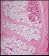
Adipose tissue in the hypodermis. Adipocytes (A) form lobules separated by dense irregular connective tissue (DICT). Several sections of the secretory part of a sweat gland (arrows) appears at the upper right corner of the micrograph.

•Two types of adipose tissue:

White (unilocular)

Brown (multilocular

58
Q

Explain Image

A
59
Q

______

Supplied with blood vessels and nerves

Adipocyte 100 µm or more

Cytoplasm is filled with single large lipid droplet (unilocular)

A

WHITE ADIPOSE TISSUE

60
Q

Explain this image

A
61
Q

Difference of brown adipose tissue vs white adipose tissue

A

Storing energy in lipid droplet in the form of triglycerides

Insulation

Cushioning to vital organs

Major endocrine organ- produces:

  • Angiotensinogen (AGE
  • Steroids
  • Leptin

Brown:

Multiple lipid droplets in the cytoplasm (hence the name)

Nucleus is round and more or less central

Called brown because mitochondria contain large amount of cytochrome oxidase which imparts brown color to cell

Oxidized fat produces heat to warm the blood vessels found in the brown adipose tissue

62
Q

Explain the image

A

Multilocular adipose tissue.

63
Q

Explain the image

A

Brown adipose tissue in which multilocular adipocytes (MA) surround blood vessel (BV). Burning the fat generates heat which is carried by the blood vessel to other part of the body. Note the unilocular adipocytes (UA) at the upper part of the micrograph.

64
Q

______

also called adventitial cells and perivascular cells

•Found around capillaries and venules

n(wrapped around capillaries)

•Surrounded by basal lamina

n(isolated from connective tissue compartment)

A

Pericytes

65
Q

Explain this image

A

Small blood vessel. Left nucleus, endothelial cell. Right nucleus, pericyte. Note basal lamina of the endothelial cell divides to surround the pericyte. CL, capillary lumen.

66
Q

Explain the image

A

Bm, basal lamina; e, endothelial cell; p, pericyte.

67
Q

______ in the wall of venules are similar to smooth muscle cells which suggests that:

A

Pericytes

68
Q

_______ principally involved in immune responses

What are the 3 types

A

Lymphocytes

  • T lymphocytes – involved in cell-mediated immunity
  • B lymphocytes – involved in production of antibodies (antibody-mediated immunity)
  • NK (Natural Killer) lymphocytes – destroy virus-infected and some tumor cells
69
Q

Explain the image

A

Left: TEM of lymphocyte (upper left corner) and mast cell (middle). Compare the size of the two cell types. Right: smear of blood showing two lymphocytes in the middle of the micrograph. The basophilic nuclei of these cells fill most of the cell cytoplasm which is visible as a thin rim.

70
Q

Prominent component of:

  • Lamina propria of GI tract, respiratory tract, urogenital tract
  • salivary glands
  • lymph nodes
  • hemopoietic tissue.
A

Plasma Cells

Portion of a chronically inflamed intestinal villus. The plasma cells are characterized by their size and abundant basophilic cytoplasm (rough endoplasmic reticulum) and are involved in the synthesis of antibodies. A large Golgi complex (arrows) is where the terminal glycosylation of the antibodies (glycoproteins) occurs. Plasma cells produce antibodies of importance in immune reactions. PT stain. Medium magnification.

71
Q

Derived from B-lymphocytes

lives 10-30 days

Relatively large

much basophilic cytoplasm

A

Plasma Cells

72
Q

What are the 3 features of plasma cells

A

•cytoplasm has strong basophilia

Due to the highly developed rER

  • Extensive Golgi complex
  • prominent nucleolus
73
Q

Explain the image

A

Plasma cells, tumor, human. The arrows point to less stained areas in the cytoplasm of plasma cells. These areas are occupied by Golgi apparatus. 400X oil immersion.

74
Q

Explain this image

A

Plasma cells in jejunum. Note the prominent nucleolus and the clear para nuclear Golgi area (arrows).

75
Q
A

TEM of Plasma cell. Note the cartwheel arrangement of the heterochromatin and the well developed rER.

76
Q

These cells rapidly migrate from blood to connective tissue

  • response to injury and inflammation
  • especially neutrophils followed by monocytes
  • their presence indicates an acute inflammatory reaction
  • Monocytes differentiate into macrophages
A

Neutrophils, Monocytes, and Eosinophils

77
Q

Explain this image

A

Named for their large eosinophilic granules in cytoplasm

▪ Nucleus bilobed

▪ Develop in the bone marrow and migrate to connective tissue (lamina propria) of the GI tract

▪ Have roles in allergic reaction

78
Q

In TEM, cytoplasmic granules reveal electron dense

  • Crystalloid body surrounded by a less electron-dense matrix
  • Contains the major basic protein

nHas strong cytotoxic effect on protozoan and helminthic parasites

n

nCell count in blood sample is high in parasitic infections and allergies

nRelease histaminase in the site of allergic reaction

•Moderate the effect of inflammatory vasoactive mediators

A

Eosinphil

79
Q

______ and mast cells are functionally related

Least numerous of WBCs

Nucleus is lobed

Cytoplasmic granules are basophilic

Originates in the bone marrow and remain in the circulatory system

•Mast cells migrate to connective tissue

Granules contain histamine, heparin and leukotrienes

A

Basophils

80
Q

What are the two images?

A

Basophil

Mast Cell

81
Q
A