Dr. Mhawi 3 Digestive System 3 Liver, Gall Bladder, Pancreas Flashcards

1
Q

What is the dual blood supply of the liver?

8.6.1

A

HEPATC PORTAL VEIN

–Carries 75% of blood supply to liver

–Carries blood from small intestine, pancreas and spleen

Essentially depleted of oxygen

§Contains nutrients

§Noxious materials

§ blood cells and breakdown products of blood cells

–From spleen

§Endocrine secretions of pancreas

Hepatic artery

–Carries oxygenated blood to liver

–Makes up remaining 25% of supply

2
Q

§Blood from hepatic portal vein and hepatic artery is _____as it perfuses hepatocytes of parenchyma

A

mixed

–Therefore, hepatocytes are never exposed to fully oxygenated blood

3
Q

What makes up the portal triad?

8.6.1

A

– branches of hepatic portal vein

– branches of hepatic artery

–draining branches of bile ducts system

4
Q

Explain the image

A

Liver, vessels injected with Prussian blue in gelatin. Green lines delineate the boundary of a liver lobule.

5
Q

What are the 4 structures of the liver?

A

§Hepatocytes

–PLATES OF CELLS

§Connective tissue (stroma)

–Blood vessels, nerves, lymphatics, travel within CT stroma

§Sinusoidal capillaries (sinusoids)

–Between plates of cells

§Perisinusoidal spaces

(of Disse) (not visible in this low magnification slide)

6
Q

What are the 3 ways to describe liver lobules

8.6.1

A

§Classic liver lobule (blue)

§Liver acinus (red)

§Portal lobule (green)

8.6.1

7
Q

Explain the image

A

Comparison of the classic liver lobule, portal lobule, and liver acinus. The area indicated in blue shows the territory of each of the three units relating to liver organization and function. The classic lobule has the terminal hepatic venule (central vein) at the center of the lobule and the portal canals containing portal triads at the peripheral angles of the lobule. The portal lobule has a portal canal at the center of the lobule and terminal hepatic venules (central veins) at the peripheral angles of the lobule. The liver acinus has distributing vessels at the equator and terminal hepatic venules (central veins) at each pole.

8
Q

What are the characteristic of the classic lobule?

8.6.1

A

§Consists of stacks of anastomosing plates of liver cells (hepatocytes)

–1 cells thick

–Plates of cells radiate from the center toward the periphery of the lobule

§Plates of cells are separated by branching system of sinusoids

–Perfuse cells with mixed portal & arterial blood

§The center of classic lobule is occupied by a large venule: TERMINAL HEPATIC VENULE

– (also called CENTRAL VEIN)

9
Q

Explain the image

A

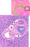
Lower panel is a higher magnification of the portal area where the branches of the hepatic artery, hepatic portal vein, and the bile duct are visible. Space of Mall is indicated by the asterisks.

  1. 6.1
  2. 6.1
10
Q

What are the characteristics of the portal lobule?

8.6.1

A

§Emphasizes the exocrine function of liver

–Major exocrine function is BILE SECRETION

§Morphologic axis of portal lobule is the i_nterlobular bile duct_ of portal triad

§Outer margins are lines drawn between the 3 central veins closest to the portal triad

§This shows the triangular block of tissue with portions of 3 classical lobules that secrete the bile which drains into its axial bile duct.

11
Q

What are the characteristics of the liver acinus?

A

§Described as diamond-shaped or oval- shaped

§Short axis defined by terminal branches of portal triad that lie along border between 2 classic lobules

§Long axis is line between the 2 central veins closest to the short axis

§Provides the best correlation between blood perfusion (heart diseases), metabolic activity, and liver pathology

12
Q

Describe the 3 zones the liver acinus?

A

§Zone 1 is closest to the short axis

§Zone 3 is farthest from the short axis and closest to the terminal hepatic venule (central vein)

§Zone 2 lies between zones 1 and 3 but has no sharp boundaries

13
Q

What are the chacateristics of zone 1?

A

–first to receive nutrients and toxins

–Last to die if circulation is impaired

–First to regenerate

–First to show degenerative changes following bile duct occlusion (bile stasis)

14
Q

What are the characteristics of zone 3?

A

–First to show ischemic necrosis (centrilobular necrosis) during reduced perfusion

–First to show fat accumulation

–Last to respond to toxins and bile stasis

15
Q

Explain the image

A

Photomicrograph of centrilobular necrosis in human liver. This photomicrograph shows a routine H&E liver biopsy specimen from an individual with congestive heart failure. Pathologic changes (referred to as ischemic necrosis) are most severe in hepatocytes in zone 3. This zone surrounds the terminal hepatic venule (central vein). This type of necrosis is referred to as centrilobular necrosis. Note the presence of multiple round vacuoles, which indicates extensive lipid accumulation. No noticeable changes are seen in the periphery of the lobule, that is zone 1 and much of zone 2

16
Q

What are the blood vessels of the parenchyma?

A

§PORTAL VEIN :

–Lumen larger than that of associated artery

–Provides venous blood to the sinusoids

§HEPATIC ARTERY:

–Thick muscular wall (typical of arteries)

–Provides arterial blood to sinusoids

Question Labeling in Gartner, 6th ed. (2014). Plate 15-3, Fig. 2, page 370.

17
Q

What are the characteristics of the central vein?

A

§CENTRAL VEIN:

–Thin wall

–Central position

–Receives blood from hepatic sinusoids

–More properly called TERMINAL HEPATIC VENULE

§Because it is the terminal venule of the hepatic portal vein system

18
Q

What are the characteristics of sinusoids and explain the image.

8.6.1

A

§SINUSOIDS:

–Wide blood vessels separating plates of hepatocytes

–Lined with thin endothelial cells

§With wide open fenestrae

–i.e., no diaphragm

§Discontinuous

–Large gaps seen between the cells

–Endothelial cells supported by discontinuous basal lamina

Liver section showing sinusoid capillaries with their endothelial cells close to the hepatocytes. The barely visible thin slit between the hepatocytes and the endothelium is the space of Disse. Kupffer cells can be seen inside the sinusoid. PT stain. High magnification. The structure of the liver sinusoid and its associated cells is illustrated in the upper right panel. Note Kupffer cells and stellate (Ito) cells.

19
Q

Explain the perisuinoidal space, Space of disse.

A

§Space situated between the hepatocytes and the endothelial cells lining the sinusoid

§Reveals irregular microvilli projecting from plasma membrane of hepatocytes

§Site of exchange of materials between blood and hepatocytes

§In adult contains plasma

In fetus contains islands of blood forming cells

Electron micrograph of a liver sinusoid in cross section. The microvilli of the hepatocytes are extending in the space of Disse. Open fenestrae are evident in the endothelial cell cytoplasm.

20
Q

Hpw does exchange work on the space of disse?

A

§Microvilli on the surface of the liver cells increase surface area for exchange of material between blood and hepatocytes

§Exchanges are accelerated due to:

–Absence of tight junctions between endothelial cells

–Large gaps in endothelial layer

–Discontinuous basal lamina

–Microvillous amplification of hepatocyte plasmalemma

21
Q

Know the image

A
22
Q

Explain the image

A

Electron micrograph of the liver. Note the two adjacent hepatocytes (red dashed line indicates their borders) with a bile canaliculus between them. The hepatocytes contain numerous mitochondria (M) and smooth and rough endoplasmic reticulum. A prominent Golgi complex (G) is near the bile canaliculus. The sinusoid is lined by endothelial cells (E) with large open fenestrae (F). The space of Disse (D) is occupied by numerous microvilli projecting from the hepatocytes

23
Q

Explain the image

A

Higher magnification of space of Disse. Note the presence of the microvilli (MV) that amplify the surface area of the hepatocyte (H) plasma membrane. RBC, red blood cell in the sinusoidal capillary (S). E, thin cytoplasm of the endothelial cell of the sinusoid. The fenestrae of the endothelial cell are indicated by the arrows.

24
Q

What are the two other cells in the space of disse?

A

–Kupffer cells

– Hepatic stellate (Ito) cells or lipocytes

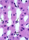
Arrows point to Kupffer cells.

25
Q

What are the characteristics of kuppfer cells?

A

§Form part of the sinusoid lining

§Derived from monocytes

§Do not form junctions with endothelial cells

26
Q

What is the function of Kupfer cells?

A

§Kupffer cells involved in breakdown of RBCs

Essential for RBC disposal

§Activity increased after splenectomy

Arrows point to Kupffer cells. The cytoplasm of these cells contains yellow pigment, hemosiderin, which results from the destruction of old RBCs. Note the binucleated hepatocyte (star).

27
Q

What are the characteristic of hepatic stellate cells?

8.6.1

A

§Normally, produce vitamin A

–Stored in lipid droplets

§And reticular fibers (regular amount)

–Support to liver parenchyma

§Converted into highly fibrogenic myofibroblasts when the liver continuously exposed to toxin (e.g., alcohol, infection with hepatitis virus)

EMs of liver at low (top) and high (below) magnification showing a hepatic stellate (Ito) cell in the perisinusoidal space. Lipid droplets in the cytoplasm of Ito cell are indicated by the asterisks.

28
Q

What is the function of hepatic stellate cells?

A
29
Q

Explain image

A

Alcoholic cirrhosis in an active drinker (A) and following long-term abstinence (B). A, Thick bands of collagen separate rounded cirrhotic nodules.  B, After a year of abstinence, most scars are gone. (Masson trichrome stain) 

30
Q

Explain the characteristics of hepatocytes

A

§Large six-sided cells

–Two sides facing space of Disse

–Four sides facing other hepatocytes

§Large central spherical nuclei

§Many cells are binucleate

–cells are tetraploid (have 4d amount of DNA)

§Each nucleus contains 1-2 well developed nucleoli

§Numerous mitochondria: 800-1000/cell

§Several small Golgi complexes per cell

§Peroxisomes

§rER

§sER

§Lysosomes

31
Q

What is the biliary tree?

A

§System of conduits of increasing diameter

§carry bile from hepatocytes to gallbladder and from there to intestine

–Inside the liver flow of bile is centrifugal (opposite to flow of blood)

§Begins at the region of central vein and proceeds to portal canal

32
Q

What are bile cannuiculi?

8.6.1

A

§Biliary tree starts with the bile canaliculi

–Canaliculus is small canal formed by apposed grooves in the surface of neighboring hepatocytes

–Form complete loop around four sides of 6-sideded hepatocyte

–Contains short irregular microvilli

–Sealed by junctional complex

33
Q

Explain the image

A

Bile canaliculi are formed when grooves on two faces of hepatocytes appose each other

34
Q

Explain the image

A

Cross section of bile canaliculus. Note the microvilli that project into the lumen.

The junctional complex (arrows) seals the bile canaliculus and prevents bile from

getting into the space between hepatocytes

35
Q

Explain how bile continues to flow to the terminal hepatic vein

8.6.1

A

§Near the portal canal bile canaliculi join to form CANALS OF HERING (cholangiole)

–Partially lined by hepatocytes

–Partially lined by cholangiocytes

§Lining the rest of the biliary tree where they become columnar

§possess microvilli and single cilium

–Canals of Hering carry bile to the bile ducts (part of the triad)

36
Q

Explain the characteristics of the gall bladder

A

§Receives diluted bile from the c_ommon hepatic duct_ of the liver through the cystic duct

§Concentrates and store bile and sends it to the duodenum

§Gallbladder removal leads to direct flow of bile from liver with few major consequences on digestion

§Wall consists of:

–Mucosa

–Muscularis

–Adventitia or serosa

37
Q

Explain the mucosa of the gall bladder

A

§Folds when the gallbladder is empty

§Consists of epithelium and lamina propria

§Epithelium

simple columnar (cholangiocytes)

–specialized for water absorption

–Closely resemble the absorptive cells of the intestine

§Well developed microvilli

§Apical junctional complexes

§apical and basal concentrations of mitochondria

§Lateral folds

38
Q

Explain the image

A

§Epithelial cells of the gallbladder actively transport Na+ and Cl- from cytoplasm to intercellular space

Electron micrographs of gallbladder epithelium. a. The tall columnar cells display features typical of absorptive cells, with microvilli on their apical surface, an apical junctional complex separating the lumen of the gallbladder from the lateral intercellular space, and numerous mitochondria in the apical portion of the cell. ×3,000. b. During active fluid transport, salt is pumped from the cytoplasm into the intercellular space, and water follows the salt. Both salt and water then diffuse into the cell from the lumen. As this process continues, the intercellular space becomes greatly distended (arrows). Fluid moves from the engorged intercellular space (arrows) across the basal lamina into the underlying connective tissue (CT) and then into blood vessels. The increase in size of the lateral intercellular space during active fluid transport is evident with the light microscope

8.6.1

39
Q

Explain the lamina propria, mucosa, and musclaris of the gall bladder

A

Photomicrograph of the Rokitansky-Aschoff sinuses in the wall of the gallbladder. This photomicrograph shows deep invaginations of the mucosa extending into the muscularis externa. These invaginations are referred to as Rokitansky-Aschoff sinuses.

40
Q

Explain the mised endo/exo gland of the pancreas

8.6.1

A

§Exocrine-

–Serous gland

–Synthesizes and secretes into duodenum enzymes needed for digestion in gut

§Proteases

§Lipase

§Amylase

§Bicarbonate

§Endocrine-

–Synthesizes and secretes into blood insulin, glucagon and somatostatin

§Regulate glucose metabolism

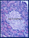
Light micrograph of pancreatic tissue. The group of lightly stained cells comprise one of too many islets of Langerhans (endocrine pancreas) responsible for secreting blood sugar-regulating hormones. This islet of Langerhans is surrounded by the acini of the exocrine pancreas.

41
Q

Characteristics of exocrine pancreas

A

§Surrounds islets of Langerhans (endocrine pancreas)

§Histologically resembles Parotid gland

–Acini formed by serous cells

§Cells exhibit zymogen granules

–Each acinus is drained by short i_ntercalated duct_

No striated ducts

§Secretes approx. 1.5 L of pancreatic juice/day

42
Q

Explain the exocrine pancreas duct system

8.6.1

A

§INTERCALATED DUCT

–Initial cells of the duct extend into the lumen of the acinus

–These cells called CENTROACINAR CELLS

§Unique to the pancreas

§These cells lack secretory granules

§Stain very lightly with eosin

–Easy to identify

Pancreatic acinus and its duct system. a. In this photomicrograph of a thin, H&E–stained plastic section, an intercalated duct can be seen beginning within a pancreatic acinus. The cells forming the duct within the acinus are the centroacinar cells. The eosinophilic zymogen granules are clearly seen in the apical cytoplasm of the parenchymal cells. ×860. b. In this schematic diagram, observe the beginning of the intercalated duct. Note the location of the centroacinar cells within the acinus. They represent the initial lining of the intercalated duct.

43
Q

Explain the image

A

Intercalated duct is encircled. Arrows pointing to the centroacinar cells.

44
Q

Explain the image

8.6.1

A

8.6.1

45
Q

Explaun the duct system from intercalted to main duct

8.6.1

A

§Intercalated ducts

–Secrete fluid rich in Na and bicarbonate

–Neutralizes chyme

§optimal pH for enzymes

§Intercalated ducts merge to form intralobular ducts

§Intralobular ducts merge to form larger interlobular ducts

§The latter join to with each other to form main pancreatic duct

Intercalated ducts (black arrow) merge to form larger intralobular collecting duct (green arrow).

46
Q

What is the black arrow pointing to?

8.6.1

A

intralobular duct

8.6.1