Dr. Mhawi 1 Digestive System 1 Tongue, Salivary Glands, and Teeth Flashcards

1
Q

What are the 3 types of oral mucosa in the oral cavity?

A
  • Masticatory mucosa
  • Lining mucosa
  • Specialized mucosa
2
Q

Masticatory mucosa if found on the ____ and ____

Has both _____ and ______ stratified squamous epithelium

A

gingiva and hard palate

keratinized and parakeratinized

In parakeratinized epithelium: superficial cells do not lose their nuclei

8.6.1

3
Q

True or False: Lining mucosa is non-kerantinized

8.6.1

A

True

4
Q

Where is specialized mucosa found?

A

dorsal surface of the tongue.

(specialized in sensation and taste)

8.6.1

5
Q

What are the 2 function s of oral mucosa?

A

•Barrier:

  • junctions between epithelial cells isolate the environment of the mouth cavity from the surrounding tissues

•Protection:

  • Oral cavity is protected from pathogens by combined actions of the epithelial cell junctions, migratory neutrophils, and saliva

Saliva contains antibodies

6
Q

Explain this image?

A

epithelial cells of the hard palate. Both keratinized and parakeratinized epithelial cells are visible.

7
Q

Explain this image

8.6.1

A

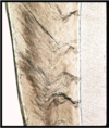
Both lips contain well-developed core of striated muscles (M)

  • Highly mobile
  • Help with ingestion and speech

Have three differently covered surfaces:

External surface

Covered by the skin

Contains hair follicles (F) and associated sebaceous glands (S), and sweat gland

Inner surface

Lined by lining mucosa (LM)

Many minor salivary glands (G) open

Vermilion zone (V)

Transitional zone between the lining mucosa and skin

Covered with very thin lightly keratinized stratified squamous epithelium

  • Rich in sensory innervations and capillaries
  • No hair follicles or salivary and sweat glands
8
Q

Explain the 3 surfaces of the tongue.

A

Composed of

Striated muscles

  • AKA lingual muscles
  • Arranged in bundles organized in 3 planes
  • Each plane arranged at right angle to the other
  • Muscle arrangement permits great flexibility

Dorsal surface

  • Covered with mostly keratinized epithelium
  • Epithelium raised into small projections called lingual papillae

Ventral surface

  • Covered with non-keratinized epithelium
  • No papillae
9
Q

Section of the Tongue

A
10
Q

______ are specialized mucosal elevations that are found on the dorsal surface

A

Lingual papillae

11
Q

What are th 4 types of lingula papillae?

A
  • Filiform papillae
  • Fungiform papillae
  • Circumvallate papillae
  • Foliate papillae

nAll contain taste buds except for filiform papillae

12
Q

Explain the function and characteristics of filiform papillae.

8.6.1

A
  • Smallest but most numerous
  • Tapered projections
  • Point toward rear of tongue
  • Composed of keratinized stratified squamous epithelium and core of l_amina propria_

Low rate of desquamation results in white coating

•No taste buds present

Filiform papillae serve only mechanical role

13
Q

Explain the function and characteristics of fungiform papillae.

A
  • Scattered among filiform and project above them
  • Covered with relatively translucent non-keratinized epithelium

Capillaries in underlying lamina propria show through it

•Papillae look red in gross appearance

•TASTE BUDS are present in epithelium on _dorsal surfac_e of fungiform papillae

14
Q

Explain the image

A

Upper panel, a photograph of the posterio-dorsal surface of the tongue. Fungiform papillae appear red due to the covering with relatively translucent nonkeratinized epithelium, that makes the underlying capillary of the lamina propria to show through. Note the presence of bulges at the base of the tongue (appear as ridges) reflecting underlying lingual tonsils in the lamina propria.

Lower panel, a micrograph of one of the fungiform papilla. Fungiform papilla projects above the surrounding filiform papillae (small solid arrows). Note the presence of the taste buds (dashed arrows) in the epithelium of the fungiform papilla.

15
Q

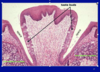
Explain the function of circumvallate papillae and the image.

8.6.1

A
  • Large, dome-shaped

•Located just anterior to SULCUS TERMINALIS

Humans have 8-12

Each papilla is surrounded by a moat-like space lined with stratified squamous epithelium

16
Q

Identify structures in the image.

A

no text.

  1. 6.1
  2. 6.1
17
Q

Id slide and characteristics.

A

circumvallate

18
Q

Explain image

8.6.1

19
Q

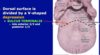
Location of papillae and sulcus termalis

20
Q

What gland is this? Explain image

A

Foliate Papillae

  • Located near the b_ase at lateral margins of tongue_
  • In the form of parallel ridges of mucosa
  • Papillae are separated by mucosal clefts
  • Small serous Von Ebner’s glands empty into clefts

Lower panel: In histological cross section, foliate papillae are visible as parallel ridges separated by mucosal clefts. SG, serous glands of Von Ebner

8.6.1

21
Q

How to differentiate lingual circumvallate and foliate papillae?

A

However, the appearance of taste buds in the apposed lateral walls of adjacent papillae helps in discriminating foliate papillae from circumvallate

NF, gustatory nerve fiber. Nuclei of the associated Schwann cells are visible.

22
Q

Explain this image.

8.6.1

A

Upper panel: Micrograph of a taste bud. The lightly stained cell of the bud are extending through the thickness of the epithelium. The taste pore (arrow) is visible in some of these buds .

Lower panel: Drawing of a taste bud, showing the taste cells and the taste pore. In addition to the taste cells, the drawing also illustrates other cell types (basal and supporting) and the nerve endings of the gustatory afferent nerve fibers.

23
Q

Explain what taste buds are and the 3 cell types.

8.6.1

A

Found in the lingual papillae of the tongue, soft palate and pharynx

Oval pale-staining structures extending through thickness of epithelium

Have a small opening at epithelial surface called TASTE PORE

•Through which fluid containing chemical substances enters

Taste bud has 3 cell types:

•Taste (sensory) cells

•Supporting cells

•Basal cells

24
Q

Explain this image and the characteristics of taste (sensory) cells.

A
  • Chemoreceptors
  • Elongated and most numerous
  • L_ightly-stained nuclei_
  • Apical surface has microvilli
  • Apically cells connected to each other or to adjacent supporting cells by t_ight (occluding) junction_
  • Basally synapse with processes (dendrites) of sensory neurons

(cranial nerves VII, IX, X)

Turnover time 10 days

Photomicrograph of a taste bud.

This high-magnification photomicrograph shows the organization of the cells within the taste bud. The sensory and supporting cells extend through the full length of the taste bud. The apical surface of these cells contains microvilli. The basal cells are located at the bottom of the taste bud. Note that the taste bud opens at the surface by means of a taste pore.

25
Explain this image
Taste bud showing sensory, supporting and basal cells. Note that the nuclei of the sensory cells are lightly stained in comparison with the supporting and basal cells Red arrow indicates the taste pore.
26
Characteristics of supporting and basal cell taste buds ## Footnote 8.6.1
Supporting cells: * Elongated and _less numerous_ * _Intensely-stained nuclei_ * Apically contain _microvilli_ * Apical _tight junctions_ With each other and with sensory cells * Do not synapse * _Turnover_ time 10 days Basal cells: •At the basal portion of the bud _Stem cells_ for _sensory_ and _supporting cells renewal_
27
What are the 4 parts of the teeth?
* Enamel: covers the crown * Cementum: covers the root * Dentin: surrounds the pulp * Pulp: central loose connective tissue
28
What are the characteristics of enamel?
_Hardest substance in body_ 98% hydroxyapatite (calcium phosphate) Hydroxyapatite arranged in _RODS or PRISMS_ nEach rod spans the enamel layer Each rod is produced by one _ameloblast_ before tooth eruption Enamel becomes _acellular_ after tooth eruption In section prepared by ground tooth technique, enamel appears striated •Striations called LINES OF RETZIUS Appear uneven Represent a record of the path ameloblast has taken during the enamel deposition Ground tooth (or bone) is a special technique by which the tooth/bone is cut thin enough (without demineralization with acids) by a sharp saw and examined with or without staining. This technique helps with the visualization of certain features in tooth (rods or prisms) and bone (layers of bone matrix in Haversian system and/or Canaliculi). 8.6.1
29
What is amelogenesis and what are its stages?
Amelogenesis is the process of enamel formation Enamel formed in two stages: * Secretory stage * Maturation stage
30
What is the secretory stage of amelogenesis? ## Footnote 8.6.1
* Ameloblasts secrete _organic matrix_ * During matrix deposition ameloblasts _move away_ from the _dentinoenamel junction_ (previous slide and red arrow in lower panel of this slide) * Movement takes _uneven course_ * Matrix deposition continues until enamel reaches _full thickness_ Upper panel: Light micrograph of the enamel formation. The enamel is deposited by the ameloblasts over pre-existing thin slab of dentin before tooth eruption. Ameloblasts move away from the dentinoenamel junction as the amelogenesis continues. The white stripe visible between the dentin and the enamel is an artifact resulted from the separation of the enamel from the dentin, most likely during the preparation of the section. Lower panel is a high magnification of the dentinoenamel junction within the boxed area in the upper panel. In this ground tooth technique, the enamel rods (prisms) appear uneven, a reflection of the uneven course the ameloblasts take during the deposition of the enamel.
31
What is the maturation stage of amelogenesis? ## Footnote 8.6.1
Ameloblasts differentiate to _transporting epithelium_ * _Remove_ organic material from matrix * _Add_ calcium and phosphate After completion of enamel formation, ameloblasts serve as _protective epithelium_ over the crown until tooth erupts Ameloblasts then _degenerate_ Explains why enamel is _not replaceable_
32
What are the characteristics of cementum?
Covers _root_ of tooth - Root fits into socket of bone (alveolus) Cementum has 50% minerals secreted by **_CEMENTOCYTES_** closely resemble osteocytes Cementum is thin layer of _bone-like_ material Has _lacunae_ filled with cementocytes, which connect with each other by _canaliculi_ Cementum is _avascular_ _Nourishment_ is provided via blood vessels in the adjacent periodontal ligament
33
What are the characteristics of dentin?
Consists mainly of * Type I collagen * Glycosaminoglycans (GAGs) * Hydroxyapatite (70%) Dentin matrix secreted by **_ODONTOBLASTS_** * Form an _epithelial layer_ over the inner surface of the dentin * During dentin deposition odontoblasts move toward the pulp cavity ni.e., opposite to the movement of the ameloblasts
34
How is dentin deposited from the odontoblast?
Dentin is deposited from _branched apical processes_ * Called odontoblast processes * Processes become longer as the odontoblast is displaced toward the pulp cavity during dentin deposition Odontoblasts are responsible for the _mineralization o_f the dentin Left) photomicrograph and (right) interpretive EM drawing of odontoblasts. Extending into the dentin from the cell apex, there is a light-staining branched odontoblast process that lacks cisternae of rER and Golgi. Secretory granules are present in this process and its branches.
35
Odontoblast processes contained in canals called \_\_\_\_\_\_
DENTINAL TUBULES A: An illustration showing the dentinal tubules (blue) within which the odontoblast processes are extended. Unmyelinated nerve fibers (yellow) extend to a short distance inside the dentinal tubules, as well. B: High-resolution SEM of dentinal tubules. Many dentinal tubules run through the dentin (De) matrix. The 3- to 4-µm-diameter processes of odontoblasts (Od) are in the tubules.
36
What are the characteristics of pulp? ## Footnote 8.6.1
Pulp is _loose connective tissue_ Highly i_nnervated and vascularized_ Myelinated nerve fibers and vessels enter pulp from the _apical foramen_ Some _afferent (sensory) nerve fibers lose myelin_ and extend short distances into dentinal tubules Transduce stimuli from tooth surface Light micrograph of cross-sectioned tooth. The section reveals the mature dentin and the lightly stained immature predentin secreted by the odontoblasts. The pulp cavity is filled with highly innervated and highly vascularized loose connective tissue.
37
What are the 4 characteristics of the periodontal ligament? ## Footnote 8.6.1
Function: _•Tooth attachment_ _•Support_ _•Bone remodeling_ •Nutrition of adjacent structures ne.g., cementum _•Proprioception_ ne.g., detection of hard particles in food
38
Explain this image.
Sections show both _dense_ and _loose connective tissue_ _Loose_ connective tissue contains blood vessels nerves, and GAGs Periodontal ligaments attaches the cementum (C) to the alveolar bone (B). D, dentin. Sharpey’s fibers are collagen fibers that are embedded in the cementum at one end (arrowheads) and in alveolar bone at the other (arrows).
39
What are the characteristics of dense connective tissue of the periodontal ligament. ## Footnote 8.6.1
Dense connective tissue consists of _bundles of type I collagen fibers_ and fibroblasts Periodontal ligament also has _OXYTALAN FIBERS_ •stain with elastic stains TEM electron micrograph of the boxed area of the periodontal ligament in the previous slide. OF, Oxytalan fibrils (very immature elastic fibers); CF, type I collagen fibrils.
40
Collagen fibers are embedded in the cementum at one end and in the alveolar bone at the other \_\_\_\_\_\_\_
As Sharpey’s fibers Top panel: Section showing the insertion of a tooth to the alveolar bone via periodontal ligament. Because this material was obtained from a young animal, the bone is being continuously reformed to adapt to the tooth’s eruption; this explains the presence of osteoclasts. The ligament is formed by oriented fibroblasts. PT stain. Medium magnification. Lower panel: Electron micrograph of Sharpey's fibers. Sharpey's fibers extend from the periodontal ligament (right) into the cementum. They consist of collagen fibrils. Sharpey's fibers within the cementum are mineralized; those within the periodontal ligament are not mineralized
41
Explain this image ## Footnote 8.6.1
Longitudinal section of demineralized tooth. The enamel (E), which is highly calcified tissue, was completely removed leaving a space (represented by the area surrounded by the dashed line). The asterisk is placed in the gingival sulcus. A, alveolar bone; C, cementum; D, dentine; G, gingiva; PL, periodontal ligament
42
What are the characteristics of gingiva? 8.6.1
Part of the masticatory mucosa Consists of * stratified squamous epithelium keratinized and * fibrous lamina propria Forms a collar around each tooth Lamina propria _tightly bound_ to the periosteum of maxilla or mandible (jaw bones) and to the crown of the tooth _GINGIVAL SULCUS_ •Normal 1 mm to 3 mm deep space between enamel and gingival epithelium (asterisk) Surrounds the crown Potentially _danger zone_ •Accumulation of bacteria leads to _gingivitis_
43
_Below the sulcus_ gingival epithelium is tightly adherent: To the _**\_\_\_\_\_\_\_**_in young individuals To the _**\_\_\_\_\_\_\_**_in adults
_enamel_ _cementum_