Disorders of the shoulder Flashcards
if pt is under 35 what shoulder conditions is common
- Glenohumeral dislocations/ instability
- Traumatic
- Atraumatic
- ACJ dislocations/ instability
- Clavicle fractures
if pt is over 35 what shoulder conditions is common
- Shoulder impingement
- Rotator cuff tendinopathy
- RC tears
- Proximal humeral fractures
- Adhesive capsulitis
- Osteoarthritis
- Glenohumeral
- acromioclavicular
Shoulder instability
- 1-2% population
- Young adults
- Male>female
- Sports and activity
- Risks
- Recurrent dislocation
- Recurrent subluxations
- Apprehension
Classification of shoulder instability
- Direction
- Unidirectional
- Multidirectional
- Timing
- Acute
- Recurrent
- Mechanism
- Traumatic
- Atraumatic
– Habitual / Voluntary
- +/- Fracture (Greater Tuberosity, Glenoid, Humeral Head)
e. g. a traumatic anterior dislocation
- Unidirectional- anterior
- Acute
- Traumatic
shoulder joint complex includes
– Glenohumeral Joint
– Sternoclavicular Joint
– Acromioclavicular Joint
– Scapulothoracic Articulation
– Thoracic Spine

Glenohumeral joint stability
- Static factors:
- Bone
- Capsule
- Labrum
- Glenohumeral ligaments
- Negative intra-articular pressure
- Dynamic factors
- Rotator cuff/LHB
- Scapula
- Mobile base
- Scapula stabilises
Multidirectional instability of the shoulder cause
- Atraumatic
- Abnormal muscle patterning
features of Abnormal muscle patterning
- no structural damage
- capsualr dysfunctional and laxity
- often bilateral

treatment of multidirectional instability of the shoulder
- Treatment:
- Prolonged physiotherapy
- Muscle re-education
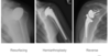
Shoulder dislocations occurs when
the humeral head is displaced from articulating with the glenoid fossa, and can be divided into three categories: anterior, posterior and inferior dislocations.
Anterior dislocations are the
most common - they make up around 90-95% of all shoulder dislocations.

cause of anterior dislocation
Anterior dislocations are usually caused by a blow to the posterior shoulder or by the arm being pushed posteriorly when the hand is placed behind the head, pushing the humeral head anteriorly to the glenoid fossa.
mechanism of trauamtic antieorr dislocation
FOOSH
presentation of anteiror dislocation
They present with an externally rotated and abducted
Pain, deformity, loss of function
pathoanatomy of trauamtic anterior dislocation
- Tear of glenoid labrum & Stretch of glenohumeral ligaments (‘Bankart’ lesion)
- +/- anterior glenoid rim fracture (‘Bony Bankart’)
- +/- posterior humeral head impaction fracture (‘Hill-Sachs’ lesion)
treatment of anteiror dislocation
Treatment:
- Manipulation under sedation
- Often undergo surgery eventually
Posterior dislocations are
far less common
causes of posterior dislocation
and are caused by epileptic seizures, electrocution, lightning strikes or a blow to the anterior shoulder, pushing the humeral head posteriorly.
presentation of posterior dislocation
They present with an internally rotated and adducted arm. There is also squaring of the shoulder and a prominent coracoid process.
- light bulb sign

Treatment for posterior dislocation
- Manipulation under anaesthetic
- Surgery to address bone defect
Inferior dislocations are very rare
cause of inferior dislocation
are caused by hyper-abduction.
presentation of inferior dislocation
They present with an abducted arm, usually with a flexed elbow (arm placed behind head).

Injuries associated with shoulder dislocations include:
Bankart lesion– tear of the glenoid labrum
Hill-Sachs lesion– dent in the humeral head caused by the lip of the glenoid fossa impacting it during the injury.
Rotator cuff muscle tear
Damage to axillary artery/nerve
acromioclavicular joint dislocation
- Common in young adults

Mechanism of Acromioclavicular joint dislocation
direct falls
Classification of Acromioclavicular joint dislocation
- Disruption of coraco-clavicular and acromioclavular ligaments
- Grade I – VII
Treatment: Acromioclavicular joint dislocation
- Usually non-operative – broad arm sling
- Surgery for pain/ instability (<5%)
clavical fracture (only bone conenction to the skeleton
80% of all clavicle fractures are mid-clavicular fractures, usually caused by a fall onto an outstretched hand (FOOSH)or blow to the shoulder.
clavicle fractures are common in
children and young adults
presentation of clavicular fracture
A displaced mid-clavicular fracture presents with a dropped shoulder and adduction of the arm. This is due to sternocleidomastoid muscle elevating the medial fragment of the broken clavicle and pectoralis major pulling the lateral fragment inferiorly.
risk of clavicle fractures
There is also a high risk with mid-clavicular fractures that they will convert to an open fracture; the sharp ends of the bone could push through the skin, which would increase the chance of infection and complicate the fracture.
management of clavicle fracture
- Usually non-operative (broad arm sling)
- Surgery for complicated cases
- Open fractures
- Neurovascular injury
Proximal humeral fractures
- Greater tuberosity fractures
-
Surgical neck of humerus fractions
- 2/3/4 parts
- Common in elderly (FOOSH)
treatment of proximal humeral fracture
- Minimally displaced- collar and cuff sling
- Displaced
- Reduction and fixation
- Shoulder replacement

Impingement Syndrome
Impingement Syndrome is the impingement of rotator cuff muscle tendons on the coraco-acromial arch.
‘a mechanical irritation of tissues between the greater tuberosity of the humerus and the coraco- acromial arch’
imingment occur due to
presentation of shoulder impingment
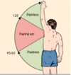
Impingement syndrome causes pain, weakness and reduced range of motion when the shoulder is flexed and/or abducted.
This condition most commonly affects the supraspinatus tendon which causes a ‘painful arc’ between 60° and 120° of abduction.
who is shoulder impingement common in
- Common in middle aged
- Pain + weakness
- Overhead/lifting/ reaching behind
diagnosis of impingment
Diagnosis involves a targeted medical history, physical examination and an X-ray.
treatment of impingement
Treatment ranges from rest and NSAIDs to manage pain, to physical therapy, to arthroscopic/open surgery depending on severity.
Adhesive capsulitis
commonly known as ‘frozen shoulder’, is a condition involving inflammation of the capsule of the glenohumeral joint.
Presentation of frozen shoulder
restricted movement and chronic pain that worsens at night and in cold weather (hence the common name of ‘frozen shoulder’).
causes of adhesive capsulitis
idiopathic (some tirggers)
- metapalsia of capsule elads to true global stiffness on passive. movment
risk factors for adhesive capsulatiis
- female
- diabetes, MI, CVA
minor trauma
natural history of adhesive capsulitis
Natural history positive
- Resolution over 2 years
- Freezing/ frozen/ thawing phases
- Does not recur
- In around 5-20% of patients, the opposite shoulder becomes affected within 5 years of the first presentation of adhesive capsulitis.
management of adhesive capsulitis
- Observant
- Physiotherapy
- Injections
- Distention arthrogram
- Manipulation under anaesthetic
- Arthroscopic capsular release
Rotator Cuff Tears
Rotator cuff tears refer to damage to any of the rotator cuff muscles: supraspinatus, infraspinatus, teres minor and subscapularis.
most common roator cuff teqar
The most common muscle to tear is supraspinatus.
Risk factors of rotator cuff muscle
include certain repetitive actions (e.g. overhead activity like painting ceilings), smoking, and a family history of rotator cuff tears.
presentation of rotator cuff tear
This injury is often asymptomatic, but the most common presentation is anterolateral shoulder pain radiating inferiorly, particularly with shoulder flexion. Rotator cuff muscles tears cause weakness of shoulder abduction, making normal daily activities such as brushing hair difficult and painful.
managment of rotator cuff tears
Management usually includes pain management and physical therapy; however, for large acute tears, or for young individuals with full thickness tears, surgery is an option.
Brachial Plexus Injuries
The brachial plexus is a bundle of nerves in the axillae that has important motor and sensory functions in the upper limbs.
- upper brachial plexus injuries
- lower brachial plexus injury
- winged scapula
Upper Brachial Plexus Injury
An upper brachial plexus injury, also known as ‘Erb’s Palsy’ or ‘Duchenne’s Palsy’, describes damage to the cervical spinal cord roots C5 and C6.
cause of upper brachial plexus injury
It is usually caused by trauma that forcefully pushes the shoulder down or shoulder dystocia (shoulders getting stuck in the birth canal) during birth.
presentation of upper brachial plexus injury
This condition presents with internal rotation and abduction of the arm at the shoulder, extension of the arm at the elbow, pronation of the forearm and wrist flexion (this positioning of the arm is sometimes called ‘Waiter’s tip’).
It also causes paraesthesia of the C5 and C6 dermatomes – lateral arm, lateral forearm and 1st and 2nd digits.
managment of upper brachial pelxus injury
Lower Brachial Plexus Injury
A lower brachial plexus injury, also known as ‘Klumpke’s Palsy’, describes damage to cervical and thoracic spinal cord roots C8 and T1.
causes of lower brachial plexus injury
It is usually caused by forced hyperextension or abduction at the shoulder (e.g. falling off monkey bars and grabbing at the last minute, yanking the shoulder), but in some (rare) cases it can be due to impingement from a Pancoast tumour (a tumour at the apex of the lung).
presentation of lower brachial plexus injury
This condition presents with a claw hand, and paraesthesia of the C8 and T1 dermatomes – 4th and 5th digits, medial border of the hand and medial forearm.

management of brachial plexus injury
Surgery can sometimes be used in this condition but there is often no treatment.

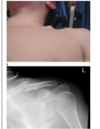
Winged Scapula
‘Winged Scapula’ is a condition that occurs with the paralysis of the long thoracic nerve, a nerve that originates from C5, C6 and C7 as they enter the brachial plexus (so it’s not technically a brachial plexus injury, but is very closely assocaited).
The long thoracic nerve normally innervates the
serratus anterior muscle, a muscle that is responsible for the rotation of the scapula to allow the arm to abduct. When this muscle is paralysed, the scapula twists out of position and protrudes through the skin.
causes of thoracic nerve injury
It can be caused by direct trauma to the path of the nerve, or can be due to over-stretching, such as in overhead weight-lifting.
presentation of winged scapula
Winging of the scapula is most visible when the shoulder is flexed and the arm is pressed against a wall (like in the image below).
Treatment of winged scapula
ranges from analgesia and lifestyle modifications (like avoiding overhead work), to surgery. It may take many years to get back to normal.
Osteoarthritic of shoulder
1) Acromioclavicular joint
Treatment
- Injections
- Excisional arthroplasty
2) Glenohumeral joint
Treatment
- Injections
- Shoulder replacement
Total shoulder replacements