Dermatology Flashcards
Diaper Dermatitis etiologies
- wearing diapers: contact dermatitis, miiaria (heat/sweat rash, blockage of eccrine sweat glands), candida (moist conditions)
- rash in diaper area as well as other areas: atopic dermatitis, seborrheic dermatitis
- affects diaper area irrespective of diaper use: scabies, bullous impetigo

management of diaper dermatitis
- frequent diaper changes every 2 hours or when soiled
- open air exposure
- topical zinc oxide or petroleum jelly
- 1% hydrocortisone cream (< 2 wk use)
- may need topical Abx

Perioral Dermatitis Clinical Manifestations
MC seen in young women; may have a history of topical corticosteroid use
papulopustules on erythmetous base which may become confluent into plaques and scales
classically spares the vermillion border

Management of Perioral Dermatitis
topical metronidazole or erythromycin
oral tetracyclines
avoid topical corticosteroids
Types I - IV of Cutaneous Drug Reactions
- Type I: IgE-mediated, ex: urticaria and angioedema. IMMEDIATE
- Type II: cytotoxic, Ab-mediated (drugs in combo with cytotoxic antibodies cause cell lysis)
- Type III: immune antibody-antigen complex. Ex: drug mediated vasculitis and serum sicness
- Type IV: DELAYED (cell-mediated) – morbilliform reactions, Ex: Erythema Multiforme
Clinical Manifestations of Cutaneous Drug Reactions
-
exanthematous/morbilliform rash: MC skin eruptions. (type IV delayed) generalized distribution of bright red macules and papules that coalesce to form plaques. Rash typically begins 2 - 14 days after medication initiation
ex: antibiotics, NSAIDs, allopurinol, thiazide diuretics -
Urticarial: (type I immediate) occurs within minutes to hours after drug administration
ex: antibiotics, NSAIDs, opiates and radiocontrast media - Erythema Multiforme (Type IV delayed) target lesions may not always be present in drug-induced EM
MC Drugs: Sulfonamides, penicillins, phenobarbital, dilantin
fever, abdominal pain, and joint pain may accompany the cutaneous drug reaction
Management of Cutaneous Drug Reactions
- dicontinue offending medication
- Exanthematous/Morbiliform: oral antihistamines
- Drug-induced urticaria/angioedema: systemic corticosteroids, antihistamines
- Erythema Minor: symptomatic therapy
- Anahylaxis: intramuscular epi
What is Lichen Planus?
idopathic cell-mediated immune response
increased incidence with Hepatitis C

Clinical Manifestations of Lichen Planus
5 Ps: Purple, Polygonal, Planar, Pruritic Papules with fine scales and irregular borders
can develop Koebner’s Phenomenon: new lesions at sites of trauma (also seen in psoriasis)
Wickham Striae: fine white lines on the skin lesions or on oral mucosa. nail dystrophy

Management of Lichen Planus
topical corticosteroids 1st line
2nd line: PO steroids, UVB therapy, retinoids. rash usually resolves spontaneously in 8 - 12 months

What is Pityriasis Rosea?
maculopapular rash with uncertain etiology (possibly associated with viral infections – HHV7)
primarily older children/young adults
increased incidence in the spring/fall
can mimic syphillis so order RPR if the patient is sexually active

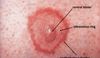
Clinical Manifestations of Pityriasis Rosea
Herald Patch** (solitary salmon-colored macule) on the trunk 2 - 6 cm in diameter
general exanetham 1 - 2 weeks later: smaller, very pruritic 1 cm round/oval salmon-colored papules with white circular (collarette) scaling along cleavage lines in a Christmas Tree Pattern
(confined to trunk and proximal extremities (face usually spared)
resolves in 6 - 12 weeks

Management of Pityriasis Rosea
none needed
pruritus: PO antihistamines, topical corticosteroids, oatmeal baths
possible UVB therapy if severe

What is the most common cause of Stevens-Johnson Syndrome/TEN?
MC after drug eruptions: especially sulfa and anticonvulsant meds
also NSAIDs, Allopurinol, antibiotics
infections are a less common cause; Ex: Mycoplasma, HIV, HSV
What is the difference between Stevens-Johnson Syndrome and TEN?
SJS = sloughing < 10% BSA
TEN = sloughing > 30% BSA, may develop skin necrosis
clinical manifestations of SJS and TEN
fever and URI Sx ► widespread blisters begin on trunk/face, erythematous/pruritic macules >= 1 mucous membrane involvement with _epidermal detachment*_ (+ Nikolsky sign)

Management of SJS and TEN
treat like severe burns: burn unit admission, pain control, withdrawal of offending med, fluid and electrolyte replacement, wound care

What is Erythema Multiforme?
acute self-limited Type IV hypersensitivity reaction
MC in young adults 20 - 40 yo
skin lesions usually evolve over 3 - 5 days and persist for about 2 weeks
Associations: Herpes Simplex Virus MC**, Mycoplasma, S. pneumo
meds: sulfa drugs, beta-lactams, Phenytoin, Phenobarbital
Clinical Manifestations of Erythema Multiforme
TARGET lesions classic**
dull, “dusty-violet” red, purpuric macules/vesicles or bullae in the center surrounded by pale edematous rim and peripheral red halo. often febrile

What is the difference between EM minor and EM major?
EM Minor: target lesions distributed acrally (distal portion of limbs–hands and feet–and head–ears and nose); no mucosal membranes involved
EM Major: target lesions with involvement of >= 1 mucous membrane (oral, genital, or ocular mucosa) < 10% BSA acrally → centrally (no epidermal detachment)
Management of Erythema Multiforme
Symptomatic
discontinue offending drug
antihistamines
analgesics
skin care
steroid/lidocaine/diphenhydramine mouthwash for oral lesions
systemic corticosteroids if severe
What are the main pathophysiological factors of Acne Vulgaris?
- increased sebum production: î androgens increase sebaceous gland activity
(MC after puberty, î androgens PCOS and Cushing’s Disease)
- Clogged sebaceous glands: due to increased proliferation of follicular keratinocytes
- Propionbacterium acne overgrowth: P. acne is part of the normal flora that overgrows in blocked pores ⇒ lipase production by P. acne which converts sebum into inflammatory fatty acids that damage healthy cells
- inflammatory response
Clinical Manifestations of Acne Vulgaris
commonly seen in areas with î sebaceous glands (face, back, chest, upper arms)
- comedones: small, non-inflammatory bumps from clogged pores
– open comedones: (blackheads) – incomplete blockage
– closed comedones (whiteheads) – complete blockage
2. inflammatory: papules or pustules surrounded by inflammation
3. nodular or cystic acne: often heals with scarring
Diagnosis of Acne Vulgaris (mild, moderate, severe)
Mild: comedones (+- small amounts of papules &/or pustules)
Moderate: comedones, larger amounts of papules and pustules
Severe: nodular (>5 mm) or cystic acne