Cranial Nerves 1 - 6 Flashcards

1
Q

The majority of cranial nerves exit the ____ surface of the brainstem, but there are exceptions. CN I and II project to _____, and CN IV exits the dorsal surface of the ___ ____.

____ exits ventrally but not in the brainstem (it has a spinal origin).

A

The majority of cranial nerves exit the ventral surface of the brainstem, but there are exceptions. CN I and II project to forebrain, and CN IV exits the dorsal surface of the caudal midbrain.

CN XI exits ventrally but not in the brainstem(it has a spinal origin).

2
Q

Taste is a part of 3 mixed nerves: ___, ___ and ___ cranial nerves.

A

Taste is a part of 3 mixed nerves: facial (CN VII), glossopharyngeal (CN IX), and vagus (CN X)

3
Q

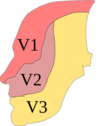
The trigeminal nerve supplies both ___ and ___ innervation to various parts of the head and neck. What are the three V’s?

Two of them, ___ and ___, are purely ___.

A

The trigeminal nerve supplies both sensory and motor innervation to various parts of the head and neck.

The three V’s are:

V1 - Opthalmic

V2 - Maxillary

V3 - Mandibular

Two of them, opthalmic and maxillary, are purely sensory.
4
Q

______ division (CN V (V1)), is asensory nerve that passes through the_______ andsupplies the eyeball, conjunctiva, lacrimal gland and sac, nasal mucosa, frontal sinus, external nose, upper eyelid, forehead and scalp.

A

Opthalmic division (CN V), is asensory nerve that passes through the superior orbital fissure and supplies the eyeball, conjunctiva, lacrimal gland and sac, nasal mucosa, frontal sinus, external nose, upper eyelid, forehead and scalp.

5
Q

_______ division (CN V (V2) ) is a sensory nerve that passes through the_____ ____. It relays sensory info from the skin of the face over the maxillam including the upper lip, maxillary teeth, nasal mucosa, maxillary sinuses and palate.

A

Maxillary division (CN V ) is asensory nerve that passes through the foramen rotundum. It relays sensory info from the skin of the face over the maxilla including the upper lip, maxillary teeth, nasal mucosa, maxillary sinuses and palate.

6
Q

The third division of the trigeminal nerveis both a ___ and ____ nerve. The _______ division (CN V - v3) passes through the ___ ___ and is both a sensory and motor nerve. The motor component supplies the muscles of _____, myohyoid, anterior belly of the digastric, tensor veli palatini and tensor tympani.

Sensation from the skin over the mandible, including the lower lip and side of the head, mandibular teeth, mucosa of the mouth and the anterior two thirds of the tongue is also relayed by CN V .

A

The third division of the trigeminal nerve is both a motor and sensory nerve. The MANDIBULAR division (CN V ) passes through the FORAMEN OVALE and is both a sensory and motor nerve. The motor component supplies the MUSCLES OF MASTICATION, myohyoid, anterior belly of the digastric, tensor veli palatini and tensor tympani.

Sensation from the skin over the mandible, including the lower lip and side of the head, mandibular teeth, mucosa of themouth and the anterior two thirds of thetongue is also relayed by CN V .

Mandibular nerve mnemonic:

“My Tensors Dig Ants 4 MoM” (Mylohyoid—Tensor Tympani + Tensor Veli Palatini—Digastric(Anterior) – 4 Muscles of Mastication (Temporalis, Masseter, Medial and Lateral Pterygoids)

7
Q

CN___:

FUNCTION: special sensory (or visceral afferent - smell).

These nerve fibers pass through foramina in the ____ of the ____ bone,pierce the dura and arachnoid of the brain and enter the olfactory bulb. In addition to enabling us to smell, ____ also induces visceral responses via the autonomic nervous system. For example, salivation is initiated in response to the aroma of food.

A

CN I - Olfactory

FUNCTION: special sensory (or visceralafferent - smell).

The olfactory nerve fibers are collectivelyknown as CN I. The fibers pass through foramina in the CRIBRIFORM PLATE of the ethmoid bone, pierce the dura and arachnoid of the brain and enter the olfactory bulb. In addition to enabling us to smell, CN I also induces visceral responses via the autonomic nervous system. For example, salivation is initiated in response to the aroma of food.

8
Q

___ innervates 4 or 6 extraocular muscles.

Is this parasympathetic or sympathetic?

FUNCTIONS: innervates 4 of the 6 extraocular muscles.

The oculomotor nerve is the chief motor nerve for the ocular and extraocular muscles. It leaves the cranial cavity and enters the __ __ __.

Within this fissure the nerve divides into the superior and inferior divisions. The inferior division also carries presynaptic autonomic fibers to the __ __. The superior division carries fibers to the __ __ and __ __.

A

CN III - Oculomotor

This supplies parasympathetic motor neurons to ciliary muscle and sphincter pupillae.

FUNCTIONS: innervates 4 of the 6 extraocular muscles.

The oculomotor nerve is the chief motor nerve for the ocular and extraocular muscles. It leaves the cranial cavity and enters the SUPERIOR ORBITAL FISSURE.

Within this fissure the nerve divides into the superior and inferior divisions. The inferior division also carries presynaptic autonomic fibers to the CILIARY GANGLION. The superior division carries fibers to the levator palpibrae and superior rectus.

9
Q

____ function is to supply motor innervation to the superior oblique extraocular muscles. This nerve passes through the___ ___ ____, where it supplies the superior oblique muscle of the eye.

Injury to this nerve inhibits the eyeball from turning out and down (inferolaterally). This is seen clinically aa ____ (aka double vision).

This nerve is the only cranial nerve to emerge dorsally from the brainstem.

A

CN IV’s (trochlear) function is to supply motor innervation to the superior oblique extraocular muscles. This nerve passes through the superior orbital fissure where itsupplies the superior oblique muscle of the eye (Remember, Live Frankly To See Absolutely No Insult).

Injury to this nerve inhibits the eyeballfrom turning out and down (inferolaterally).This is seen clinically as DIPLOPIA (doublevision).

CN IV is the only cranial nerve to emerge dorsally from the brainstem.

10
Q

____ nerve function is to supply motor innervation to the lateral rectus extraocular muscles. This nerve arises between the ___ and the ___ (___ junction) on the brain and wil ltravel through the __ ___ ___ to innervate the lateral rectus muscle. The action of this muscle is to ___ the eye.

A

CN VI’s (abducens) function is to supply motor innervation to the lateral rectus extraocular muscles. The abducens nerve arises between the pons and the medulla (PMJ) on the brain, and will travel through the superior orbital fissure to innervate the lateral rectus muscle. The action of this muscle is to ABDUCT the eye.

In this picture, the bad eye is the RIGHT eye. She cannot ABDUCT her right eye.

11
Q

___ nerve:

General sensory function: __ and __ divisions

General motor function: ___ divison.

Also, it acts like a trail finder for all parasympathetic pathways

A

CNV nerve:

General sensory function: V1 (opthalmic) and V2 (Maxillary) divisions

General motor function: V2 (Mandibular) divison.

Also, it acts like a trail finder for all parasympathetic pathways

12
Q

The oculomotor nucleus originates at the level of the __ __ in the __-__. The muscles it controls are the striated muscle in the levator palpebrae superioris and all extraocular muscles except for the superior oblique muscle and the lateral rectus muscle.

The Edinger-Westphal nucleus supplies ____ fibers to the eye via the ___ ganglion, and thus controls the sphincter pupillae muscle (affecting pupil constriction) and the ciliary muscle (affecting accommodation).

A

The oculomotor nerve nucleus originates at the level of the superior colliculus in the mid brain. The muscles it controls are the striated muscle in levator palpebrae superioris and all extraocular muscles except for the superior oblique muscle and the lateral rectus muscle.

The Edinger-Westphal nucleus supplies PS fibers to the eye via the ciliary ganglion, and thus controls the sphincter pupillae muscle (affecting pupil constriction) and the ciliary muscle (affecting accommodation).

13
Q

The olfactory nerve enter the skull through the ___ ___ of the ___ bone to synapse in the Olfactory Bulb.

A

These olfactory nerves enter the skull through the Cribriform Plate of the Ethmoid Bone to synapse in the Olfactory Bulb.

14
Q

The ___ ____ is formed by neural processes of neuroepithelia on upper lateral and septal wall of the nasal cavity

Neural processes traverse perforations of the cribriform plate of ethmoid bone and synapse in olfactory bulb.

Third order connections of olfactory bulb form olfactory tract that ends in the orbital frontal cortex and temporal lobe cortex

A

The olfactory nerve is formed by neural processes of neuroepithelia on upper lateral and septal wall of the nasal cavity

Neural processes traverse perforations of the cribriform plate of the ethmoid bone and synapse in olfactory bulb.

Third order connections of olfactory bulb form olfactory tract ends in the orbital frontal cortex and temporal lobe cortex

15
Q

Sympathetics come in and will go to the eyelid itself because there is a region on the eyelid that has smooth muscles (tarsal muscles). These muscles are not associated with the levator palpebrae superiororis muscle (which are innervated by which cranial nerve?)

The tarsal muscle is separately innervated by _____ fibers that originate in the ___ ___ cord.

The levator palpebrae superioris muscle elevates and retracts the upper eyelid.

Since smooth muscles located in the eyelid are innervated by sympathetics, how does this relate to Horner’s syndrome?

A

Sympathetics come in and will go to the eyelid itself because there is a region on the eyelid that has smooth muscles (tarsal muscles). These muscles are not associated with the levator palpebrae superiororis muscle, which are innervated by CN III.

Smooth muscle that is located in the eyelid is innervated by sympathetics.How does this relate to Horner’s syndrome?

Horner’s syndrome is a combination of symptoms that arises when a group of nerves known as the sympathetic trunk is damaged.

The most common clinical signs of Horner’s Syndrome are: Drooping of the eyelid on the affected side (ptosis) The pupil of the affected eye will be constricted (miosis), or smaller than usual. The affected eye often appears sunken (enophthalmos)

16
Q

1) ___ nucleus of V (a relay for pain and temperature is analogous to the anterolateral pathway for the body)
2) ___ nucleus – (a relay for fine touch is analogous to the dorsal column medial lemniscus pathway for the body)
3) ____ nucleus of V – (a unique nucleus specialized for proprioception of chewing, eye and tongue muscles, this is the ONLY case where sensory neurons are found in the CNS and not in a peripheral ganglion).

A

1) Spinal nucleus of V (a relay for pain and temperature is analogous to the anterolateral pathway for the body)
2) Principle nucleus – (a relay for fine touch is analogous to the dorsal column medial lemniscus pathway for the body)
3) Mesencephalic nucleus of V – (a unique nucleus specialized for proprioception of chewing, eye and tongue muscles, this is the ONLY case where sensory neurons are found in the CNS and not in a peripheral ganglion).

17
Q

The first cranial nerve provides the ultimate form of neural _____ – neuron replacement. Most neurons cannot be replaced. The nasal mucosa is a hostile environment. The olfactory epithelium (like all epithelia) has the property of replacement so that when the epithelium is damaged (by chemical or physical trauma) it can be regenerated or replaced. Neurons in the olfactory epithelium are always being replaced by ___ ___.

A

The first cranial nerve provides the ultimate form of neural plasticity – neuron replacement. Most neurons cannot be replaced. The nasal mucosa is a hostile environment. The olfactory epithelium (like all epithelia) has the property of replacement so that when the epithelium is damaged (by chemical or physical trauma) it can be regenerated or replaced. Neurons in the olfactory epithelium are always being replaced by stem cells.

18
Q

The olfactory epithelium is specialized with chemoreceptive neurons that have axons that travel through the cribriform plate of the ethmoid bone to the olfactory bulb. Loss of olfaction the sense of smell is called ____.

A

Note the olfactory epithelium is specialized with chemoreceptive neurons that have axons that travel through the cribriform plate of the ethmoid bone to the olfactory bulb. Loss of olfaction the sense of smell is anosmia

19
Q

The lens projects an image that is both ____ and ___ ___ on the retina. The temporal retina sees the _____ visual field and the nasal retina sees the _____ visual field. The upper retina the lower visual field and the lower retina the upper visual field.

A

The lens projects an image that is both reversed and upside down on the retina. The temporal retina sees the nasal visual field and the nasal retina sees the temporal visual field. The upper retina the lower visual field and the lower retina the upper visual field.

20
Q

The projection of axons from the ganglion cells in the ____ retina crosses at the ___ ___, close to the ___ gland.

In contrast, the projection of the axons on the ____ retina (receiving light from the ____ fields) remains ipsilateral and does not cross at the optic chiasm.

This means information from the ____ visual fields (____ visual fields) must cross in front of the pituitary gland at the optic chiasm while the sensory information from the more ____ or in the ____ visual fields does not cross at the chiasm.

This is important in cases of pituitary tumors where crossing fibers can be more selectively effected by tumor growth.Note the pathway of nasal (crossed) and temporal axons (uncrossed) at the chiasm. The right visual field (from both eyes) projects to the left thalamus and this information is then relayed to the left visual cortex.

A

The projection of axons from the ganglion cells in the nasal retina crosses at the optic chiasm close to the pituitary gland.

In contrast, the projection of the axons on the temporal retina (receiving light from the nasal fields) remains ipsilateral and does not cross at the optic chiasm.

This means information from the temporal visual fields (lateral visual fields) must cross in front of the pituitary gland at the optic chiasm while the sensory information from the more nasal or in the medial visual fields does not cross at the chiasm.

This is important in cases of pituitary tumors where crossing fibers can be more selectively effected by tumor growth.Note the pathway of nasal (crossed) and temporal axons (uncrossed) at the chiasm. The right visual field (from both eyes) projects to the left thalamus and this information is then relayed to the left visual cortex.

21
Q

The right visual field (from both eyes) projects to the ____ thalamus and this information is then relayed to the ___ visual cortex.

A

The right visual field (from both eyes) projects to the left thalamus and this information is then relayed to the left visual cortex.

22
Q

The bitemporal (in the temporal visual field of both eyes) hemi-anopia (half of the visual field is blind) is due to the fact that fibers from the nasal retina see the temporal visual field in each eye and these fibers must cross at the optic chiasm. The fibers from the temporal retina see the nasal visual field in each eye and these fibers do/do not cross at the chiasm so they will be more likely spared with a pituitary tumor.

A

The bitemporal (in the temporal visual field of both eyes) hemianopia (half of the visual field is blind) is due to the fact that fibers from the nasal retina see the temporal visual field in each eye and these fibers must cross at the optic chiasm. The fibers from the temporal retina see the nasal visual field in each eye and these fibers do not cross at the chiasm so they will be more likely spared with a pituitary tumor.

23
Q

CN III: oculomotor nerve innervates AR and the __ __ ___(which opens the eyelid). Is this parasympathetic or sympathetic innervation?

What is oculomotor palsy? The eye is parked __ and ___ with a ____ (dilated) pupil.

How does this happen?

A

CN III: oculomotor nerve innervates AR and the levator palpebrae superioris (opens eyelid). This is parasympathetic innervation. Sympathetic innervation supplies the tarsal muscles.

Oculomotor palsy: eye is parked down and out with a mydriatic (dilated) pupil

CN III palsy causes ptosis (droopy eyelid), because the levator palpebrae muscle is no longer innervated. The eye is out because CN IV (LR6) is still pulling it out. It will be down because CN IV (SO) is yanking the back of the eye causing the eye to go down.

24
Q

Lateral gaze palsy is from a defectve __ nerve.

A

Abducens nerve (CN VI).

SO4LR6AR3

25
Q

The position of the motor neurons in the nuclei for cranial nerves III and IV are in the midbrain and the motoneurons for cranial nerve VI are in the lower pons.

The nucleus for cn III is under the superior colliculusThe nucleus for cn IV is under the inferior colliculusThe nucleus for CN VI is in the ___ ____ near the ____.III and IV arise from the _____.VI from the ___ ___ ___.The SO (superior oblique) is innervated by ____The LR (lateral rectus) is innervated by CN ___AR (all the rest of the extraocular muscles) are innervated by ____Note that III and VI exit the brain in the midline above (III) and below (VI) the pons while ____ is the only cranial nerve to exit on the dorsal surface. All three nerves enter the orbit through

A

Note the position of the motor neurons in the nuclei for cranial nerves III and IV are in the midbrain and the motoneurons for cranial nerve VI are in the lower ponsThe nucleus for cn III is under the superior colliculusThe nucleus for cn IV is under the inferior colliculusThe nucleus for cn VI is in the caudal pons near the pontine medullary border.III and IV arise from the midbrainVI from the pontine medullary borderThe SO (superior oblique) is innervated by CN IVThe LR (lateral rectus) is innervated by CN VIAR (all the rest of the extraocular muscles) are innervated by CN IIINote that III and VI exit the brain in the midline above and below the pons while IV is the only cranial nerve to exit on the dorsal surface. All three nerves enter the orbit through

26
Q

The ____ nerve innervates the bulk of the eye muscles: superior and inferior rectus, medial rectus, and inferior oblique. If this nerve is damaged, the action of the remaining two muscles (superior oblique and lateral rectus) pulls the eye ___ and ____. The nucleus is located under the superior colliclus in the midbrain, and the nerve fibers exit ventrally, in the ___ fossa.

A

The CN III (oculomotor) nerve innervates the bulk of the eye muscles: superior and inferior rectus, medial rectus, and inferior oblique. If this nerve is damaged, the action of the remaining two muscles (superior oblique and lateral rectus) pulls the eye down and out. The nucleus is located under the superior colliclus in the midbrain, and the nerve fibers exit ventrally, in the interpeduncular fossa.

27
Q

The __-___ nuclues is the source of the parasympathetics to the eye. It constricts the pupil and accommodate the lens. The fibers travel in the ___ nerve, so damage to that nerve will also produce a dilated pupil.

A

The Edinger-Westphal nuclues is the source of the parasympathetics to the eye. It constricts the pupil and accommodate the lens. The fibers travel in the __ nerve, so damage to that nerve will also produce a dilated pupil.

28
Q

All sensation from the face and mouth is covered by the mixed _____ nerve. A branch of the this nerve is injected with anesthestics by your dentist when you have your cavity filled. This nerve also carries motor fibers to the muscles of mastication (chewing). The most prominent of these is the __ muscle, the hard knot in your cheek when you clench your teeth.

A

All sensation from the face and mouth is covered by the mixed trigeminal nerve. A branch of the trigeminal is injected by your dentist when you have your cavity filled. The trigeminal also carries motor fibers to the muscles of mastication (chewing). The most prominent of these is the masseter muscle, the hard knot in your cheek when you clench your teeth.

29
Q

The ___ nucleus of the trigeminal nucleus is a thin ribbon of cells that runs along the fourth ventricle and cerebral aqueduct, just outside the periaqueductal grey in the midbrain

A

The mesencephalic nucleus is a thin ribbon of cells that runs along the fourth ventricle and cerebral aqueduct, just outside the periaqueductal grey

30
Q

The motor nucleus is located in the _____ and is often hard to see. The best landmark is the presence of trigeminal nerve fibers streaking through the adjacent middle cerebellar peduncles (MCP). The fibers appear as a hand gripping a pale egg. The pale egg is the motor nucleus.

A

The motor nucleus is located in the mid-pons, and is often hard to see. The best landmark is the presence of trigeminal nerve fibers streaking through the adjacent middle cerebellar peduncles (MCP). The fibers appear as a hand gripping a pale egg. The pale egg is the motor nucleus.

31
Q

The spinal nucleus of V is easiest to see in the _____ medulla, although it extends throughout the entire medulla. Here it bears some resemblance to the dorsal horn of the spinal cord, both functionally and anatomically. Just like the dorsal horn, it receives ____ afferents. The adjacent spinal tract of V is analogous to Lissauer’s tract, as it is carrying those same pain afferents before they synapse.

A

The spinal nucleus of V is easiest to see in the caudal medulla, although it extends throughout the entire medulla. Here it bears some resemblance to the dorsal horn of the spinal cord, both functionally and anatomically. Just like the dorsal horn, it receives pain afferents. The adjacent spinal tract of V is analogous to Lissauer’s tract, as it is carrying those same pain afferents before they synapse.

32
Q

Identify:

A

spinal V tract

33
Q

Identify:

A

Trigeminal motor nucleus (mid pons)

34
Q

Identify

A

Trigeminal Principle nuclelus (mid pons) and more lateral to the motor nucleus

35
Q

Identify:

A

Mesencephalic tract (in pons)

36
Q

Identify

A

Substantia nigra

37
Q

When the eye is completely moved as far as it can go to the temporal side it is _____. Failure to completely abduct the eye (move it laterally) is due to weakness or paralysis of the ___ ___ muscle due to a defect in cranial nerve __.

A

When the eye is completely moved as far as it can go to the temporal side it is abducted. Failure to completely abduct the eye (move it laterally) is due to weakness or paralysis of the lateral rectus muscle due to a defect in cranial nerve VI (abducens)

38
Q

The ___ nerves passes through the superior orbital fissure to reach all of the extraocular eye muscles EXCEPT the superior oblique and the lateral rectus.The parasympathetic component of the nerve (preganglionic parasympathetics) comes from the _____ nucleus which synapse on the ___ ___.

A

The oculomotor nerves passes through the superior orbital fissure to reach all of the extraocular eye muscles EXCEPT the superior oblique and the lateral rectus.The parasympathetic component of the nerve (preganglionic parasympathetics) comes from the edinger-westphal nucleus which synapses on the ciliary ganglion.

39
Q

Accommodation of the eyes occurs bilaterally in response to a change in focal distance of the target. The eye accommodates to a closer visual target by ______ the pupil and at the same time _____ the lens. The lens becomes more _____ and bends the light to converge on the retina

  • The pupillary light reflex occurs when a light is shined in the eye. The pupil of both eyes _____. (parasympathetic reflex).
  • In both accommodation and the pupillary light reflex, the response of the contralateral eye is said to be a ‘consensual’ response.
  • The lens is suspended in front of the _____ chamber of the eye by suspensory ligaments (zonular fibers) of the ciliary body. The ciliary smooth muscle is a circular sphincter. When its diameter is reduced, the suspensory ligaments ____, and the elastic properties of the lens allow it to resume a more spherical shape. Contraction of the smooth ciliary muscle is sphincter-like reducing the diameter of the ring of the ciliary body and thereby ____ the tension on the zonular fibers. As a result of this traction on the circumference of the lens, its natural property is to become more _____.The more spherical the lens becomes the more it is able to accommodate bending light to focus on the object for ___ vision. Accomodation is under _____ control by the oculomotor nerve. The parasympathetic ganglion is the ciliary ganglion and the postganglionic fibers travel on short ciliary branches of the nasociliary nerve to the eyeball. When the ciliary muscles relax, the tension of the zonular fibers increases and this stretches out or flattens the lens again.•In the iris, the pupillary sphincter is circular ____ muscle. The diameter of the pupil is reduced by the pupillary constrictor muscle. Unlike the lens, there is also a dilator pupillae muscle that is innervated by _____ input to expand the pupil.•Both the pupillary sphincter and the ciliary muscle are innervated by postganglionic parasympathetic fibers from the ciliary ganglion. They travel to the eye on ___ ___ nerves. The preganglionic parasympathetic fibers arise from the ______ nucleus associated with the oculomotor nucleus. They course with CN III and take its ___ division to a nerve root to the ciliary ganglion.
A

Accommodation of the eyes occurs bilaterally in response to a change in focal distance of the target. The eye accommodates to a closer visual target by constricting the pupil and at the same time relaxing the lens. The lens becomes more spherical and bends the light to converge on the retina.

•The pupillary light reflex occurs when a light is shined in the eye. The pupil of both eyes constricts (parasympathetic reflex). •In both accommodation and the pupillary light reflex, the response of the contralateral eye is said to be a ‘consensual’ response.

The lens is suspended in front of the vitreous chamber of the eye by a suspensory ligaments (zonular fibers) of the ciliary body. The ciliary smooth muscle is a circular sphincter. When its diameter is reduced, the suspensory ligaments relax and the elastic properties of the lens allow it to resume a more spherical shape. Contraction of the smooth ciliary muscle is sphincter-like reducing the diameter of the ring of the ciliary body and thereby reducing the tension on the zonular fibers. As a result of this traction on the circumference of the lens, its natural property is to become more spherical. The more spherical the lens becomes the more it is able to accommodate bending light to focus on the object for near vision. Accomodation is under parasympathetic control by the oculomotor nerve. The parasympathetic ganglion is the ciliary ganglion and the postganglionic fibers travel on short ciliary branches of the nasociliary nerve to the eyeball. When the ciliary muscles relax, the tension of the zonular fibers increases and this stretches out or flattens the lens again.•In the iris, the pupillary sphincter is circular smooth muscle. The diameter of the pupil is reduced by the pupillary constrictor muscle. Unlike the lens, there is also a dilator pupillae muscle that is innervated by sympathetic input to expand the pupil.•Both the pupillary sphincter and the ciliary muscle are innervated by postganglionic parasympathetic fibers from the ciliary ganglion. They travel to the eye on short ciliary nerves. The preganglionic parasympathetic fibers arise from the Edinger-Westphal nucleus associated with the oculomotor nucleus. They course with CN III and take its inferior division to a nerve root to the ciliary ganglion.

40
Q

The smooth muscle that controls the spherical”’ness” of the lens is called the ___ muscle. When it is stimulated by _____ innervation it actually causes the lens to become ____ spherical in order to bend light from near objects to focus on the retina (as in reading)

A

The smooth muscle that controls the spherical”’ness” of the lens is called the ciliary muscle. When it is stimulated by parasympathetic innervation it actually causes the lens to become MORE spherical in order to bend light from near objects to focus on the retina (as in reading)

41
Q

Postganglionic sympathetic fibers on the ___ __ nerves innervate pupillary ____ muscles. They arise from ___ ___ ganglion and travel to the orbit with the plexus on the ___ artery and its branches.

A

Postganglionic sympathetic fibers on the short ciliary nerves innervate pupillary dilator muscles. They arise from superior cervical ganglion and travel to the orbit with the plexus on the carotid artery and its branches.

42
Q

Parasympathetic root to eye: Preganglionic parasympathetic fibers from __-____ nucleus in the midbrain travel with CN ____. Postganglionic parasympathetic fibers from the _____ ganglion innervate ciliary muscles via ___ ___ nerves and constrict pupil.

A

Parasympathetic root to eye: Preganglionic parasympathetic fibers from edinger-westphal nucleus in the midbrain travel with CN III. Postganglionic parasympathetic fibers from the ciliary ganglion innervate ciliary muscles via short ciliary nerves and constrict pupil.

43
Q

Parasympathetic innervation to smooth muscle in the eye arises from the ____ ganglion. The ganglion is associated with the _____ nerve and the inferior division of the____ nerve.

A

Parasympathetic innervation to smooth muscle in the eye arises from the ciliary ganglion. The ganglion is associated with the nasociliary nerve and the inferior division of the oculomotor nerve.

44
Q

_____ Palsy:

Eye has a pupil that is mydriaticand“Down and Out”

This is due to loss of parasympathetic tone and paralysis of most of the extraocular muscles AR3.

A

Oculomotor palsy:

Eye has a pupil that is Mydriatic, “Down and Out” This is due to loss of parasympathetic tone and paralysis of most of the extraocular muscles AR3.

45
Q

Pupillary responses or the lack of pupillary responses can provide significant input regarding the neurological state of your patient.

Pupillary constrictors are innervated by the _____ fibers. Preganglionic parasympathetic fibers coming from EW nucleus, travel via the ___ ____ and synapse in the ___ ganglion.

Postganglionic parasympathetic fibers travel to the eye via ___ ___ ___.

Pupillary Dilators are innervated by ___ ____. Preganglionic fibers arise from T1/T2 and synapse in the ___ ____ ganglion and the postganglionic fibers travel up the internal _____ artery into the carotid plexus.

A

Pupillary responses or the lack of pupillary responses can provide significant input regarding the neurological state of your patient.

Pupillary Constrictors are innervated by the parasympathetic fibers. Preganglionic parasympathetic fibers coming from EW nucleus, travel via the oculomotor nerve and synapse in the ciliary ganglion.

Postganglionic parasympathetic fibers travel to the eye via short ciliary nerves.

Pupillary Dilators are innervated by sympathetic fibers. Preganglionic fibers arise from T1/T2 and synapse in the superior cervical ganglion and the postganglionic fibers travel up with the internal carotid and into the carotid plexus.

46
Q

_____ innervate smooth muscle (tarsal muscle) component of Levator Palpebrae.

A

Sympathetics innervate smooth muscle (tarsal muscle) component of Levator Palpebrae

47
Q

In ____ ___, the sympathetic innervation of the face is damaged. There is decreased sweating on the face, the eyelid droops, and the pupil does not dilate.

A

In Horner’s syndrome, the sympathetic innervation of the face is damaged. There is decreased sweating on the face, the eyelid droops, and the pupil does not dilate.

48
Q

The III cranial nerve is in the midbrain region of the _____ colliculus. CN III exits via the ____ fossa, and between the ___ and ___ of the circle of willis.

An aneurysm of either of these vessels and frequently of the posterior communicating artery can impinge on the nerve and result in __ ___symptoms.

A

The III cranial nerve is in the midbrain region of the superior colliculus. CN III exits via the interpeduncular fossa, and between the SCA and PCA of the circle of willis.

An aneurysm of either of these vessels and frequently of the posterior communicating artery can impinge on the nerve and result in oculomotor palsy symptoms.

49
Q

Which nerve passes through the superior orbital fissure to reach the superior oblique muscles?Where is this nerve’s nucleus?

A

Trochlear (SO4) Nucleus is under the inferior colliculi in the mid brain.

50
Q

Where does the abducens nerve exit? The exit is near the ___ ___ ____.

A

The exit of the nerve is in the lower pons near the pontine medullary border. It will abduct the eye via the lateral rectus.

51
Q

The optic (II) nerve, branches of the oculomotor (superior and inferior) nerve, nasociliary nerve, ophthalmic artery, abducens nerve, and optic nerve travel into the orbit surrounded by the ___ tendinous. The ____ ophthalmic vein, ____ nerve, lacrimal nerve, trochlear nerve will enter the orbit outside this common tendon ring to via the ___ __ fissure.

A

The optic nerve, branches of the oculomotor nerve, nasociliary nerve, ophthalmic artery, abducens nerve, and optic nerve travel into the orbit surrounded by the annulus tendineus. The superior ophthalmic vein, frontal nerve, lacrimal nerve, trochlear nerve will enter the orbit outside this common tendon ring to via the supraorbital fissure.

52
Q

_______ tract joins the anterolateral pathway and the ___ ___ joins the medial lemniscus to the thalamus.

A

Trigeminothalamic tract joins the anterolateral pathway and the trigeminal lemniscus joins the medial lemniscus to the thalamus.

53
Q

Motor axons from the trigeminal motor nuclei are only found in what division?

A

Mandibular

54
Q

Trigeminal ganglion cells with vibration and fine touch info from the face synapse in the ____ nucleus. This relay nucleus sends the vibration info to the thalamus via the crossing __ __.

A

Trigeminal ganglion cells with vibration and fine touch info from the face synapse in the principal nucleus. This relay nucleus sends the vibration info to the thalamus via the crossing trigeminal lemniscus

55
Q

The cells of origin (primary neuron) for pain and temperature in the body are in the DRG. They enter and immediately synapse in the dorsal horn in the __ ___. The secondary neuron immediately crosses at that spinal segment and ascends through the brainstem to the ____ nucleus of the ____ thalamus. Fibers ascend in the spinal cord and brainstem in the ____ pathway. Thalamic fibers travel to the ___ central gyrus – the primary sensory cortex.

A

Note the cells of origin (primary neuron) for pain and temperature in the body are in the DRG. They enter and immediately synapse in the dorsal horn in the substantia gelatinosa. The secondary neuron immediately crosses at that spinal segment and ascends through the brainstem to the VPL nucleus of the dorsal or caudal thalamus. Fibers ascend in the spinal cord and brainstem in the anterolateral pathway. Thalamic fibers travel to the post central gyrus – the primary sensory cortex.

56
Q

Ophthalmic: V1

This is the sensory limb of the eye blink reflex. Note the ___ ciliary nerves carries the postganglionic parasympathetics for ____ in the consensual light and accommodation reflexes.

The long and short ciliaries carry the sympathetic postganglionics (from the SCG) for ___.

A

Ophthalmic: V1 know these branches be able to draw themThis is the sensory limb of the eye blink reflex. Note the short ciliary carries the postganglionic parasympathetics for miosis in the consensual light and accommodation reflexes.The long and short ciliaries carry the sympathetic postganglionics (from the SCG) for mydriasis.

57
Q

The branches of V2 (___) carry ____ postganglionics to the nasal mucosal glands and to the lacrimal gland.

A

The branches of V2 (maxillary) carry pterygopalatine postganglionics to the nasal mucosal glands and to the lacrimal gland.

58
Q

Maxillary Branches?

A

Superior Alveolar (Upper teeth)

Infraorbital

Zygomatic

Nasal Air Sinus

Septum Palate -

Greater & Lesser Palatine

Max flies on superior alveolAIRlines and goes IN(fra) Orbit. The plane goes in zigzags (zygomatic) and he starts to feels signs of naus(eous)al and sepsis. He is then dropped off at Greater and Lesser Palastine.

59
Q

Branches of mandibular (V2)

What are the four muscles of mastication?

A

Buccal

Lingual

Inferior Alveolar

Mental

4 Muscles of mastication:

  1. Mylohyoid
  2. Ant. Digastric
  3. Tensor Tympani
  4. Tensor Veli Palatini

Manz goes to Bucca di beppo with Ling. Ling tells mans that she has an inferior complexity to elves. Mans thinks she is mental.

60
Q

Branches of Opthalmic:

Frontal

Supraorbital

Supratrochlear

Nasociliary -Long & Short Ciliary -Ant. &Post. Ethmoidal -InfratrochlearLacrimal

A

Frontal

Supraorbital

Supratrochlear Fergie has a super track called supraorbit Nasociliary

The tall and short silly clowns go to the children and say

“YOU’RE INFRA A TREAT!! WE’VE GOT ETHNIC FOOD BEHIND YOU AND IN FRONT OF YOU! -Long & Short Ciliary -Ant. &Post. Ethmoidal -InfratrochlearLacrimal

61
Q

Which cranial nerves have parasympathetic nerves??

A

3, 7, 9, 10

On March 3rd, 1910, Paris had an earthquake

62
Q

The nerves passing through the superior orbial fissure can be remembered with the mnemonic, “__ ___ ___ ___ ___ ___” - for what?

A

The nerves passing through the fissure can be remembered with the mnemonic, “Live Frankly To See Absolutely No Insult” - for Lacrimal and Frontal divisions of the ophthalmic nerve (V1), Trochlear nerve (IV), Superior division of the oculomotor nerve (III), Abducens nerve (VI), Nasociliary branch of the ophthalmic nerve (V1) and Inferior Division of the oculomotor nerve (III)

63
Q

____ ___ muscles are controlled by parasympathetic fibers that originate from the Edinger-Westphal nucleus (accessory oculomotor nucleus), which travels along the oculomotor nerve (CN III), synapse in the ciliary ganglion, and then enter the eye via the short/long ciliary nerves. It’s function is to constrict the pupil when exposed to bright light (pupillary reflex) during accomodation.

A

Pupillary sphincter muscles are controlled by parasympathetic fibers that originate from the Edinger-Westphal nucleus, which travel along the oculomotor nerve (CN III), synapse in the ciliary ganglion, and then enter the eye via the short ciliary nerves