CNS/PNS Flashcards
Like other organ-systems, the nervous system contains various cell types with neurons being the most important. Unlike other organ systems, the cell bodies of neurons have long processes called axons that allow one neuron to communicate with hundreds or thousands of other neurons.
These can synapse with nearby neurons, so-called interneurons, or project vast distances through the spinal cord and peripheral nerves. Most neurons have an inhibitory influence and suppress information so that only the important stuff gets the attention it deserves.
The nervous system can be divided into three major components:
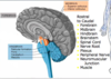
the Central Nervous System or CNS,
the Peripheral Nervous System or PNS and
the Autonomic Nervous System or ANS.
What does the CNS include?
The CNS includes the brain, brainstem, cerebellum and spinal cord.
What does the PNS consist of?
The PNS consists of sensory nerves whose cell bodies originate in ganglia that lie outside of the CNS and motor nerves whose cell bodies lie in the anterior horn cell of the spinal cord and certain cranial motor nuclei in the brainstem.

Accurate diagnosis of nervous system disorders critically depends on determining the anatomy of the lesion. Does the lesion lie on the right or left side of the body? Is it rostral or caudal along the so-called “neuro-axis”. In other words, is the lesion located in the cerebral hemispheres, deep structures of the brain, the brainstem, the cerebellum, the spinal cord, nerve root, nerve plexus, peripheral nerve, neuromuscular junction, or in the muscle? An accurate anatomical diagnosis will limit the differential diagnosis to a manageable few disorders, and that will improve the efficiency and efficacy of the care you deliver to the patient.
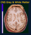
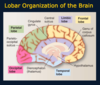
The surface of the brain consists of creases and folds, called ____ and _____ that are unique to every individual, much like fingerprints, but common features permit identification of important brain topography.
gyri and sulci
What separates the frontal lobe from the parietal lobe?
The central sulcus

What divides the temporal lobe from the frontal and parietal lobes? Lobes are large areas of the brain that house specific functions
Sylvian fissure

The frontal lobe contains many executive functions such as what?
deciding what you want to do next, what thoughts you want to express in words or writing, where you want to move your eyes to view the world, where you want place your arm, manipulate an object or move your leg. Think of it as a “to do” or the motor part of the brain.
What does the parietal lobe do?
The parietal lobe is the area that receives and interprets sensory information from principal modalities such as fine touch, position sense, vibration, pain and temperature. It determines the texture, shape, temperature and other characteristics that allow you to identify an object held in your hand but also to know where your hand is in three dimensional space.
Think of it as an area of the brain that interprets the physical environment around you.
What does the temporal lobe do?
The temporal lobe has several important functions. In the superior temporal gyrus lies the primary auditory cortex (called Heschl’s gyrus) that processes sounds. It is shown by the asterisk. Just posterior to it lies “Wernicke’s area” that interprets the processed sounds as words and mediates the comprehension of language.
Another important function of the temporal lobe is memory (part of the Limbic system)

What are the main roles of the occipital lobe?
it also interprets the environment but is devoted to a special sense: vision.
The cerebellum consists of two lobes or hemispheres, right and left. What do they do?
Unlike the cerebral hemispheres that govern the contralateral side of the body and sensory world, the cerebellar hemispheres receive and organize motor information about the limbs on the same or ipsilateral side of the body. Their function is to smooth out anticipated movements so movements are coordinated with what the rest of the body is doing.
The medial view of the brain shows the four major subdivisions of the cerebral hemispheres, the frontal, parietal, temporal, and occipital lobes, plus the limbic lobe that contains elements of the limbic system. What does the limbic system do?
It is involved in emotion, learning and in the formation of new memories. (older memories are ok)
There is no obvious boundary between the parietal and occipital lobes on the lateral view of the hemisphere but in the medial view, the ______ clearly demarcates the two lobes.
parieto-occipital sulcus

This slide shows the major lobes of the brain from a lateral, medial, superior and inferior (base of the brain) views. Note the interhemispheric fissure. This is the other major fissure in addition to the lateral or Sylvian fissure.

What is the concept of hemispheric dominance?
The left hemisphere is devoted to language and calculation, which defines hemispheric dominance, and the right hemisphere is devoted to visual-spatial processing. Most individuals are left hemispheric dominant and right handed but about two-thirds of left handed individuals are also left hemispheric dominant.
The remainder exhibit language function in both hemispheres, and only a small minority actually house language in the right hemisphere.
The cortex can be divided into what?
primary and association cortex.
What are the four types of primary cortex?
one motor and three sensory: auditory, visual and somatosensory.

The primary sensory cortex receives the most direct connections from its sensory organs in the periphery. Each has a neighboring association area that processes the raw sensory data to give meaning to it.
For example, the primary visual cortex detects basic features of the visual world such as edges, light, dark, color, location, direction of movement, and so on. The nearby association cortex takes this information and recognizes faces, objects and makes sense of the visual world.
Most of the cerebral cortex consists of association cortex which can be unimodal or heteromodal. Unimodal means that only one sensory or motor modality is involved. Heteromodal association cortex manages information from multiple sensory modalities and involves higher levels of information processing. It is this heteromodal cortex that is most developed in humans when brains are compared among different animal species.
These two drawings nicely depict the flow of information from the primary sensory cortex to association cortex (left) and to higher order association cortex (right) that is heteromodal cortex. (However, notice for motor how the flow is reversed- info first in association cortex then to the primary motor cortex) How is sound processed?

Sound is detected in its basic qualities such as pitch, frequency, tone in primary cortex centers and that information is processed by neighboring association neurons into words, for example, and these are given meaning in Wernicke’s area nearby.
T or F. Comprehension of language in Wernicke’s area is heteromodal in so far that language can be heard (auditory), read (visual), and felt (Braille for the blind).
T.
Where does the primary motor cortex lie? How does it work?
in the precentral gyrus and it uses the corticospinal tract to carry information to the spinal cord to produce an intended movement.
Flow of information here is reversed when compared to sensory information flow but the general scheme applies. Complex planning begins in the region with “executive function”, the so-called prefrontal association cortex that receives input from multiple sensory and other areas of the brain. It sends a motor command to the premotor cortex, that receives input from the cerebellum and basal ganglia and then conveys the information to the primary motor cortex for execution. Some of this information, however, is also sent from association cortex to the spinal cord via the corticospinal tract.
What is visual agnosia?
A lesion to the association area produces problems with information processing. For example, visual agnosias involve the inability to recognize objects although its features can be described in detail.
If you take a photo of someone you recognize and look at it upside down, the recognition disappears although you can describe the details just as well. That is a visual agnosia.