CM-Derm Flashcards

1
Q

what are the 3 layers of the skin and? what is contained in each?
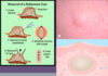
A

1. epidermis

  • stratified squamous epithelium
  • melanocytes, langerhans cells, merkels cells

2. dermis

.3-3.0 mm

-papillary layer

3. subcutaneous layer

2
Q

what is the function of the skin and nails? (5)

A
  1. physical barrier- prevents toxins, organisms, trauma
  2. temp regulation
  3. protection against UV radiation
  4. synthesis of Vitamin D
  5. sensation
3
Q

what are the four types of hair?

A
  1. lanugo- fine, covers fetus
  2. vellus- peach fuz
  3. intermediate- vellus and terminal hair
  4. terminal-scalp, beard, axilla, pubic area influenced by hormones
4
Q

what are the 3 phases of hair growth?

A
  1. anagen-growth phase, 3 years
  2. catagen- degenerative stage, weeks
  3. telogen-resting phase, varies by body site
5
Q

what are the four functions of hair?

A
  1. protection
  2. regulation of temp
  3. evaporation of perspiration
  4. sensation
6
Q

what is really important thing to do when examining the patient?

A

UNDRESS THEM

oh la la..NOT!..be professional

7
Q

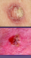
what are some things you want to take note of when looking at lesion? (6)

A
  1. how many are there?!
  2. type
  3. size
  4. color
  5. palpation (consistency, mobility, temp, and moisture)
  6. margination
8
Q

when there are multiple lesions, what are the three things you want to take note of?

A
  1. arrangement/configuration
  2. confluence
  3. distribution/location
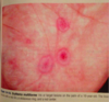
9
Q

Macule lesion

explain

A

A macule is a flat, distinct, discolored area of skin less than 1 cm wide that does not involve any change in the thickness or texture of the skin.

10
Q

papule

explain

A

A well-circumscribed, elevated, solid lesion, less than 1 cm. Usually dome shaped.

11
Q

plaque

explaination

A

A well-circumscribed, elevated, superficial, solid lesion, greater than 1 cm in diameter. Usually “plateau-like” with a flat top.

12
Q

bullae explaination

A

A raised, circumscribed lesion (> 0.5 cm) containing serous fluid above the dermis.

13
Q

crust explaination

A

Varying colors of liquid debris (serum or pus) that has dried on the surface of the skin.

14
Q

pustule explaination

A

A small (< 1 cm in diameter), circumscribed superficial elevation of the skin that is filled with purulent material.

15
Q

wheel explaination

A

Transient, circumscribed, elevated papules or plaques, often with erythematous borders and pale centers. These lesions are due to dermal edema and usually resolve within twenty-four hours

16
Q

ulcer explaination

A

A lesion with greater than 50% surface area ulceration.

17
Q

what are four examples of shapes you can use to describe the skin lesions?

A
  1. round
  2. oval
  3. annular
  4. serpiginous
18
Q

atrophy explaination

A

Thinning or depression of skin due to reduction of underlying tissue.

19
Q

what are four patterns you can use to describe the distribution of lesions?

A
  1. symmetrical
  2. exposed areas
  3. sites of pressure
  4. random
20
Q

what is vitiligo

A

loss of pigmentation

21
Q

explain what grouped lesions for disseminated would look like?

A
22
Q

explain the locations that are common for…:

  1. acne vulgaris
A
23
Q

explain the location of atopic dermatitis

A
24
Q

explain the site for photosensitive eruptions?

A
25
where is the rash for pityriasis rosea?
26
where do you see the rash for psoriasis?
27
where is the rash for seborrheic dermatitis?
28
what type of test is this?
woods lamp
29
what type of test is this?
punch test
30
what do you use Tzank smear for?
HERPES SIMPLEX
31
what are 5 general categories you can use for skin treatment?
1. topical 2. systemic 3. cyrosurgery 4. electrocautery 5. excisions
32
what are 3 key things to remember about sunscreen use?
1. **at least 30 SPF** 2. **UVA and UVB protection** 3. alive every two horus
33
what is the percent increase in risk if you go tanning for melanoma?
75% so don't go tanning.... ## Footnote ....just to look good!
34
what are four general bacterial sources that can cause bacterial infections?
1. coagulase-negative staph 2. staph aureus and group A, B, and G strep 3. break in stratum cornium method of infection 4. MRSA
35
atopic dermatitis ## Footnote what age group is this found it? what are the three characteristics in a patient that make them higher risk for this? what are three things you might see on this patient? what type of hypersensitivity is this? where is this most commonly found (2) places and 4 others? what must you differentiate from? what is the diagnosis made from?
chronic relapsing skin disoder that starts in children and goes through adulthood ALLERGY, ALLERGIC RHINNITIS, ASTHMA, **PERIORAL PALOR, DENNIE MORGAN LINES, ALLERGIC SHINERS** **type 1 IgE mediated hypersensitivity** -dry skin, puritis with "itch scratch cycle" flexor creases (antecubital and popliteal) most common neck, eyelids, forehead, face, and dorsum of hands and feet, *dermatographism* characteristic can be either atopic or contact (touching something you're allergic to) \*make sure to culture for **S. aureus** and make sure not secondary infection and **HSV** in crusted lesions\* **DX: IgE serum levels**
36
atopic dermatitis ## Footnote what does this come from? what are the three stages and what is characteristic of the rashes at each?
**allergic reaction** **acute:** pruritis, redness, *vesicle formation, oozing, crusting* **subacute:** pruritis, redness, *parched, or scaled* **chronic**: pruritis, skin thickening, *hyperpigmentation, excoriation, fissuring*
37
atopic dermatitis ## Footnote what are 6 possible treatment options for this?
**1. avoid irritants** 2. skin lubrication/emollients (moistureizers) **3. topical corticosteroids** **4. antihistamines** 5. calcineurin inhibitors (tacrolimbus + pimecrolmus) 6. UVB phototherapy is effective
38
according to the green pance book, what can the rash from atopic dermatitis look like?
papules and plaques with or without scales can have edema, erosions, or crusts goodluck.
39
if someone has atopic dermatitis what do you worry about?
since so itchy, they scratch a lot and can lead to secondary infection from s. aureus! watch out for this!!
40
for atopic dermatitis....what can you do to figure out what is causing it?
PATCH TESTING!!!
41
what are 6 things that can contribute to atopic dermatitis exacerbation?
1. milk 2. eggs 3. fish 4. skin hydration 5. time of year 6. stress/clothing
42
seborrheic dermatitis ## Footnote what does this occur in? what are the four most common places? what is the most common agent? what does this cause in infants and what does this cause in adults? what do the infected areas look like? what are the 7 treatment options for this?
subacute/chronic **inflammation** of the areas with **increased SEBACEOUS GLANDS** **_body folds, face, scalp, genitalia_** caustitive agent: pityrosporum ovale (yeast) "cradle cap" in infants, "dandruff" in adults _scattered **yellow gray** **scaly macules and papules** with **GREASY** LOOK!!!_ sticky crusts with fissures found behind the ears!!! Tx: 1. OTC dandruff shampoo 2. cold tar shampoo 3. **Ketoconazole** 4. cradle cap: **olive oil compresses, shampoo with sulfur, or ketoconazole shampoo** 5. UV radiation **6. shampoos with selenium sulfide** **7. topical steroids** hydrocorisone
43
stasis dermatitis ## Footnote what causes this? what two populations of people is it worse in? the incompetency leads to what **5 things**? what does the pt complain of? what **3 things** do you see before the skin changes? what are two ways to make the DX and what do you see?
chronic rash on lower legs **secondary to venous insufficiency** worse in pregnancy and women *see papules, scales and crusts* valvular incompetency leads to **edema, dermatitis, _brown stippled hyperpigmentation_, fibrosis, _ulceration_** (30%) pt complain of aching in legs that is worse with standing, relieved by walking typically see caricose veins, superficial phlebitis, and venous thrombus *before skin changes* DX: 1. doppler studies, sonography or venography will confirm chronic insufficiency 2. biospy-show dilated tortuous veins, edema, and fibrin deposition
44
what are the treatment options for stasis dermatitis? (6)
**1. manage edema** **2. topical corticosteroids** **3. wet compress for oozing and crusting (caution with ulcers)** 4. compression stockings 5. sclerosis of varicose veins to prevent dermatitis 6. vascular bypass, endothelial thermal ablation or stenting (these are only mildy effective)
45
dyshidrosis ## Footnote what type of dermatitis is this and where does it occur? what age group? what is it commonly associated with? what is the **funny food it is associated with looking like** and what other characteristic is common? what are four triggers? what does it transform in to in late disease (5)? what two things are used to diagnose it?
acute/chronic puritic **vesicular dermatitis on _palms and soles_** **purititc, clusters of small vesicles in clusters "tapioca like appearence" on palms/soles** triggers: *sweating, emotional stress, warm/humid weather, metals* Late disease: papules, scaling, lichenification, erosions ruptured from vesicles, painful fissures DX: culture and KOH to rule out dermatophytosis
46
dyshridrosis ## Footnote what are the 5 treatment options?
1. **burrows solution and antihistamines** (control itch) **2. topical high steroids** 3. systemic steroids if severe **4. intralesional triamcinolone** 5. fissures treated with: collodoin
47
lichen simplex chronicus ## Footnote what happens to the skin and what is that cause from? what is this a long term manifestation of? what do you see as a manifestation on the skin? what are the 6 areas on the body this is common in? what does a biopsy show?
development of **epidermal hyperplasia 2nd to physical trauma of scratching and rubbing** long term manifestation of atopic dermatitis from ALL THAT ITCHING **skin becomes extremely sensitive _to touch including trauma, touch, rubbing or scratching_** leads to skin thickening from the increased rubbing and scratching well circumscribed thick, firm, plaques that are highly puritic even light touch causes this patients to itch a ton! most common areas: nuchal area, scalp, ankles, lower legs, upper thighs, and exterior forearms DX: KOH to rule out fungal infection, BIOPSY SHOWS _HYPERPLASIA AND HYPERKERATOSIS!!_
48
lichen simplex chronicus what are the four treatment options?
interupt the scratch itch cycle ## Footnote 1. topical steroids 2. intralesional triamcinolone 3. antihistamines (ESP AT NIGHT!!) 4. occusive dressing with out without tar prep/steroids
49
how does lichen simplex chronicus appeare on black skin?
follicular pattern of smaller papules...not plaques like on white skin ## Footnote SO TRICKY!
50
nummular dermatitis ## Footnote when does this occur? who is it most common in? what do the rash look like and where is it most common? what are the four treatment options?
puritic inflammatory disorder in young adults (white males) and elderly in **fall and winter but exact cause unknown** grouped _vesicles that coalesce to form_ coin shaped plaques with erythematous base with clearly demarkated boarders most common on _extremities_ TX: moisterizers & topical steroids for class I and II tar baths or UVB phototheryapy
51
contact dermatitis: non allergic ## Footnote what is this caused by? (3) what do the acute and chronic skin presentations look like?
comes from contact with an irritating substance **direct toxin on the skin** *common causes: abrasive, cleaning agents, caustic agents* **Acute**: erythema, vesicles, erosions, crusting **chronic**: papules, plaques, crusts
52
contact dermatitis: allergic ## Footnote what cells initiati this? what are 5 common causes? what is the difference between the acute and chronic skin presentations?
t-cell mediated, occurs in those that have become sensitized * common causes: medication, jelwrey, rubber, disinfectants, cosmetics, plants* acute: erythema, vesicles, eroisions, crusting chronic: papules, plaques, crusts \*\*can do patch testing after dermatitis to figure out the cause\*\*
53
autosensitisation dermatitis "ID REACTION" what is this? what causes this? what are the two presentations of the rash? what is the treatment?
dermatitis occuring at a **site distant from primary dermatitis** caused by the release of _cytokines from sensitization at primary site_ rash: maculopapular/papulovesicular Tx: corticosteroids
54
asteatotic dermatitis ## Footnote when is this common and who is it common in? where are the three places it shows up? what should you avoid and what should you do? what is last resort treatment?
pruritic dermatitis occuring in the **WINTER,** COMMON IN ELDERLY *legs/arms/trunks* avoid hot showers!!! take some baths with oils and skin lub....god that sounded dirty. Tx: corticosteroids if lesions are inflammed
55
cellulitis ## Footnote where does this infection occur? what are the 3 organisms most likely to cause this in adults? what are the 3 organisms likely in kids? what are the 5 symptoms the patient will experience? what is the strange animal you can get this from? what do you do to DX this?
acute, spreading inflammation of the **dermis and subcuteous tissues**, occurs from breaks in the skin adults: **s. aureus, group A strep can get it from _cat bite pasturella mulicida_** children: **hae. influenzae (periorbital), strep pneumoniae, s. aureus** expanding, red, swollen and tender, FEVER!! HEAD!! LYMPAHDENOPHATHY!! EDEMA!! NOT sharply demarkated...pt feels **ILL** DX: culturing and drainage of discharge by needle aspiration
56
cellulitis ## Footnote what are the four treatment options? and what is really important to do when treating a cellulitis patient?
1. rest/heat 2. **begin treatment ASAP with abx that cover haemophilis influenzae, strep and staph!** 3. **dicloxacillin or cephalosporin**, if allergic to penicillin use **erythromycin** 4. if severe give **first generation cephalo IV!!** really important to mark the boarders so you can tell if it is improving or not!! may need surgery if necrotizing infection develops
57
erysipelas ## Footnote what aged people is this common in? what are the two places this commonly appeares? what is the #1 causing organism? what does the rash look like and what is it characterized yet? when does it disappeare? what is the DX and TX?
variant of cellulitis in ELDERLY on FACE AND EXTREMITIES **group A beta strep** painful macular rash with well defined margins, vessicles, FEVER, **rapid onsetand progression**"_fiery red face*"*_ * rash desquamates in 5-10 days* * DX:* bacterial culture TX: treat with antibiotics depending on organism!
58
impetigo contagiosa ## Footnote what two age groups is this common in? what two environmental things is it common with? where do you typically see this and is there a family hx? what **key** description of the lesions? what are the **3** organisms that commonly cause this? what are the three tx options?
common in INFANTS AND CHILDREN!! higher rates with poor hygiene and malnuitrition **HIGHLY CONTRAGIOUS!!! COMMONLY ON FACE!!! usually have family history of this!** **THICK CRUSTED "HONEY" colored macules and papules! starts as one, and spreads** **#1- staph aureus** **#2-GABHS** **#3-streptococcus pyoderma** **TX:** 1. _bactroban_ 2. _cephalexin_ or _dicloxacillin_ if severe and need systemic
59
bullous impetigo ## Footnote what organism causes this? who is it most common in? what three places do you see this rash most commonly present? what do you worry about as a SEVERE COMPLICATION?
Staph group II, **staphlococcus with endotoxin** **STAPH AUREUS** * most common in children/infants* overall not common * face, neck, and extremities* erythema that progresses to epidermal sloughing, worry about progression to scalded skin syndrome!!! TX: 1. bactroban 2. cephalexin or dicloxacillin if systemic needed
60
folliculitis ## Footnote what organism is most common for causing this? what is the second most common? what three areas does this most commonly effect? what are the two types of presentations you see? what are the two treatments?
**staph aureus infection of the hair follicles!** commonly seen on _butt, thighs and beard!_ "superficial hair" can be *pseudomonas* PAPULES AND PUSTULES Tx: antiseptic cleaners mupirocin
61
faruncles ## Footnote what organism causes this most often? what does it present as? what do you want to culture it for? what are the two treatment options?
abcess formation from **STAPH AUREUS** deeper hair infection of the hair folicle than folliculitis, tender nodule with _1 central plug_ **raised tender warm and fluculent** want to culture it for MRSA Tx: incision and drainages! antibiotics
62
carbuncles ## Footnote what organism is the most common cause of this? what is the presentation? how do you differentiat it from a farbuncle? what are the two treatment options?
abcess formation caused by **STAPH AUREUS!!** **raised tender warm and flutulant** _larger and deeper than farbuncle, more painful, "interlocking farbuncles with multiple openings!"_ TX: incision and drainage and antibiotics
63
infectious intertrigo ## Footnote what three organisms can cause this? tx and dx?
strep group A, B, or G Staph pseudomonas dx: culture and treat with specific antibiotics
64
erythrasma ## Footnote what bacteria causes this? what does the rash look like? what is the interesting thing you use to diagnose this? what are the two treatment options?
_corynebacterium minutissimum_ lesions in intertriginal areas, _tan or pink_, sharply demarcated DX: _coral red fluorescnece on woods lamp_ Tx: benxyl peroixide and topical antibiotics
65
necrotizing fascitis ## Footnote when does this occur? what is necrotized? what is the most common cause of this?
occurts after minor trauma extensive necrosis of subcutaneous tissue _group A strep_
66
condyloma accuminatum ## Footnote what virus strains cause this? what are the four descriptions used to describe these lesions? what is the treatment?
"genital wart" from _HPV 6 and 11_ lesion type: **1. papular** **2. cauliflower** **3. keratotic** **4. flat topped** Tx: no cure but can be removed, tough it a lot of them
67
herpes simplex ## Footnote what is a hint that an outbreak is going to occur? what is the important description of these? what are the two different types; where are they found and what percent of the population has them? how do you dx it and what do you see? what are the four treatment options?
**prodromal phases:** 24 hours before outbreak, get burning and tingling "painful grouped vesicles on erythmatous base!" HSV1: oral lesions **85%** population infected; transmitted via saliva, **outbreak triggered by random things** HSV2: genital herpes (more common and detrimental in women! more likely to have complications like ulcers and necrotic tissue), **25%** population infected DX: _tzank smear, geimsa stain shows **GIANT MULTINUCLEATED CELLS**_, can also check for antibodies for this​ TX: supportive therapy suppressive therapy *foscarnet* **Acyclovir, valacyclovir, famcyclovir**
68
acute hepetic gingivostomatits ## Footnote what virus causes this? where does this tend to effect? how often are the outbreaks and who are they common in? what are three things you might find in this patient? explain the maturation of the vesicles?
HSV-1-trigeminal nerve predilection, eruptions 2x a year common in 6 months-5 years CHILDREN abrupt onset **fever, anorexia, red mucosa** **vesicles appear on gums, lip, tongue** **_vesicles colase to form ulcers or plaques_**
69
acute herpetic pharyngotonsillitis ## Footnote what virus causes this? who is it the most common in? what are four symptoms you see with this? what do the lesions look like?
more common in HSV1 than HSV2 primarily in ADULTS **fever, malaise, headache, sore throat** _vesicles on posterior pharynx and tonsils that RUPTURE to form ulcers_ (may have grayish exudate)
70
_Herpes Simplex-2_ what does this cause? what percent of the population is infected with this? where does this typically have predilection for? what do the lesions start and finish as? what percent of people will have reactivation in the first 12 months? how many reactivations will they have in their lifetime?
causes **genital lesions** 25% of the population infected with this **asymptomatic shedding and painful eruptions can occur** _sacral root ganglion predilection_ _VESICLES_ rupture to form _ULCERS_ reactivation in 90% occur in the first 12 months!! 30% have 6 episodes in their lifetime!!
71
where does Herpes simplex virus tend to hide?
dorsal root ganglion ## Footnote this is why it reactivates
72
what are the _5_ complications you worry about from herpes simplex virus?
**1. herpetic withlow** (vesicles on the fingers) **2. herpes gladiatorum** (disseminated cutaneous infections common ing wrestlers) **3. keratoconjunctivitis** (dendritic corneal ulcers) **4. HSV or CNS ENCEPHALOPATHY!!** YIKES!! causes change in mental status and headache **5. infection during pregnancy** can infect the child
73
what is herpetic whithlow?
herpes lesion on the FINGERS
74
molluscum contagiosum ## Footnote what virus causes this? describe what they look like with the FOUR key descriptors? what two populations is this common in? what are the 5 most common areas? what is the treatment 7 options?
poxviridae virus "pox virus" "dome shaped" flesh colored with waxy appearence with _central umbilication_ 2-6mm in *_children_* and *_sexuallly active adults_* HIGHLY CONTAGIOUS! viral disease of skin and mucosal membranes *face, trunk, **_abdomen_**, thighs, **_genitals_*** Tx: usually self limiting and lasts 3-6 months if therapy needed, lesions have to be destroyed individually **1. DOC curettage** **2. cryrotherapy** **3. electrodessication** **4. acid, Retin A, Kerolytic pain, Imiquimod cream**
75
varicella-zoster virus ## Footnote explain the differences seen between the primary and secondary eroptios of this virus? how are they descirbed? what sign do you watch out for? what is the order of the lesion developement? where does it begin and where does it spread to?
VARICELLA-ZOSTER virus **_varicella (chicken pox):_** 1st exsposure **vesicles on a erythematous base "DEW DROPS ON A ROSE PETAL"** describe the different stages macules-\>papules-\>vesicles "dew drops on a rose petal"-\>pustules-\>crusts \*\*appeare in crops!\*\* **BEGIN ON FACE AND TRUNK AND SPREADS TO EXTREMITIES** **_Herpes zoster (shingles)_**: VZV reactivation along a **Dermatone in THORACIC OR LUMBAR REGIONS**, reactivation from **ganglionic satelite cells****!** **-****Hutchinson's sign:**lesions on the nose mean lesions in the eye since**trigeminal nerve involvement CN #5**
76
what are the two complications you worry about with herpes zoster virus reactivation (shingles)?
1. **eye involement herpes zoster opthalmicus:** look for _hutchinson's sign_ which is lesions at the end of the nose, if seen here likely it is already in the eye since it follows along the _trigeminal nerve or CN 5_ 2. **ear involvement herpes zoster oticus**: look for _ramsay hunt syndrome_ if lesions are seen on the ear, likely in the canal since it follows _facial nerve or CN 7_
77
how long can the post herpetic neuraligia with shingles last? what is a thing you worry about if eldery?
\>3 months...so give these people some pain meds ## Footnote occurence likelyhood is greater if over 60!
78
what is the treatment options for varicella zoster virus? (4)
1. **acyclovir, valacyclovir** 2. **pain management for post therapeutic neuralgia** 3. **tricyclate antidepressants** 4. **corticosteroids**
79
what can you do to prevent varicella-zoster virus? (2 options)
VACCINATION!! **child:** vaccinated 1-2 years old for varicella **adult:** Zostavax single dose \>60yrs...basically literally a booster of varicella, becuase it is the same virus, just marketed differently to apeal to elder adults! \*\*can't give if allergic to gellatin, neomycin, pregnant, or immunocomprimised!\*\*
80
verrucae ## Footnote what are they caused by? what is the most common? what are the 5 types of warts that can present? what are the three things that help you diagnose this?
caused by _100+ serotypes of HPV!_ _verrucae vulgaris mos common!_ *firm papules, skin colored, **_vegitation_**s, _red brow spots_* **1. skin warts:** flat/superficial **verrucae vulgaris** **2. plantar warts:** deep, "tiny heads of colliflower" **3. oral cavity warts or in pharynx:** can be life threatening if block the airway **4. angiogenital warts:** squamous epithelium of external genitalia CONDYLOMATA **5. cervical warts:** HPV 16 and 18 cause dysplasia Dx: microscope: **hyperplasia, hyperkeratosis,** **koiliocytic squamous cells present!** immunofluorescence-\>see HPV
81
verrucae ## Footnote what are the 6 treatment options??
1. spontaneous regression common 2. salicyclic acid plasters, cyrosurgery, electrocautery 3. imiquimod 4. intralesional interferon 5. surgical excision 6. ketolytic agent
82
erythema infectiosum
_human parvovirus B19_ fifth disease, red face"slapped cheek", arthropathy pink lacey rash with slapped face appearence, arthropathy is common in older children and adults infectious disease associated with arthropathy! spreads by respiratory droplets! \*\*\*CAUTION!!...this can cause an aplastic crisis in sickle cell patients\*\*\*
83
roseola infantum ## Footnote what virus causes this? what unique thing does the fever correlate with and how long does it last? what is interesting about the rash location and progession? who is this common in?
_herpes virus 6 or 7_ _fever for 4 days, this resolves before you get the pink macular rash!!\*\*_ during this time though the child still appeares well, doens't seem sick! children only childhood exanthem that starts on the trunk and migrates to the face!!
84
measles ## Footnote what virus causes this? what type of rash? what are the 3 things you should relate to this? what can you see in the mouth and what do they look like? where does the rash start? what is the treatment?
_paramyxovirus_=maculopapular rash **URI prodrome with 3 C's:** **COUGH, CORYZA, CONJUNCTIVITIS** FEVER, COUGH, ANOREXIA **_KOPLIK SPOTS IN THE MOUTH:_** small red spots in the buccal mucosa with _blue/white_ paler _center_ **_Brick red rash on skin begining at the hairline!!_** Tx: supportive and antiinflammatories!!
85
rubella (german measles) ## Footnote what virus is this caused by? how long does the rash last? what is the important thing to consider if the woman is pregnant and what 3 things can it cause? what do you see for lymphadenopathy? what is the buzz word rash?
_togavirus_ rash lasts _3 days!! pink maculopapular rash head to toe TERATOGENIC!_ can see lymphadenopathy **posterior cervical and posterior auricular** can see transient joint pain and photosensitivity in young women **TERATOGENIC IN 1ST SEMESTER:** congenital syndrome, sensineural deafness, **"BLUEBERRY MUFFIN RASH!"**
86
what is the most common fungal infection?
SUPERFICIAL fungal infections
87
irritant dermatitis ## Footnote what causes this? what things can cause this?
Results from contact by irritating substance **Direct Toxic effect on skin** **Occurs in anyone exposed to causative agent** Common Agents- **Abrasives, cleaning agents, caustic agents** Rash Acute--erythema-Vesicles-Erosion-Crusting Chronic--Papules-Plaques-Crusts
88
what are the three most common mucocutaneous fungal infections?
1. candida species: require warm humid environment 2. malassezia species: humid environment with **lipids** for growth 3. dermatophytes: infect keratinized epithelium, hair follicles, and nail apparatus
89
what are the 6 dermaphytes that infect the epidermis?
**Tinea corporis** **Tinea cruris** **Tinea pedis** Tinea manuum Tinea facialis Tinea incognito
90
what are the two dermaphytes that infect the hair and hair follicles?
Tinea capitis Tinea barbae
91
what dermaphyte infects the nail appartus?
Tinea unguium (aka onychomycosis)
92
what is the most common dermaphyte?
trichophyton rubrum most common, although 10 species
93
what percent of people experience at least 1 yeast infection and which one is most common?
70% have 1 infection tinea pedis is usually the most common "athletes foot"
94
how are dermaphytes usually spread?
from one person to another, usually from fomites this means that it touches an object then another person touches it and then you get it from the contact with the fomite
95
for hair and nail infections...what type of treatment is required?
for hair and nails, need to use SYSTEMIC treatment!!!! topical won't work for these!!
96
explain what allows the dermaphytes to live?
they metbolize and live on **KERATIN!!!** the fungus attacks the skin, nails and hair where **keratin is the major structual protien, leads to infection!**
97
what are RF for dermophyte infection? (5)
1. atopic diathesis/dermtitis (cell mediated immune deficiency for **T. rubrum**) 2. topical immunosuppression from topical corticosteroid ( **tinea incognito)** 3. systemic immunosuppresion 4. sweating 5. high hummidity
98
if you suspect a dermaphyte infection, how do you identify it? 3 options, explain what you would see on the first
if its scaly, scrape it!!! 1.KOH most common method _multiple septated, tubelike structures (hyphae or mycelia) and spore formation_ 2. culture 3. biopsy if KOH and cultures are negative
99
what are the four of the 9 classes of arthropods that can cause skin reactions?
**Arachnida** (4 pairs of legs) – mites, ticks, spiders, scorpions) **Chilopoda** (centipedes) **Diplopoda** (millipedes) **Insecta** (3 pairs of legs) – **lice**, **bedbugs**, ants, bees, wasps, hornets, catepillars, butterflies, moths, fleas)
100
what is a dermaphyte?
a superficial fungal infection that can affect the hair, nails, and skin
101
what are the three most common dermophytes in the world? what is the most common one in the industerial world?
trichophyton, microsporum, and epidermophyton **trichophyton rubrum is the most common one in the industerial world**
102
explain how a fungal infection (dermatophytosis) is named? what are the names of the body for the different infections? (8)
tinea means fungal infection...so thats the first part the second is the area of the body: pedis: foot cruris: groin corporis: trunk, legs, arms and neck barbae: beard unguinum: nails manuum: hand facialis: face capitis: head
103
what are 6 drugs that can cause drug induced alopecia?
1. thallium 2. vitamin A 3. retenoids 4. antimitotic agents 4. anticoagulants 6. OCP
104
why is psoriasis a primary care disorder?
because there are so many comorbidities, and we manage those ## Footnote Psoriatic Arthritis Obesity Metabolic syndrome Vascular disease (CVD, CeVD, PVD) Malignancy Autoimmune disease Nonalcoholic Fatty Liver Disease (NAFLD) COPD OSA (Obstructive Sleep Apnea) Parkinsonism Psychiatric Disorders Alcohol abuse
105
what are the give drugs that can exacerbate psoriasis?
1. ETOH 2. beta blockers 3. lithium carbonate 4. antimalarials 5. interferon
106
explain the theory behind biologics used for psoriasis?
The newly developing psoriasis lesion is precipitated by an autoimmune reaction where there is a **genetic defect inherent in the interaction between keratinocytes, macrophages and T-lymphocytes** For reasons that are poorly understood, the **macrophages** appear in the **epidermis** and proceed to **identify the keratinocytes as foreign antigens.** These antigens are then presented to **T-lymphocytes which form large populations which then incite the inflammation of psoriasis.** _(Large populations of activated T-Cells are found in bases of Psoriasis plaques.)_ ***Psoriasis is a TH1-Cell mediated disease*** “Biologic” Psoriasis drugs target and disable particular steps along the pathway of T-Cell activation in target tissues (skin keratinocytes) which ultimately results in clinical disease improvement in most patients
107
what are the five biologics that can be used to treat psoriasis?
1. etanercept 2. alefacept 3. adalimumab 4. infliximab 5. efalizumab
108
what can the suffix of biologics tell you? - ximab - zumab - umab - cept
- ximab: chimeric monoclonal antibody - zumab: humanized monoclonal antibody - umab: human monoclonal antibody - cept: receptor-antibody fusion protein
109
stevens johnson's syndrome ## Footnote what is this thought to be a sever form of? what percent of the BSA? what does it look like? what are the two most common drugs that cause this? what are the **3** complications you worry about? what does the patient **present with** and then what do they look like **4 days later?** what is the treatment? what is the _special sign_?
thought to be a severe form of *erythema multiforme but unknown cause* mucotaneous blistering reactions most often caused by _drug rxn sloughing **especially sulfanamides and anti convulsants**_ **worry bout complications!! infections, fluid loss, and electrolyte imbalance** pt presents with: fever, photophobia, sore throat with mucosal inflammation and cutaneous lesions on trunk 4 days later: **diffuse erythema, necrotic epidermis, wrinkled surfaces, sheet like loss of epidermis, raised flaccid blisters nikolsky sign** **tx: send them to the burn unit for care, fluid care and electrolyte imbalance!! treat it like a severe burn!**
110
what are the drugs that can cause steven johnson syndrome or toxic epidermal necrolysis?
sulfonamides, anticonvulsants, aninopenicillins, quinolones, cephalosporins, tetracyclines, phenobarbital, phenytoin, allopurinol, corticosteroids
111
what percent of pts with SJS have mucosal ulcers?
90% have mucosal ulcers anywhere from the mouth to the anus and are painful!!
112
what are five things that SJS can cause that you worry about?
1. infections 2. fluid loss 3. electrolyte imbalance 4. tubular necrosis 5. erosion in lung and gut
113
how long does skin regrowth take for SJS?
3 weeks!!
114
Toxic epidermal necrolysis (TEN) what is this a severe form of? what is the difference between this and SJS? what are the presentations? what percent of sloughing? what are three things you worry about? how do you tx?
severe form of SJS, unknown cause but immune mediate \*\*\*Only difference, HIGHER TEMP AND MORE SLOUGHING\*\*\* life threatening **diffuse erythema, necrotic epidermis, wrinkled surfaces, sheet like loss of epidermis, raised flaccid blisters nikolsky sign** **sloughing \>30% BSA,** mucotaneous blistering reactions most often caused by drug rxn worry about infections, fluid loss, and electrolyte imbalance TX: send to burn unit and treat like severe burns!!!
115
what are two keys lab keys a patient with SJS or TEN will have?
anemia and lymphopenia
116
necrotizing fascitis ## Footnote what are the 3 bacteria common for causing this? what three populations is it most common in? what are **6** characteristics of the pt? what is the **triad**? what is a important thing to _monitor_? what are the two treatment options?
**polymicrobial or group A strep** or **clostridial** salterwater necrotizing fascititis: **virbrio** **DIABETICS, ALCOHOLICS, IV DRUG USERS** rapidly progressing erythema, tissue crepitus, HIGH TEMP, tachycardia, hypotension, altered mental status triad: elevated WBC, elevated BUN, hyponatremia (not present in all but heighten suspicion) \*\*\*\*MONITOR RENAL FUNCTION BECAUSE THIS IS A HALLMARK OF THE DISEASE\*\*\*\* TX: **1. aggressive surgical debridement-**do US, CT, MRI to determine the amount that needs to be taken out **2. aba to cover ALL pathogens- carbapenem of b-lactam/b-lactamase inhibitor with clindaymycin and one against MRSA like vancomycin**
117
diabetics account for what percent of necrotizing facititis cases?
20-30%
118
what is the mortality rate for necrotizing facititis?
25-70%
119
erythema multiforme ## Footnote what type of hypersensitivity is this? what two other things can it effect? what are the three main causes? 5 2 1 explain the characteristic rash appearance for this? what also accompanies this? what are the 3 treatments?
**type IV hypersensitivity rxn** (delayed cell mediated), \*\*\*can effect lungs and eyes\*\*\* causes: 1. drugs-**sulfonamides, phenytoin, barbituates, penicillin, allopurinol** 2. infections: _HERPES SIMPLEX (#1), mycoplasma_ 3. idiopathic Rash: macular-\>papular then vesicles and bullae form in the center of the papules get target or iris lesions "dusty violet" red, with mucosal lesions that erode and are painful TX: 1. avoid trigger 2. control herpes outbreak: acyclovir 3. systemic corticoids if severe
120
what are the two different types of erythema multiforme?
**erythema multiforme minor:** _target lesions_ distributed acrally, _no mucosal membrane lesions_ **erythema multiform major:** _target lesions_ that _involve \>1 mucous membrane, NO EPIDERMAL DETACHMENT!!_ (oral, genital, ocular mucosa)
121
what percent of people are effected by acne? what are the common age groups for females and males?
85% of young people are effected females: 10-17 yrs old males: 14-19 years old
122
what is acne a disorder of? what are 3 key factors?
pilosebaceous unit ## Footnote key factors: follicular keratinization, androgens, propionibacterium
123
what is the rate limiting factor in acne disease instigation?
sebaceous gland activity!!!
124
what is the pilosebaceous unit?
long and narrow tube AKA pore with single hair follicle the epithelium lining at the enterance of pore is _stratum corneum_ this is prone to **overkeratinization "clogging" if right factors are present**
125
sebaceous glands ## Footnote where are the most common locations? what do they secrete? what are they highly sensitive to? what do these do to the size of the pore?
most dense on **face, chest, back, and upper arms and secrete triglyceride rish oil!** around the infundibulum of the pore, each pore has one sebaceous gland _highly sensitive to hormonal stimulation by **androgens**_ circulating testosterone reaches the skin (dihydrotestosterone) and increases the size and oil production of SG
126
explain the hormal influence on acne? (3)
1. hormonal influences during puberty- increase sebum production and keratinization (both men and women), majority resolve post puberty 2. increase cortisol production secondary to prolonged stress increase angdrogen production which increases SG size and secrection 3. middle aged women with pre-mestrual acne are often showing a sensitivity to a brief surge in testosterone produced by the ovaries to initiate meses
127
what are two other structures in the pilosebaceous unit besides SG?
1. **apocrine sweat glands:** communicated with infundibulum, *c**lear sweat with cytoplasmic components** caues odor and phermones* **2. eccrine sweat glands:** positioned outside of the unit, next to it, secrete **clear errine sweat**
128
what cause goosebumps?
arrector pili muscle contracting it connects the infundibulum with the skin surface
129
what are three characteristics that can make a person predisposed for acne?
1. increased sebum production from sebaceous glands 2. overgrowth of keratin producing cells lining the pore walls 3. keratinization leads to the formation of a sticky plug, blocks the superficial aspect of the pore forming a **microcomedo**
130
open comedone ## Footnote what is it? how does it happen?
"blackhead" ## Footnote forms when the pore wall is able to dilate in response to the increased pressure created by trapped sebum, creating a non-inflammed lesion
131
closed comedone ## Footnote what causes it? what is it?
"whitehead" ## Footnote a semi-ferm dome shaped papule which is also non inflammed forms when trapped sebum pushes up against the non-dilated opening of the pore
132
what is the role of neutrophils in propionibacterium acnes? what do they do and what does this lead to?
when inflammation occurs they are attracted and attach to follicular wall 1. release hyrolyses which weaken follicular wall 2. wall eventually ruptures 3. spillion out highly irritating FFA's in to surround tissue
133
what is the only acne that is not inflammed?
comedonal acne
134
how is acne graded?
**severity: 1-4**, higher grades have a higher overal lesion count, more inflammation, larger lesions, etc **type:** **comedonal, pustular, papularpustule, nodular, nodulocystic** Ex: grade 1 comedonal Ex: grade 3 nodulocystic acne
135
isotrentinoin ## Footnote how does this drug work? what makes this so tightly regulated? what percent are cured? what do you have to do to be able to prescribe it?
drastically attenuates activity of sebaceous glands and rate of keratinization in epidermis **EXTREMELY TERATOGENIC** **can be permanent cure for 85% of patients on 6 month course** **now subject to federal regulatory aparatus called _i-pledge_**
136
if pt presents with acne, and isn't in typical age range... pre-pubescent, pubescent, middle aged women then what must you consider?
occupational acne!!!! workplace exsposure to light oils (machine oil, cosmetics) which occlude pores leading to comedogensis and acne
137
pomade acne ## Footnote what does this come from? what parts of the body are effected? how long does it take to clear once the instigator is stopped?
unique form of acne **secondary to chronic use of oily hair mousses, gels, tonics by susceptible individuals** _always_ distributed on _upper forehead/hairline/anterior scalp with fairly clear border_, **lower face unaffected** stop use of hair product, acne clears in 2-6 weeks
138
what are three things you need to rule out when treating a patient with resistant acne vulgaris?
1. in women: exclude **adrenal and ovarian dysfunction** 2. exclude **gram negative folliculitis**
139
what should you do about resistant acne?
REFER
140
what percent of fair skinned indiviausl does roceasea occur in?
10%
141
what is facial flushing and what disease is it commonly assocaited with? (3 characterists of facial flush)
ROSACEA 1. greater baseline blood flow 2. larger more numerous blood vessels 3. vessels more superficial
142
what are the three main classifications of candidal infections based on location?
1. **cutaneous (diaper dermatitis and candidal intertrigo)** ## Footnote **2. mucosal candida of the mouth and pharynx** **3. vulvovaginal**
143
what is the most common species of candida?
candida albicans
144
Cutaneous candidal infection 1. candidal intertrigo 2. diaper dermatitis what is the characterists of these? where are the places you would find these? what are the two TX options?
**patches and pustules on a ERYTHEM MATOUS BASE _beefy red_ the erode and confluent with "_SATELITE LESIONS_"** painful with puritis **Candidal intertrigo**: *axillae, groin, intergluteal, cleft* **diaper dermatitis:** irrritabiltiy with urination, defication, changing diapers, *genital region, inner aspect of thights and butt* *TX:* **keep dry, antifungals nystatin, imidazole powder**
145
oropharyngeal candidiasis what does this look like and what in what population can this look completely different? what is the key characteristic of this? what are the 2 treatment options?
thrush, _white curd_ like plaques that can be _scraped off_ ## Footnote what to find the percipitating cause and treat that, then treat with oral antifungals \*\*\*\*\*KEEP IN MIND, PEOPLE IN DENTURES CAN APPEARE BIRGHT RED INSTEAD OF WHITE CURD LIKE\*\*\*\* Tx: nystatin oral fluconazole or itraconazole suspension, swish and spit or swallow
146
vulvovaginal candidiasis what percent of females can get this and what can it come from? what are 5 RF for this? what does it look like and what is an important symptoms? what are two treatment options?
**75% of females get this at least once,** \>20% are colonized with C.albicans normally, so this is a overgrowth of normal biota RF: age extremes, pregnancy, DM, corticosteroids, HIV white cottage cheese like discharge/plaques, burning while peeing, and puritis TX: topical/intravaginal azoles or oral fluconazole
147
what can vulvovaginal candidiasis be closely related to?
some women just get it right befor their period because of the pH change that allows microbiota to grow better
148
balanitis candidiasis ## Footnote who is this common in? who must you treat? treatment?
common in **uncircumsized men** **erosions with white plaques under foreskin** treat sexual partner TX: topical nystatin, warm soaks to alievate itching/burning
149
what are 6 RF for candidiasis?
1. diabetes 2. pregancy 3. obesity 4. HIV/AIDS 5. moisture 6. IUD
150
what do you use to diagnose candidiasis? what do you see?
KOH \*\*\*see pseudohyphae and budding yeast\*\*\*
151
general dermaphyte infections "ringworm...but not a worm" what does the rash look like? where might you see other symptoms? what are the treatments? what is important to insure successful treatment? what do you need to monitor? and what can't you do?
**erythematous _ANNULAR_ patch with distinct boarders and _central clearing_, usually fine scale covers patch** itching, stining maceration or peeling fissures are common _between the digits_, _nail discoloration and onchyologys of the nail bed and plate_ Tx: keep area dry with powder, topical antifungals and then systemic if don't work _compliance and monitor is very important with these!! NEED TO MONITOR LIVER FUNCTION AND CANT DRINK WITH GRISEOFULVIN_
152
kerion
boggy inflammatory plaque studded with pustules can appear in any of the dermaphyte infections but most common in **tinea capitis**
153
tinea corporis (dermaphyte) ## Footnote what population of people do you worry about in this? what do you see on the hair? what does the rash look like? what do you treat with and for how long? how long should the treatment continue?
**_worry about this in wrestlers**_, _**broken hair shafts seen as black dots_** SEEN ON BODY!!! scaling, sharp, _**annular** ring shaped **plaques** with c**entral hair clearing with scales**_ Tx: topical antifungals, *usually responds within 4 weeks* \*\*\*\*continue treatment 1-2 weeks after clinical clearning\*\*\*
154
tinea capitits (dermatophyte) ## Footnote who is it most common in? what two things will you see? what is the treatment?
most common fungal infection in **children** causes **_ALOPECIA_**, can have presence of **_KERION_** TX: oral griseofulvin
155
what is the most common dermaphyte?
Trichophyton rubrum
156
Tinea pedis (dermatophyte) ## Footnote what is this called? where are the two most common presentations? what does the lesion look like? how long can it last? how can ou prevent it?
"**Athletes foot"**, most common 20-50s two most common presentations: 1. **in between the twos** **2. mocasin presentations with sole and heels** erythema, scaling, maceration or bullae formation with burning or stinging \*\*\*can last months to years to lifetime\*\*\* Prevention: shower shoes, keep it dry antifungal powder for tx
157
what are three risk factors for athletes foot?
1. hot/humid climate 2. occlusive footware 3. hyperhidrosis
158
tinea versicolor aka pityriasis versicolor what organism causes this? what does it look like on the person? what does it look like under the microscope? what color does it fluoress? what type of yeast is this? when do people usually notice this? where is it most common? what are the 3 treatmetion options and how long does it last?
malassezia furfur "velvety" hyopigmented macules 4-5 mm _"spaghetti and meatballs"_ on KOH hyphae and spores, green/yellow flurescence on woods lamp, **_THIS IS A YEAST!!! lipophilic_** yeast that lives in te keratin of skin and hair follicles but can overgrow, NOT CONTAGIOUS **people usually get this in the summer when they go in the sun and get tan but get blotchy areas that don't tan** most common on _upper trunk and shoulders_ Tx: **ketoconazole shampoo, selenium sulfide, oral ketoconozole in extreme cases** \*\*dyspigmentation persists months after successful treatment\*\*
159
explain how tinea versicolor presents in different skin types?
untanned skin: light brown macules tanned skin: hypopigmented macules brown/black skin: dark brown macules \*\*since this is hypopigmentation, it dependswhat the backdrop looks like to determine the color\*\*
160
when does tinea versicolor taper off in prevalance?
5th or 6th decade
161
what are 4 risk factors for tinea versicolor?
1. sweating 2. tropical climate 3. aerobic exercise 4. cocoa butter application
162
angular chelitis ## Footnote what are four RF for this? what do you see? what are the two causes you need to differentiate between and who are they most common in?
associated with increase moisture and saliva at the commisures RF: thumb sucking, sagging face, lip lickers, dentures **erythema and masceration at the commissures** KOH for _candidiasis (_seen in older person) and **culture** **_staph aureus_** (seen in atopic dermatitis)
163
tinea cruris (dermaphyte) ## Footnote what is this? where is it common and what does it spare? what is it always associated with? what does lesion look like? what do you tx with?
"**JOCK ITCH'** dermaphyte infection in **upper thigh, groin and extend to butt** _scrotom and penis rarely involved!_ always associated with tinea pedis, weird!! large scaling with well demarked plaques with _central clearning_ Tx: antifungal
164
exanthematous/mobilform rash ## Footnote when can this happen? what does it show up as? what are the **5** most common drug causes? what do you treat with?
most commmon skin eruption following drug admin ## Footnote generalized **bright red macules and papuples that colasce to form plaques** _abx, NASAIDS, allopurinol, thaizide, diuretics_ TX: oral histamines
165
urticaria and angiodema drug rxn what type of hypersenstivity is this? when does it develope? what four causes are this? what are the two treatment options?
**Type I IgE mediated** 2nd most common type, occurs within _miniutes to hours_ drugs: abx, NSAIDS, optiates, and radiocontrast Tx: systemic corticosteroids and anithistamines depending
166
what are the three reactions you can get from drugs?
1. exanthematous/mobiliform rash (MOST COMMON) 2. urticaria and angiodema 3. erythema multiform
167
lichen planus ## Footnote what is this? where are the 5 most common locations of this? what is the pneumonic to remember this? what does this rash look like? where are they most common? what can you see on the nails ? what happens if you itch?
acute or chronic inflammatory dermatitis in adults *UNKNOWN CAUSE MOST MOST COMMONLY FOUND ON THE **WRIST!!!!*** **_5 P'S_** **PURITIC** **PLANAR** **POLYGONAL** **PURPLE** **PAPULES** Flat topped shiny lesions, **_lacy_** papules with fine white lines on surface called **_wickman striae_** \*\*lacy lesions of the oral mucosa most common and can have _longitudal splintering of the glands, lesions in hair can cause alopecia_\*\* KOEBBNER PNENOMENON PRESENT (itch, the more it itches)
168
lichen planus ## Footnote how do you diagnose this? what is there a link with so you should test for? what are three treatment options?
dx: punch biopsy \*\*\*\*\*\*\*\*link with HEP C so want to check for this too!!!\*\*\*\*\* Tx: topical steroids cyclosporine mouthwash for oral lesions may need systemic cyclosporine, corticosteroids, or retinoids for severe
169
pityriasis rosea ## Footnote what might this be caused by? what are two things that preceed the systemic rash? what does it look like and what pattern does the rash take? what is the treatment
unknown, thought **herpes virus 7**, fall/spring, teenagers and young adults, **URI prodrome first 1-2 weeks prior that leads to the herald patch** herald patch, salmon colored (solitary round salmon plaque with scales on trunk) which preceeds a _widespread symmetrical papular eruption_ by 1 week this new rash is _salmon colored and follows the the natural skin folds making a christmas tree distribution_ Tx: self limiting 6-8 weeks, UVB may be helpful if first week
170
should you use steroids with acne?
Oral and topical Steroids makes acne worse… let them know that if someone has to go on steroids could make their acne worse
171
Psoriasis ## Footnote length of cell cycle? turnover time? whats reduces whats increased? differentiation?
1.**CELL CYCLE IS SHORTENED TO 36 hours!!!** **2. INCREASED TURNOVER TIME TO 4 DAYS** **3. GRANULAR LAYER IS REDUCED WITH MASSIVE OVER PRODUCTION OF KERATIN, THIS CREATES THE THICK WHITE SCALY PLAQUES YOU SEE** 4. **poor differentiation of cells** \*\*\*\*\*\*BASICALLY MASSIVE INCREASE IN THE SPEED AND NUMBER OF CELLS\*\*\*\*\*\*
172
psoriasis is the combination of....
1. hyperproliferation of epidermis 2. concommitant inflammation and vascular changes 3. perfect combination of genetic and environmental pressures
173
how much does the growth fraction increase in psoriasis?
60-100% and population of proliferating cells is doubled
174
epidermal turnover in psoriasis is....
28x normal
175
psoriasis ## Footnote what does this look like for lessions? where is it most common? what else is commonly assocaited with this? what sign might you see? what do you get an increase of? what can this lead to that you worry about?
papulopustular disease with scaly papules and plaques Raised annular plaque with thick silver/white scales on extensor surfaces elbows/knees, nail pitting, _auspitz sign!!_ **keratin hyperplasia from T cell activation** most common on _scalp and extensor surfaces like elbows and knees_, but can be anywhere extensive disease have _nail involvement with tiny pits, ridges, and seperated from nail bed (25%)_ **_CAN LEAD TO PSORATIC ARTHRITIS_**
176
psoriasis ## Footnote what is auspitz sign that is characteristic?
when you scrape off the white/silver plaques leaving many pinpoint bleeders in the inflammed tissue below
177
what pnenomenon do you see with psoriasis?
koebner phenomenon ## Footnote scratching makes it itch more, gets worse!
178
is there a genetic component of psoriaisis?
1/3 genetic
179
psoriasis leading to psoratic arthritis...what are the characteristics of this?
occurs in 5-10% of patients Labs: elevated ESR, UA, decreased iron "sausage digits" with pencil in cup deformity, stiffness relieved after 30 mins activity \*\*LEADS TO HEART DISEASE\*\*\*
180
psoriasis treatment options for: mild mod severe
**mild:** 1. topical corticosteroids 2. vitamin D **calcipotriene** 3. topical coal tar salycyclic acid prep and occulsive dressing to remove scales **mod:** 1. Retnoids- tazarotene gel **severe:** 1. UVB/PUVA 2. MTX (immune agent) 3. cyclosporine (immune agent)
181
what percent of patients with psoriasis actually get treatment?
25%
182
chronic stable plaque psorisis ## Footnote where are the three areas this is most common? what is it called when person get its it from repetitive trauma? what is a worry with this?
most common subtype of psoriasis ## Footnote _trunk, scalp, and extremities of extensor surfaces_ "**koebner pnenomenon"- occurs in areas with repetitive trauma, tile setter etc.** ITS REALLY UNPREDICABLE, CAN BE STABLE AND THEN ALL THE STUDDEN GET A LOST WORSE INTO 20-30% BSA WITHOUT REASON (life/stress) relatively fixed, and stable pink erythematous scaling plaque
183
inverse psoriasis
goes in areas other than the ones excpect.... ...can find on the penis and groin area
184
guttate psoriasis ## Footnote acute or chronic? what preceeds this and what do the lesions look like? what do you want to make sure you test a patient with this for?
**acute/ SUDDEN** papulosquamous eruption strep or viral URI, _"rain drop/red paint splatter"_ appearance _brightly_ erythematous round scaling disseminated papaules often persons first experience with their psoriasis, want to do **throat culture for strep and lymphatic and treat with empiric abx**
185
_guttate psoriasis_ what is the difference in presentation between young and older individuals? what are treatment options?
**older:** pt has chronic stable plaque psoriasis gets URI and gets guttate flare **younger:** gets guttate psoriasis flare post URI which sets them up for the _progression to chronic plaque psoriasis_, so not ideal for younger pts because acute progess can set them up for long term issues TX: penicillin, erythromycin, azithromycin, topical steroids, UVB \*\*spontaneously resolve within 4-6 weeks, can develop into chronic plaque psoriasis\*\*
186
palmoplantar pustular psoriasis (PPP) ## Footnote is this serious? what does it look like and where is it? what can cause it in the pt? what are the three treatment options and which one is the most important?
abrupt and _LIFE THREATENING_ condition characterized by _widespread pustules that coalasce to form lakes of pus, accompanied with fever and malaise_ **DEBILITATING chron eruption of PALMS AND SOLES** **painful and deeply seeded** \*\*can lead to depression in pt\*\* TX: 1. GO TO STRONGEST TOPICAL STEROID WITH OCCULSIVE DRESSING AND PLASTIC WRAP 2. retinoids and UV light
187
what is the most severe complication from psoriasis?
cardiovascular disease
188
should you ever use corticosteroids in psoriasis?
no because it can cause breakouts and precipitate pustular psoriasis during taper... ...corticosteroids are less effective....don't use them because of the risk
189
acanthosis nigricans ## Footnote what type of disorder is this? what are three things it is assocaited with? how does it appear and what does it eventually look like? what is the treatment?
**hyperpigmentation disorder**, acquired or hereditary commonly associated with **obescity, endocrine disorders (insulin resistance), and paraneoplastic syndroms** develops insidiously _initially darkens and appears dirty but becomes **thick, velvety with accentuated lines**_ Tx: None you gotta fix the underlying condition!
190
1st degree burn ## Footnote what is damages and what does it look like?
minor damages to epidermis erythema, tenderness, _absence of blisters!!_
191
2nd degree burn ## Footnote what are the two types? what do they extend to? what characteristics do they have?
**1. superficial partial thickness burn** - goes to _papillary dermis_ - thinned walled _blisters, most blanche with pressure_, painful **2. deep superficial burns** - extend to _reticular dermis_ - _thicker_ walled _blisters_ which may *rupture*, mixture of erythema and pallor, painful with pressure application
192
3rd degree burn ## Footnote what does this destroy, what does the skin look like, what does this person lack
destroys the **epidermis and dermis** including **dermal appendages** skin appears **white and leathery or charred** **dry _without presence of sensation_**
193
4th degree burns ## Footnote what does this involve? pain?
destroy skin subcutaneous tissue, fascia, muscle or bone significant charring, with **_exposure of muscle fascia or bone_** **_EXTENSIVE DAMAGE TO NERVES_ resulting in little to NO PAIN!!**
194
5th degree burn
results in amputation of body part
195
what are the most common complications from burns? (4)
1. infection...duh, no skin (#1) ## Footnote 2. inhalation injury 3. neutrogenic shock from pain 4. renal/multisystem failure
196
what should you NEVER do for a burn! (2)
1. never immediately put water in it...could be a chemical burn and water could acitvate the chemicals and make it worse....NEED TO IDENTIFY CAUSE FIRST! 2. NEVER USE ICE!!!!!!!!!!! use only mild soap and water if indicated
197
if chemical burn...what do you do for: non water activated chemical phosphorus hydrofluoric acid
1. **IF NON WATER ACTIVATED SUBSTANCE IDENTIFED:** irrigate profusely for _20 mins with running water_ 2. phosphorus use **copper sulfate** 3. hydrofluoric acid: copious lavage _30 mins!_
198
parkland formula used for burns ## Footnote what is this? and what is really really really important to measure in burn victums?
used to determine the amount of fluid needed for " aggressive intravenous resusitation" with _lactated ringers_ (preferred) \*\*need to monitor urine output as measure of circulation and hemodynic stability
199
what can chronic healing burns undergo?
malignant transformation to _squamous cell carinoma_ ## Footnote _\*\*\*\*Majolin ulcer\*\*\*\*_
200
what is important to do with burn victums?
wrap fingers and toes individually because as healing they can grow together and cause major avoidable issues for pt
201
what is the power of 9's used for burn victums?
allows you to quickly calculate the percent of a persons body is burned. _lund and browder chart_ head=9% each arm=9% front of leg=9% back of leg=9% front of torso=18% back of torso=18%
202
what are 4 labs you want to monitor with burn victums?
1. hematocrit 2. electrolytes 3. BUN 4. creatine
203
what is the treatment for burns?
sulfadiazine most common burn ointment ## Footnote unfortunately you just have to wait and watch....make sure they don't get infection...skin grafts are possible but it just takes time... :/
204
hidradentitis suppurativa
occurs in areas with **apocrine glands** like **axilla, angiogenital, and scalp leading to potential scarring and fibrosis** females: axilla more common men: angiogenital more common **TENDER INFLAMMATORY NODULES OR ABCESSES, open comedomes and _sinus tracts_ may drain _purlulent_ fluid** TX: 1. trimcinoloine 2. drainage of the abscess and excision of the sinus tracts 3. antibiotics until lesions resolve \*\*should consider psychological support for anogenital because it can be stressful\*\*\*
205
lipomas ## Footnote what are these a tumor of? what are there 4 characteristiscs? what are a subset of these you do worry about and what do you need to do about them? what is a treatment option? what can cause this from long term use?
**benign adipose subcutaneous tumors** neoplasm of _mature fat cells_ no harm to the patient SOFT, NONTENDER, LOBULATED, MOVEABLE MASSES under the skin, can be only one or a whole bunch of them and can be up to 6 cm! **angiolipomas: _have vascular component_,** should be excised, these are *tender in cold temp and with compression* Tx: lipomas are fine and only should be removed in irritating area or for cosmetic reasons. if soft and not connected to connective tissue can do LIPOSUCTION!! -angiolipomas with vacular component need to be removed chronic steroids can cause uneven and abnormal fat distributions= lipomas
206
cysts
**epidermal inclusion secondary to traumatic implantation of the _epidermis into the dermis_** epidermis grows in the dermis with accululation of keratin within the cyst cavity "dermal node" and most common on palms, soles, and fingers!!! this is firm and immobile so it is differnt then a Lipoma cause Lipoma's are soft and very mobile. Tx: excise that bad boy
207
melasma what does this cause? what two locations is it most common, what are 3 things that can cause it and one specific mediation? who is this most common in? what are 3 treatment options?
"black spot" acquired hyperpigmentation of sun exposed areas and can be associated with OCP, pregnancy, meds _diphenyludantoin_ or idiopathic ## Footnote most common: **MALAR** and **FRONTAL FACE** viewing with a woods lamp accentuates hyperpigmented macules **\*\*90% FEMALES\*\*** Tx: 3% hydroquinone/trentoin gel hydroquinone glycolic acid in cream
208
what is really important in melasma?
1. wear sunscreen!!!! ## Footnote 2. NEVER USE MEH OR ETHERS, causes permanent loss of melanocytes!!
209
pilonidal disease ## Footnote what is this? where does it occure? what are 2 RF? what is the treatment?
**piliodonal cysts in an abcess in the _sacroccygeal cleft_** assocaited with subsequent _sinus tract formation_ \*\*painful fluctulant area of the sacrococcygeal cleft\*\* MALES RF: _obese, hirsute_ Tx: surgical drainage and follicle removal may be required with unroofing of the sinuses
210
diabetic ulcers ## Footnote what do these look like and where do you find them? do they feel them? what are the three treatment options and what is the benefits of option #3?
deep punched out lesions over **malleoli and plantar surfaces** of feet or toes \*\*painless because of neuropathies\*\* Tx: 1. **lifestyle changes** 2. **wet to dry dressings** or hydrogels because wounds heal better if kept wet 3**. hydrocolloids and enzymatic preps** - maintain moisture - promote debridement - improve rates of epithelization
211
stasis ulcers ## Footnote what does this occur from? what preceeds it? what does it look like? what makes the pain better? what are the two treatment optons?
occur from **chronic venous flow stasis** preceeded with stasis dermatitis **wide but not deep with irregular, undulating edges** \*\*elevation of the leg relieves pain\*\* Tx: 1. elevation and compression stockings to promote venous return 2. whirlpool limb and unna boot
212
arterial ulcers ## Footnote what is something you might see on exam with this? what are 3 treatment options?
painful \*\*pulses are diminised or absent and distal area is cold\*\* Tx: **1. lifestyle changes** 2. wet to dry dressings or hydrogels because wounds heal better if kept wet **3. hydrocolloids and enzymatic preps** - maintain moisture - promote debridement - improve rates of epithelization
213
decubitis ulcer ## Footnote what is this caused by? what 2 locations are most common? what are 3 possible complications? what is the most important part of TX and 5 ways to achieve this? if ulcer develops what should you use?
imparied blood supply caused by **localized pressure** Most common in _hip and sacrum_ complications: osteomyelitis, bacteremia, and sepsis Tx: **_PREVENTION IS KEY!!_** reposition, massaging prone area, minimize friction, airmattres, good nuitrition -use moist sterile gauze if ulcer developes
214
explain the four stages for decubitis ulcers?
**stage 1:** _nonblanching_ erythema of the skin **step 2:** necrosis, superficial or partial thickness involving the epidermis and or dermis forming a shallow ulcer _dermis, blister, or abrasion_ **stage 3:** deep necrosis, crater ulcer with full thickness skin, can extend down to the fascia _subcutaneous layer_ **stage 4:** full thickness ulceration with _necrosis to muscle, tendon, or bone_
215
urticaria ## Footnote what are the most common causes of this? what percent of the population experience this? the release of what 3 things cause this? what does the rash look like? what are the diffrence between acute and chronic?
most commonly from: food, drugs, heat, cold, stress, or infection *15-20% of the population* **blancheable hives or wheals on skin caused by release of _histamines, bradykinin, and kallikrein_ from mast or basophils** \*\*the release of these cause the capillaries to get leaky and the fluid causes the swelling\*\* acute: mins to hours IgE mediatd, treat with **H1 antihistmaine, diphenhydramine, or fexofenadine, EPI FOR ACUTE!** chronic: \>6 weeks, lesions that wax and wayne, idiopathic cause probs stress TX: H2 first, then H1 if no response Can differentiate from other because they: A) have **dermatographism**! hives can form after firmly stroking or scratching the skin. you can literally write the person’s name on their arm B) hives will BLANCH; perform **diascopy**: if you press the center, it will turn white
216
what percent of the causes of urticaria aren't identified?
80% oh man!
217
darrier's sign ## Footnote what is this and what causes it?
localized urticaria appearing where the skin is rubbed HISTAMINE INDUCED
218
systemic scleroderma ## Footnote what is this? what are the **5 common presentations**? what is the **#1 think you worry about in this?** what test do you do in the lab? what are the treatments?
thickening and harderning of the skin **via collagen deposition** 1. raynauds (75%) 2. vascular changes in nail bed 3. GI dysmotility "watermelon stomache" 4. puffy hands 5. fixed face \*\*\*\*\*WORRY ABOUT PULMONARY FIBROSIS AND ACUTE RENAL FAILURE\*\*\*\*\*\*\* DX: ANA-SPECKLED Tx: treat system effected renal-ACE inhibitors raynauds-calcium channel blockers Gi: promotility lungs: cyclophosphamide
219
CREST Syndrome what is the pneumonic to remember the symptoms and what do you need to monitor annually in these patients?
LIMITED SCLERODERMA **C- calcinosis** of joints leading to puffy hands **R- raynauds** **E-Esophageal dysmotility** **S-sclerodactyly** of _MCPs_ **T: telangiectasis** \*\*complication=pulmonary hypertension so need to get _annual PFT/DLCO to make sure no lung fibrosis_\*\* Tx: symptoms
220
what do you need to avoid in schleroderma pts because it can cause a RENAL CRISIS?
high dose corticosteroids. don't do it!
221
alopecia ## Footnote what percent have family history? what happens in this? what does the hair look like? and what about new hairs? what are the two categories of alopecia? what are the two treatment options
cause unknown can be assocaited with autoimmune like **SLE** 10-20% have family hx, usually toung disruption of normal hair cycle where hair follicles prematurely enter inactive phase exclaimation mark point hair pulls out easily from head, new hairs often gray or white **1. androgenic alopecia:** male pattern baldness, TX: minoxidil or finasteride **2. alopecia arata:** exclaimation pointhairs, *alopecial totalis or universalis* TX: **1. intralesional triamcinolone for small lesions** **2. systemia corticosteroids for large regions**
222
alopecia totalis alopecia universalis what do these mean?
alopecia totalis=entire scalp alopecia universalis=entire body
223
onychomycosis (tinea unguium) what fungus causes this? where is it most common? what does the look like? what MUST you do to dx this? what is the treatment for this? what percent are cured vs. clinical improvement?
_trchophyton rubrum_ most common in _urban areas_ infection of 1 or more fingernails or toe nails **opaque, thickened, discolored nails with subungual keratinzation, cracking nail** all dx must be confirmed with lab, NEVER DO on just clinical alone!! KOH This is because you must do _SYSTEMIC TREATMENT for 12 weeks **terbinafine**_ \*\*\*\*\*only cured in 30-50% but 75% improve clinically\*\*\*\*\*
224
what percent of dystrophic nails are fungal infections
50%
225
how is onycomycosis transmitted? how long can spores survive?
person to person spores survive 5 years in environment
226
what are four RF for onchomycosis?
1. exzema 2. DM 3. immunosuppresion 4. occusive footwear
227
explain the three types of onychomycosis?
**distal and lateral subungual onychomycosis (DLSO):** infection begins at nail fold and extends **subungually**, *_always associated with tinea pedis_* **superficial white onychomycosis (SWO):** invades surface of the dorsal nail **proximal subungual onchmycosis (PSO):** pathogen enters nail fold, cuticle area and invades underlying nail matrix and nail, _associated with immunocomprimsed states_
228
paronychia ## Footnote what is this? what is the difference in the acute/chronic pathogen causes? what might this form that is a medical emergenct? what is the TX for mild, mod and severe?
acute infection of lateral or proximal nail fold, pain my extend into the proximal nail fold and eponychium **acute: staph aureus** **chronic: candidia** may form a felon _soft tissue infection of the pulp space, it can rupture and cause osteitis or osteomyelitis so MUST DRAIN THIS!!!!! THIS IS AN EMERGENCY!!!!!!_ TX: mild: local wound care, warm soaks mod: topical antibiotics like bacitracin +/- topical corticosteroids severe: oral antibiotics with or without excision/drainage with finger web block
229
peduculosis capitis ## Footnote how many legs does this have? who is it most common in? what do the eggs look like? can this cause infectious disease? what are the 4 treatment options and WHAT MUST YOU REMEMEBER TO DO??
"Head Lice" **6 legs 1-2 mm** common in overcrowding or poor hygiene, children! lice lay **white oval egges at base of hair follicle**, only survive few hours off scalp CANT CAUSE INFECTIOUS DISEASE THANK GOD TX: 1. permerthrin DOC 2. lindane/ivermectin (oral) DOC 3. special combes and petroleum jelly 4. wash all bedding and clothes, and put the in the dryer, or put them in a bag for 14 days \*\*\*\*\*\*\*KEY: MUST REAPPLY IN 7-10 DAYS TO KILL ANY NEWLY HATCHED EGGS AND LICE\*\*\*\*\*
230
pediculosis corporis ## Footnote where doe these live? can these transmit disease? what do you need to do? treatment?
**live in the seams of the clothing, NOT THE BODY!!** **_THEY DO TRANSMIT INFECTIOUS DISEASE_** dispose of infected clothing and bedding!! puritis!! Tx: **permethrin**
231
pediculosis pubis ## Footnote what is this? what does it look like? what is the treatment? who else must you treat?
"CRAB LICE/PUBIC LICE" _brownish gray specks_, with puritis for months TX: permethrin cream MUST TREAT SEXUAL PARTNER!!!
232
bed bugs ## Footnote what organism causes this? where are they? how do they attack you? what is the treatment?
_cimex lectularius_ ## Footnote HIDE IN THE CREVICES OF WALL AND MATTRES, COME OUT AND BITE YOU THEN RUN BACK pupular urticaria, _bites appeare on skin exposued while sleeping!!_ Tx: apply bug reppelant to skin and permethrin spray to clothing \*\*\*must treat bedding since this is where they hide\*\*\*
233
scabies ## Footnote what is the parasite that causes this? how many legs? where is this a major concern? where is the itching characteristic? what do you see on the skin? what is pathoneumonic if you see on the skin? what are the four treatment options and what must you do??????
sarcoptes scabei 8 legged mites, MAJOR COCERN IN LESS DEVELOPED COUNTRIES ## Footnote _webbed spaces between fingers and toes, wrists, around belt line or at edges of socks, **EXTREMELY PURITIC**_ burrows, papules or nodules on scrotom glands or penile shaft are indicative, brown dots visible microscopically...thats the poop TX: **1. 1% lidane, 5% permetherin lotion** 2. ivermectin (oral) **2. antihistamies for itching** 3. topical corticosteroids for itching \*\*\*\*\*\*MUST TREAT EVERYONE AROUND THEM REGARDLESS OF SYMPTOMS!!!\*\*\*\*\*\*
234
how many cases of scabies are there world wide a year?
300 million
235
what must you educate the pt about the tx of scabies?
puritis and dermatitis lasts several weeks after treatment because the feces is still irritating in the burrows of the skin... ## Footnote gross I'm so itchy now thats disgusting
236
what is the secondary infection most common with scabies?
group A strep infection
237
what can you do when scraping the skin to get sample of scabies to facilitate yield?
drop of mineral oil helps facilitate yield, random!!!
238
where is the prevalence of scabies nearly 100%?
South and central america!!!
239
black widow spider bite ## Footnote what spider species is this? what does this cause? what does the lesion look like with which key thing? what are the four treatment options?
**_latrodectus​ mactans_** _neurological overstimulization_ (muscle aches, spasms, rigitity), OUTSIDE THE HOUSE, target lesions with _diaphoresis around effected site_ Tx: 1. muscles spasms: **diazapam** 2. pain: **opoids** 3. ridgity: **calcium gluconate** **4: antivenom** (requires horse serum senstivity testing first and reserved for elderly or infants!)
240
brown recluse spider bite ## Footnote what species of spider causes this? where are they most likely found? when does the pain start? what are 3 things/apparences the lesions takes? what symptoms does the patient get? WHAT DOES THIS LEAD TO? what are two treatment options?
_loxosceles reclusa_ ## Footnote most bites happen in the morning when somone puts on their clothes that have been lying on the ground MOSTLY INSIDE THE HOME **pain 3 horus after bite:** **1. infarct on skin with rapid blood coagulation** **2. "sinking" macule pale grey in color and eroded in the center with halo appearance (pance)** **3. _"red halo"_ hemmoragic bullae that undergoes eschar formation (pearls)** fever, headache, malasie and arthralgia leading to NECROSIS Tx: oral corticosteroids with consideration of early excision at bite site
241
spider bites are manages with....
local care and analgesics!
242
cherry angiomas
benign vascular neoplasm most \>30 have them electrocauter, laser surery, cryrosurgery
243
what are the ABCDE's of moles?
A-asymetry B-Border C-color D-diameter **\>6mm** E-evlolving/changing
244
epidermoid (sebaceous) cyst ## Footnote or called Epithelial Inclusion cyst where does this cure? how large can it grow? what three places is it located? same as Pilar cysts but where do you see these?
epithelial lining of the hair follicle assymptomatic may grow to max of 3 cm usually face, neck, upper trunk Pilar cysts on SCALP make sure if removing.. remove capsule as well!
245
dermatofibroma ## Footnote what does it look like? cancerous? what does it do with palpation?
pink, dome shaped BENIGN **pucker with paplpation** no tx need she seemed to like this one....so know it!!!
246
skin tags ## Footnote begign? what can it look like?
begign growths can have many may be flat or pedunculatted
247
cutaneous horn ## Footnote what does it look like? what is it made of? who is it more common in?
**hard conical shaped** compacted keratin **more common in fair skinned people** **related to sun exposure** _base may be benign or malignant_
248
aquired nevomelanocytic nevi ## Footnote what types of cells are this in? how large are they usually? whar are the three types of nevi?
benign skin tumors from **melanocytes derived nevus cells** acquired macules, papules, or nodules onset: childhood, adolescense, symetrical, macules, papules, nodules, several or many _Stops enlarging in ADOLESCENCE!_ **usually** **Junctional:** located in dermal/epidermal junction, macules or slightly raised **compound:** in pappilary dermis, raised lesions **dermal:** fibrotic, dermal
249
indications for removal of nevi (5) things
**1. site**: scalp, mucous membranes, anogenital area **2. growth:** rapid change in size **3. color:** varigated **4. border:** if irregular border **5. erosions** **6. symptoms: itching, hurt, or bleed**
250
dyplastic nevi ## Footnote what is characteristic of these? what do they look like? what is this a possible precursor for?
moles that _continue to develop later in life unlike acquired nevomelanocytic nevi_ early adolescent onset, irregular, assymetric, continued but limited growth in adulthood, maculopapular pigmented, curcumscribed irregular borders, maculopapular and can have distinct or indistinct borders **_possible precursor for superficial spreading melanoma (SSM)_**
251
what are 3 common skin cancers?
**non melanotic:** - basal cell carcinoma - squamous cell carcinoma **melanotic:** -malignant melanoma
252
what is the most common form of cancer?
skin cancer!!!
253
what percent of melanomas are caused by UV light?
65-90%
254
hypertropic scars ## Footnote what are these? what can you treat with? what do you need to be really careful about?
pigmented darker skinned individuals and pink in lighter skinned intralesional steroids be carefule when removing it because it can come back as a full blown keyloid!!!
255
what can predispose a pt for cancer? (7) things!
**1. family history** **2. sunburns** **3. sun exposure** **4. PRIOR HISTORT OF SKIN CANCER #1 RISK** **5. immunosuppresion** **6. chemical exsposure** **7. _fair skin, blond hair, blue and green eyes_**
256
if you tan in a tanning bed before age 30 how much do you increase your risk of melanoma by?
75% ## Footnote HOLY CRAP I NOW REGRET TANNING IN HIGHSCHOOL!!!!! NOOOOOO
257
venous malformation ## Footnote wat do they look like? what are they classfied by? how do they grow?
appear at birth as _flat, irregular, red to purple patches_ classified by vessel type \*\*\*grow in proportion to the patient\*\*\*
258
what does hair growing in a mole mean?
it is healthy!!!!! non cancerous!! key!
259
keratocanthoma ## Footnote what is this a variant of? what does it look like?
variant of _squamous cell carcinoma,_ rapidly growing epithelia lesions **_DOME SHAPED NODULE WITH CENTRAL CRATER!!!_**
260
what is the most common skin cancer? which is most dangerous?
basal cell carcinoma most common ## Footnote melanoma most dangerous
261
bullous pemphigoid (vesicular bullae) what typ of rxn is this? whater is it most comon? what type of hypersensitivity is this? what does it attack an what does it cause? what forms? what do you treat with?
**autoimmune blistering of the skin** most common in axillae, thighs, groin, abdomen _Type II HSN_ on the _basement membrane_ of the skin causing _subepidermal blistering_ prodrome: urticarial papular lesions **then gets BULLAE containing serous or hemorrhagic fluid, collapses and crusts** tx: topical steroids, systemic prednisone, azathioprine
262
acne vulgaris what are the four causes?
**1. increased sebum production** seen with puberty and increased androgens **2. clogged sebaceous glands** **3. _propionibacterium_ acne overgrowth** - anaerobic bacteria that is part of normal flora in the PS unit - digeste the sebum, but when large plug stimulates baterial for form _lipase_, this breaks down sebum in to fatty acids which spart _inflammation process_ causing neutrophil attach to follcular wall **4. inflammatory response**
263
what must you do with all carcinomas?
MUST REMOVE THE WHOLE THING!!!!!!!!!!!!!!! ALL BORDERS!!!! think basal and squamous!!!
264
Acne Vulgaris ## Footnote what are the three general types?
1. **comedomes** **2. inflammatory pustules/papules** **3. nodular or cystic acne**
265
acne vulgaris comedomes what are the two types? explain what causes each.
1. open "blackheads" ## Footnote - infundibulum: hyperkeratosis, comecyte cohesiveness - androgen stimulations and sebum secretion 2. closed "whiteheads" - accumulation of shed keratin and sebum - formation whorled lamellar concentrations
266
acne vulgaris inflammatory pustule/papule what is the cause of this? (2) what causes the pustule and what causes the papule?
mild inflammation caused by **1. propionbacterium acnes** **2. sebaceous lobule regression** **pustule:** inflammed _comedo_ fills with purlulent material that pools at the surface **papule:** inflammed comedo enlarges into red papule without pooling or purlulent material at the top
267
acne vulgaris nodule formation/cystic acne what is this marked with? what happens under the skin? what are 3 things you can see with nodules?
marked inflammation with _scarring_ nodular: _follicular walls of papules/pustules **rupture**, spills into the surrounding tissue_ \*\*\*\*nodules increase scarring, can become infected, and form communications of "sinus tracts"...which is so disgusting it makes me want to puke\*\*\*\*
268
what are the ratings for acne severity? (3) what are the meds associated with each?
mild: comedones +/- small amounts of pustules **retinoids, azelaic acids, salicyclic acid** moderate: comedones larger amounts of pustules and papules **topical trentoin, erythromycin, clindamycin** severe: nodular or cystic **oral minocycline, tetracycline, doxy, Isotrentoin**
269
what are the treatment options for acne vulgaris? (6) options
1. benzyl peroxide 2. topical antibiotics (clindamycin, erythromycin, dapsone) 3. azeleic acid-unique plant derived compound that has anti-bacterial and anti-comedogenic properties 4. salycyclic and glycolic acids gels and washes 5. topical retenoids-adapalenes (differin), tretoin, and tazarotene 6. oral antibiotics minocycline, tetracycline, doxy! these are different than the topical abx!! 7. isotretinoin-drastically attentuates acivitt of sebaceous glands and rate of keratinization in epi ONLY DERM CAN DO THIS HIGHLY TETRAGENIC!!!! \*\*\*\*\*notice there is a difference between oral and topical antibiotics\*\*\*\*\*\*\*
270
if acne is attributed to endocrine disorder, what do you want to make sure you check for labs?
1. testosterone 2. FSH 3. luteinzing hormone
271
rosacea ## Footnote what is this caused from? who is this most common in? what can be triggers for this? what are **4 unique presentations to this?** what distinguishes this from acne? what are the 5 treatment options?
conrtoversy if cause is from inflammation or infectious most common in fair skinned individuals triggers: **hot/cold,, HOT DRINKS, hot baths, spicy foods, ETOH,** emotions **_flushing and telangiectasis_** are key features of disease!!! causes _**phyma** (enlargement of random area of the body), **symmetrical presentation**_ can have facial burning or stining, dry appearance, occular manifestifestations \*\*\*absense of comedones, distinguishes it from acne\*\*\* TX: 1. topical metronidazole sodium, sulfacetamide, sulfur, erythromycin 2. systemic abx: minocycline, doxy, metronidazole 3. isotrentinoin 4. ivermectin (anihelminthic) 5. surgery for phyma
272
erythmatotelangiectatic rosacea what is it
facial flushing with telangiectasis, central face edema, _SPARES PERIORBITAL AREAS_
273
papulopustular rosacea (PPR)
central face erythema with papules and pustules, less often stinging SPARES THE PERIORBITAL AREA
274
Phymatous rosacea ## Footnote what is it and where does it occur?
follicular orficies thicken!!!! get nodularities that are disfiguring! more common in men get a rubery thickening of skin of the _nose, chin, forehead, eyelids, or ears_
275
actinic kerratosis "solar keratosis" what does this have the potential to develop into? what type of condition is this? what causes this? who is it more common in? what does it feel like and what cells are increasing? what are the 3 treatment options?
potential to develop ino _squamous cell carinoma_ ## Footnote **thickening of the horny layer of the epidermis, _premelignant condition_** caused by _sun exposure_ -more common in fair skinned individiuals. can also develop into cutaneous horn!!! **rough dry "sandpaper" appearence from _hyperkeratinization**_ and _**plaques_** TX: topical 5-fluorouricil, **cryrosurgery**, laser
276
seborrheic keratosis ## Footnote who is this most common in? is this cancerous? what causes this? what **are the 3 key buzz workds for this**? what are the 4 possible treatment options?
MC in fair skinned and ELDERY from _sun exposure_ **_BENIGN_** **_thickening of the keratin (keratinozation) of the horny layer of the epidermis_** "velvety wart apperance" "greasy" "stuck on appearence" benign plaque beige-brown-black TX: since begnin don't need to do anything but if bother the patient can... 1. cryrosurgery 2. electrodessication 3. 5-fluorocil 4. ecurettage
277
difference bewteen **solar lentigo and seborrheic keratosis?**
the difference: solar lentigo is a subcategory of seborrheic keratosis and solar lentigo is the browning part and seborrheic keratosis is the stuck on part so here you can see the brown patch=solar lentigo and stuck on part seborrheic keratosis
278
vitiligo ## Footnote what type of condition is this and what is destroyed? what are two things this can be associated with? what does the condition present as? what are the 3 treatment options? what do you want to be aware of in these patients?
**_AUTOIMMUNE_** destruction of the **melanocytes** associations: *thyroid disease, pernicios anemia* 30% have family hx **macules of hypopigmentation that can occur focally, generalized pattern etc** tx: **1. SUNSCREEN** **2.** costmetic coverup 3. repigmentation by DERM \*\*caution can cause depression in patients ebcause it effects them socially\*\*
279
basal cell carcinoma ## Footnote how does this cancer most commonly present? fast or slow? why does the pt ususally seek help? where is it most common? what is the treatment?
MOST COMMON SKIN CANCER _pearly boarder (most common presentation), smooth nodule with telangectasis!!! KNOW THIS_ spreads **SLOW, easily treated _they can itch or bleed and this is why pt seeks help because it won't heal!!_** ear, face, neck most common!!! Can come in these 3 other presentations, but not as important: Flat scaly lesions Flesh colored or Brown Morpheaform-scar like Tx: complete eradication 1. excise 2. curettage 3. cryrotherapy 4. surgery 5. mohs
280
more pics of basal cell carcinoma
281
Melanoma ## Footnote how aggressive? what is it most commonly associated with? metastasis? what colors can it be? what abou borders? how does the pigment spread? what about lesion type? what is the treatment?
**VERRYYYYY AGRESSIVE!!!!** **80% ASSOCIATED WITH UV RADIATION!!!!** (can be in eye and anus) **HIGH METASTASIZEEEEEEE RATE!!!!!** **_BLACK OR DARK BROWN, SOMETIMES BLUE WITH MULTIPLE COLORS_** **_IRREGULAR BORDERS_** **_OUTWARD SPREADING PIGMENT_** **_CAN BE MACULAR TO NODULAR OR FLAT_** _Tx:_ _\*\*\*\*\*MUST EXCISE ALLL OF IT THROUGH MOHS OR FULL THICKNESS EXCISIONAL BIOPSY!!!\*\*\*\*\*_
282
what are the four types of melanoma and who/where would you find them?
**1. Superficial spreading malignant melanoma** \*\*\*\*MOST COMMON\*\*\* single melanoma upper back and legs mostly adults plaque irregular GREAT IRREGULARITY always growing **2. lentigo maligna melanoma** in eldery **3. nodular malignant melanoma** **4. acral lentiginous melanomas** **\*\*\***In palms, soles, nail beds\*\*\*
283
what is the most common melanoma type?
superficial spreading melanoma
284
where are the 5 places melanoma likes to metastasize?
1. lymph nodes 2. skin 3. liver 4. lungs 5. brain
285
NAIL AND GENITAL MELNOMA
286
LENTIGO MALIGNA
287
what determines the prognostic factor for melanoma mets? what are four regional places with worse prognosis?
the depth of the cancer=breslow's depth ## Footnote \*\*deeper=worse prognosis\*\* worse prognosis if on upper back, upper arm, neck, or scalp
288
what is key in melanoma?
early detection!!!
289
squamous cell carcinoma ## Footnote what happens in this? why do patients often seek help? metastasis? what are two things that commonly preced it? what are the treatments?
2nd most common type of skin cancer ## Footnote originates in the **keratinocytes of the mucosa and skin KERATINIZATION** _patients often seek help because they bleed or itch and they don't seem to heel (same as basal)_ red firm nodules, scaly with crust, sometimes bleed **METASTASIZES** often preceeded by **actinic keratosis or HPV** Tx: excision, curettage, cyrotherapy, radiation, mohs,
290
squamous cell carcinoma account for what percent of skin cancers?
20 %
291
WHAT KIND OF SQUAMOUS CELL'S ARE THESE?
Solar Keratotic Squamous Cell Carcinoma
292
although melanoma accounts for 3% of skin cancer it accounts for \_\_\_\_\_% of deaths?
66% ## Footnote wow.
293
hemangioma ## Footnote what is this and who is it in? growth pattern? where do you see this?
30% have this at birth **vascular tumor, benign** predilection for head and neck growth for 6-12 months then dreases 10% a year after that
294
dysplastic nevi can turn into...
**_MELANOMA_**
295
bowen's disease what does this develop into? what does it look like? what is the treatment?
develops into _squamous cell carcinoma_ _slightly raised pink patch with scaling_ Tx: cyrosurgery, curettage, 5-florouricil
296
actinic keratoses can turn into...
**_squamous cell carcinoma_**
297
kaposi sarcoma ## Footnote who is this most common in? local/systemic? what cells does it effecT? what virus is it associated with? where do you see most symptoms? what are 3 thearpy choices? What are the two different types? Who are these types in? what does it look like in each type? and what is the treatment for each type?
most common in **HIV/Immunosuppresed patients** **multfocal systemic tumor of endothelial cell origin** **connective tissue cancer associated with** **HHV-8(human herpes virus 8)** *\*\*Get mainly GI symptoms\*\** can pretty much take any visual form TX: HAART therapy, chemo/radiation * 2 types, who does it present in, how does it present, tx:* 1) **Classic KS:** elderly men of Mediterranean origin without AIDS. Purple plaques or nodules on the lower limbs. Treatment is by cryotherapy, electrocoagulation, or radiotherapy. 2) **AIDS-related form:** is the most aggressive form, with widely disseminated purple papules or plaques on the skin, mucous membranes, and viscera. Treatment is by chemotherapy. \*\*\*Kaposi Sarcoma is a cancerous disease that affects the lungs, intestines, and mouth & these skin lesions should clue you into perhaps a bigger process going on inside the body
298
lyme disease ## Footnote what bacteria causes this and in what type of tick? what does the rash look like in the first stage? what about second stage? what two test do you use to diangose? what med for treatment? WHAT ARE 3 OTHER SYMPTOMS YOU CAN SEE WITH THIS?
_borellia burgdoferi_, IXODES TICK!!!! ## Footnote Stage 1: erythma migrans, erythematous plaque at bite site in 70-80% of people, "bullseye lesion" but can just be traveling erythema without rings within 30 days of bite, if on scalp won't always get the bullseye, may just get 1 linear streak, FEVER AND FLU LIKE SYMPTOMS!! stage 2: secondary lesions \*\*neurological, muskuloskeletal, and cardiac symptoms\*\* DX: ELIZA and western blot Tx: Doxycycline
299
Rocky mountain spotted fever ## Footnote what bacteria causes this? which 3 states is it most common in? where does the rash start and where does it travel to? what is it made up of and what do we worry about? what is the tx?
**Rickettsia ricketsi** this leads to disseminated erythematous and hemmoragic macules and papules typically appear on ankes or wrists before becoming systemic! \*\*the hemmoragic leads to necorsis in extremeities because tissues aren't getting blood. since rash happens at wrist and ankles first, see necrosis here first\*\* **Carolinas, tennesse, Oklahoma** Tx: doxycycline
300
when are the onsets for these: 1. nevomelanocytic nevi 2. dyplastic nevi 3. superficial spreading melanoma
1. nevomelanocytic nevi**=****childhood/adolescence** 2. dyplastic nevi= **early adolescence** 3. superficial spreading melanoma**=****any age, most in adulthood**