Clinical Genetics: Chromosomal abnormalities II Flashcards
What are the different strutural chromosomal abnormalities?
-
Translocations
- Reciprocal
- Robertsonian
- Inversion
- Deletion
- Duplication
- Rings
- Isochromosomes
- Microdeletions/Microduplications
Why do structural chromosomal abnormalities occur?
- Because DNA double strand breaks occur throughout the cell cycle
- These double strand breaks are generally repaired through DNA repair pathways
- However, Mis-repair leads to structural abnormalities

What is a reciprocal translocation?
- Physical exchange of two chromosomal segments between non-homologous chromosomes
- Mechanism is called Non-Homologous End Joining (NHEJ)

What are the chromosomes formed as a result of translocation called?
- Derivative chromosomes - structurally rearranged chromosome
What is the difference between a balanced and an unbalanced translocation?
- Balanced = have the right amount of each chromosome just maybe not in the expected place
- Unbalanced = too much or too little of a particular chromosome

What are the risks of being a carrier of a balanced translocation and an unbalanced translocation?
- Carriers of unbalanced translocations at significant risk of chromosomal disorder
- Carriers of balanced translocations at risk of producing unbalanced offspring
- In rare cases balanced translocations can lead to severe conditions such as Chronic myeloid leukaemia (CML)
How does a balanced translocation lead to development of chronic myeloid leukaemia?
- ABL gene is a proto-oncogene on chromosome 9
- BCR gene (breakpoint cluster region) on chromosome 22
- When balanced chromosomal translocation occurs between chromosomes 9 and 22 you form the philadelphia chromosome
- On philadelphia chromosome BCR and ABL are brought together to form new BCR-ABL1 fusion gene (now an oncogene)
- This results in uncontrolled tyrosine kinase activity which results in cancer in the individual

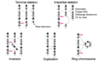
How are carriers of unbalanced translocations produced?
- Just before meiosis I homologous chromosomes line up next to each other
- If you have derivative chromosomes, because they have genetic material of 2 different chromosomes, they struggle to find and line up with their homologous pair
- They only way for them to do this is to form pachytene quadrivalents
- This means you get an increase in the no. of ways those 4 chromosomes are seperated which can result in a loss of genetic material within the resulting gametes
- E.g. If chromosomes are seperated along horizontal blue line
- One daughter cell will have a gain in yellow chromosome and a loss of the end of the purple chromosome;
- The other daughter cell has a loss of the end of the yellow chromosome and gain of the purple chromosome.

What are the clinical results of the unbalanced reciprocal translocation?
- Many lead to miscarriage (hence why a woman with a high number of unexplained miscarriages should be screened for a balanced translocation)
- May lead to Learning difficulties, physical disabilities
- Tend to be specific to each individual so exact risks and clinical features vary
What is a robertsonian translocation?
- Occurs when two acrocentric chromosomes break at or near their centromeres, and the fragments are joined together again possibly forming a chromosome with just the two sets of long q arms meaning there’s a loss of the satellites (short p arms).

What chromosomes can be affected by robertsonian traslocation?
- Only affects chromosomes 13, 14, 15, 21 and 22 as these are the only acrocentric chromosomes
Why would a carrier of a balanced robertsonian translocation only have 45 chromosomes?
- Person would have 46 chromosomes and then robertsonian translocation of acrocentric chromosome results in formation of chromosome with 2 sets of long q arms
- Short p arms don’t form new chromosome as they are lost so result is loss of 1 chromosome
Why is the loss of a chromosome as a result of a robertsonian translocation not as damaging as a loss of a chromosome due to non-disjunction (monosomy)?
- p arms encode rRNA (multiple copies so not deleterious to lose some)
How can someone be an unbalanced carrier of a robertsonian trasnlocation?
- If 46 chromosomes present including Robertsonian then must be unbalanced
What are some common robertsonian translocations?
- Robertsonian translocations 13;14 and 14;21 relatively common.
- 21;21 translocation leads to 100% risk of Down syndrome in fetus
What are the consequences of robertsonian translocations?

What are the 2 different mechanisms that can lead to trisomy 21?
- Trsomy 21 can be due to non-disjunction during meiosis
- Trisomsy 21 can also be due to a robertsonian transloaction between chromosome 21 and another acrocentric chromosome, e.g. 14.
What are some other structural chromosomal abnormalities/changes?
-
Terminal deletion - Deletion of the end of a chromosome
- Only way the chromosome can be made stable is if a new telomere is added; without the telomere the cell will die
- Interstitial deletion - Deletion of the middle of a chromosome
- Inversion - 2 breakpoints in a chromosome and section that’s cut out is repaired but placed upside down
- Duplication - Section of a chromosome is replicated
- Ring chromosomes - Ends of chromosomes (telomeres) are broken off and because new teolmeres aren’t added the rest of the chromosome forms a ring structure
What are the consequences of a deletion?
- Causes a region of monosomy which results in:
- Haploinsufficiency of some genes (don’t have 2 copies of a gene)
- Monosomic region has phenotypic consequences
- Phenotype is specific for size and place on deletion
What are some conditions caused by an interstitial deletion of a gene?
- Prader-Willi
- DiGeorge Syndrome
- Cri du chat
What techniques can be used to visualise microdeletions/microduplications?
- High resolution banding
- Array CGH
- NOTE: large structural abnormalities can be seen with G-banding and FISH
Explain how array CGH works
- Patient DNA and control DNA are extracted from samples
- Patient DNA labelled with Cy3, green and control DNA labelled with Cy5, red
- They are then mixed together and hybridised to the microarray
- Patient and control DNA compete to hybridise to the microarray
- Each spot on the array is then scanned to identify the colour of fluoresence it produces

Give some examples of microdeletion syndromes

How are microdeletions/microduplications formed?
- Unequal crossing over/non-allellic recombination
- Crossing over of homologous chromosomes that aren’t lined up properly

