Clinic: Images deck Flashcards

1
Q
A

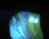
anterior subcapsular cataract

2
Q
A

Posterior subcapsular cataract

3
Q
A

EKC

4
Q
A

AMD

5
Q
A

BRVO

6
Q
A

Ischaemic CRVO

7
Q
A

Non ischemic CRVO

(variable dilated, tortuos veins; retinal heamorrhages; retinal thickening; OD oedema)

8
Q
A

BRVO

9
Q
A

CRAO

10
Q
A

BRAO

11
Q
A

L ONH with inferior notch and a CDR of 0.8
(This is a cfp by the way so not flipped)
(this particular patient from pharma also had a superior arcuate VF defect and was diagnosed with NTG, Diurnal IOP was identified as 21, then put on xalatan with a target pressure of 15)

12
Q
A

R: sup rim vessel bayonetting (blue arrow), inf rim thinning (white arrow), subtle RNFL loss (black arrow)
L: sup rim thinning (blue arrow), inf rim thinning (white arrow)
NB: This was a patient with early asymptomatic glaucoma (pharma june 2020)

13
Q
A

GCC thickening from a significant R ERM

14
Q
A

Fundus: R inf RNFL thinning L sup/inf RNFL thinning
OCT: R Inf-temp wedge-like RNFL defect L sup/inf-temporal wedge-like RNFL defect
VF (24-2): were not concordant with structural findings
(this patient from pharma also had heavily pigmented TM, posterior iris bowing, and bilateral krukenberg spindle - so everything together suggested a structural pigmentary glaucoma, and pt was referred to ophthalm for consideration of tx. IOP was R 15 L 19. CCT was 600ish OU)

15
Q
A

Choroidal naevi with drusen at the macula (left) and midperiphery (right)

16
Q
A

Choroidal naevus with a central pigmented area and a surrounding halo. Are associated with reduced rate of transformation to choroidal melanoma

17
Q
A

papilloedema

18
Q
A

Recent superior retinal detachment
[H33.011-013]

19
Q
A

CRVO

20
Q
A

BRVO

21
Q
A

Stage 3 Hypertensive Retinopathy (due to presence of the haemorrhages)

22
Q
A

atrophic/dry amd

23
Q
A

glaucomatous optic atrophy

24
Q
A

Subretinal haemorrhage in exudative/wet AMD

25
Full thickness macula hole
26
Degenerative drusen temporal to the macula (H35.361-363). This is actually an asymptomatic 20-30yo genetic problem.
27
Macula Pucker secondary to ERM [H35.371-373]
28
Retinitis Pigmentosa
29
Central serous chorioretinopathy (CSCR. H35.711-713)
30
Asteroid hyalosis [H43.21-23]
31
Recent onset Retrobulbar Neuritis (a week ago or less probably. H46.11-13). Trololol
32
Epiretinal membrane
33
Vitreous degeneration
34
Choroidal naevus temporal to the macula
35
Corneal infiltrates seen in adenoviral keratoconjunctivitis (so we're talking EKC, PCF) [B30.0 - decision maker plus]
36
Metallic foreign body embedded in the peripheral cornea
37
Band keratopathy. Legit. It doesn't look like it but still. Normally looks white-ish. (H18.423 ). Btw did you know band keratopathy can occur more peripherally ssometimes rather than central.
38
Hyphema secondary to blunt ocular trauma
39
Posterior synechia in chronic iridocyclitis [https://www.youtube.com/watch?v=oOrWDt1o1gk]
40
Temporal pallor of the optic disc in partial optic atrophy [H47.291-293]
41
Recurrent corneal erosion occurring in the pupillary region
42
CL induced exposure keratopathy causing a peripheral staining pattern
43
corneal bulging or rupture of descemets membrane secondary to post lasik ectasia
44
Congenital optic pit with associated macular edema
45
Sudden corneal opacification in acute corneal hydrops (i.e. in keratoconus)
46
Railroad track image characteristic of posterior polymorphous dystrophy
47
EBMD
48
corneal guttata (indicative of a corneal endothelial dystrophy like Fuchs)
49
50
Lash collarettes associated with anterior blepharitis. [a ring-like formation around the lash shaft]
51
22yo F patient, morning headaches.
mild papilloedema LE with few week old haemorrhage