Chapter 6 Flashcards

1
Q

hematopoiesis occurs via a stepwise maturation of CD34+ hematopoietic stem cells converting into either myeloid stem cells or lymphoid stem cells

Myeloid stem cells can transform into erythroblasts, myeloblasts (neutrophils, basophils,eosinophils), monoblasts (monocytes), and megakaryoblasts

Lymphoid stem cells can become either B or T lymphoblasts

A

Cells mature and are released from the BM into the blood

2
Q

A normal WBC is 5-10 K/uL

low WBC= leukopenia, high WBC= leukocytosis

A
3
Q

What are the main causes of neutropenia?

A
  1. Drug toxicity (eg. chemo with alkylating agents)-damage to stem cells
  2. Severe infection (e.g. gram-negative sepssi)- increased movement of neutrophils into tissues results in decreased criculating neutrophils

As a tx, GM-CSF or G-CSF may be used to boost granulocyte production, therbu decreasing risk of infection

4
Q

What are the main causes of lymphopenia?

A
  1. Immunodeficiency
  2. High cortisol state (e.g. exogenous steorids or cushing syndrome)
  3. Autoimmune
  4. Whole body radiation- lymphopenia is the first cell sign to go with radiation
5
Q

What are some causes of neutrophilic leukocytosis?

A
  1. Bacterial infection or tissue necrosis- induces release of marignated pool and BM neutophils, including immature forms (immature cells are marked by decreased Fc receptors (CD16)
6
Q

Basophilia is classically seen in ______

A

CML

7
Q

Causes of lymphocytic leukocytosis?

A
  1. Viral infections- T lymphocytes undergo hyperplasia in response to virally infected cells
  2. Bordetella pertussis infection- products lymphocytosis- promoting factor, which blocks circulating lymphocytes from leaving the blood to enter the lymph node
8
Q

What causes infectious mononucleosis?

A

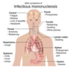
EBV infection that results in a lymphocytic leukocytosis comprised of reactive CD8+ T cells (CMV can also cause)

During this, EBV is spread via saliva and primarily infects the oropharynx, resulting in pharyngitis, the liver, resulting in hepatitis with hepatomegaly and elevated LEs, and B cells

9
Q

Again, in Infectious mono the main immune response is via CD8 T cells, resulting in:

A
  • Generalized LAD due to T-cell hyperplasia in the lymph node paracortex
  • Splenomegaly due to T-cell hyperplasia in the PALS
  • High WBC count with atypical lymphocytes (reactive CD8 cells) in the blood
10
Q

How is Infectious mono diagnosed?

A

Monospot test for screening- detects IgM Abs that cross-react with horse or sheep red blood cells (heterophile Abs). Usually positive 1 week after infection

A negatve monospot test suggests CMV as a possilbe cause of IM

Definitie diangosis made via serologic testing for the EBV viral capsid antigen

11
Q

What are the complications of infectious mono?

A

Increased risk of splenic rupture- avoid contact sports

Rash is exposed to ampicillin

Dormancy of vius in B cells elads to increased risk for both recurrence and B-cell lymphoma, especially if immunodeficiency develops

12
Q

What is acute leukemia?

A

A neoplastic proliferaiton of blasts; defines as the accumulation of 20+% of blasts in the BM

Incresaed blasts crowd out normal hematopoiesis, resulting in an acute presentation with fatigue, anemia, thrombocytopenia (bleeding), or neutropenia (infection)

The blasts then enter the blood stream, resulting in a high WBC count.

13
Q

Acute leukemia is divided into:

A

acute lymphoblastic leukemia (ALL) and acute myelogenous leukemia (AML)

14
Q

What is ALL?

A

Neoplastic accumulation of lymphoblasts (20+%) in the BM

lymphoblasts are marked by positive nuclear staining for TdT, a DNA polymerase (TdT is absent in myeloid blasts and mature lymphocytes)

15
Q

Who gets ALL?

A

Mostly children, associated with down syndrome (usually arises AFTER 5 yo)

16
Q

What are the subtypes of ALL?

A

B-ALL and T-ALL

17
Q

B-ALL

A

-Most common subtype, usually marked by lymphoblasts (Tdt+) that express CD10, CD19, and CD20

Excellent response to chemo, requires prophylaxis to scrotum and CSF

18
Q

What is the prognosis of B-ALL based on primarily?

A

Cytogenetic abnormalities

t(12:21) has a good response; common in children

t(9:22) has a poor prognosis (PHiladelphia); common in adults

19
Q

T-ALL

A

Marked by lymphoblasts (TdT+) that express markers ranging from CD2 to CD8 (e.g. CD3,CD4, CD7). Dont express CD10

Usually presents in teenagers as a medistinal (thymic) mass (called acute lymphoblastic lymphoma becuase the malignant cells form a mass)

20
Q

What is acute myeloid leukemia?

A

Neoplastic accumulation of immature myeloid cells in the BM- Myeloblasts are usually positive for MPO

Crystal aggregates of MPO may be seen as Auer rods

21
Q

Who gets AML?

A

Most common in older adults; 50-60+ yo

22
Q

Subclassification of AML is based on cytogenetic abnormalities, linegae of immature myeloid cells, and surface markers. What are the man subtypes?

A
  1. Acute promyelocytic leukemia (APL)
  2. Acute monocytic leukemia
  3. Acute megakaryocytic leukemia
23
Q

Describe Acute promyelocytic leukemia (APL)

A

– Marked by a t(15:17) which involves translocation of the retinoic acid receptor (RAR) on chrom 17 to chrom 15; RAR disruption blocks maturation and promyelocytes accumulate

  • Increased risk of DIC
  • Tx with all-trans-retinoic- acid (vitA derivative), which binds the altered receptor and causes the blasts to mature (and eventually die)
24
Q

Describe acute monocytic leukemia

A

Proliferaiton of monoblasts; usually lack MPO

Blasts prolifeate in the gums (below)

25
Describe acute megakaryocytic leukemia
Proliferation of megakaryocytes; lack MPO Associated with Down syndrome and usually presents BEFORE 5 yo
26
Note that AML may also rise from pre-exisiting dysplasia (myelodysplastic syndromes), especially with prior exposure to radiation/alkylating agents
Myelodysplastic syndromes usually present with cytopenias, hyperellular bone marrow, and increased blasts less than 20%
27
What are chronic leukemias?
Neoplastic proliferation of mature ciruclating lymphocytes; marked by high WBC count usually insidious in adults and in older adults
28
What are the main chronic leukemias?
1. Chronic lymphocytic leukemia (CLL) 2. Hairy cell Leukemia 3. Adult T-cell leukemia/lymphoma (ATLL) 4. Mycosis Fungoides
29
Describe Chronic lymphocytic leukemia (CLL)
Neoplastic proliferation of naive B cells that coexpress CD5 and CD20; most common leukemia overall Increased lymphocytes and SMUDEG cells on smear
30
What are the main complications of CLL?
1. Hypogammaglobinemia-infetion is the most common cause of death in CLL 2. Autoimmune hemolytic anemia 3. Transformation to diffuse large B-cell lymphoma (RIchter transformation)-marked clinically by an enlarging lymph node or spleen
31
Describe hairy cell leukemia
Proliferation of MATURE B cells marked by hair cytoplasmic processes Cells are positive for tartrate-resistant acid phosphatase (TRAP)
32
What are the clinical findings of hairy cell leukemia?
Splenomegaly due to accumulation of ahiry cells in red pulp dry tap on BM aspiration due to marrow firbsosis LAD usually absent
33
How is hairy cell leukemia tx?
2-CDA (cladribine), an adenosine deaminase inhbiitor
34
Describe Adult T-cell leukemia/lymphoma (ATLL)
-Proliferation of mature CD4 T cells Associated with HTLV-1; most common in Japan and Caribbean Clinical features include rash, generalized LAD, and lytic bone lesions with hypercalcemia
35
What is Mycosis fungoides?
A neoplastic proliferation of mature CD4 T cells that infiltrate the skin, producing localized skin rash, plaques, and nodules. Aggregates of cells in the epidermis are called **Pautrier microabscesses** ## Footnote **Spread to blood prodcues Sezary syndrome**
36
Sezary syndrome
37
What are myeloproliferative disorders?
Neoplastic proliferation of mature ells of myeloid lineage; disease of late adulthood (50-60 yo) Results in high WBC count with hypercellular BM
38
Complications of myeloproliferative disorders?
1. Risk of hyperuricemia and gout due to high cell turnover 2. PRogression to marrow fibrosis or tranformation to acute leukemia
39
What are the main myeloproliferative disorders?
1. Chronic myeloid leukemia (CML) 2. Polycythemia Vera (PV) 3. Essential Thrombocytopenia (ET) 4. Myelofibrosis
40
Describe Chronic myeloid leukemia (CML)
Neoplastic proliferation of ature myeloid cells, especially granulocytes and their precurosrs, **Basophils are characteristically increased** ## Footnote **Driven by t(9:22) translocation (Philadelphia chromosome) which generates a BCR-ABL fusion protein with increased tyrosine kinase activity**
41
Tx of Chronic myeloid leukemia (CML)?
Imatinib, which blocks tyrosine kinase activity
42
How does Chronic myeloid leukemia (CML) present?
Splenomegaly common- enlarging spleen suggest progressiont accelerated plase of disease- tranformation to acute leumeia usually follows shortly after Can transform to AML (2/3 of cases) or ALL (1/3 or cases) since mutation is in a pluripotent stem cell
43
How is CML distinguished from a leukomoid rxn (reactive neutrophilic leukocytosis)?
- Negative leukocyte alkaline phosphatase (LAP) stain (granulocytes in a leukemoid rxn are LAP positiive) - Increased basophils (absent with leukemoid rxn) - t(9:22) present
44
What is PV?
A neoplastic proliferation of mature myeloid cells, especially RBcs (grnaulocytes and platelets also increased) Associated with a JAK2 mutation
45
How does PV present?
Cinical symptoms are mostly due to hyperviscosity of blood: - blurry vision and HA - increased risk of venous thrombosis (e..g hepatic vein, portal vein, and dural sinuses) - Flushed face due to congestion (plethora) - Itching, especially after bath due to histamine release
46
How is PV tx?
phlebotomy and hydroxyurea (death occurs within 1 yr without tx)
47
PV must be distinguished from reactive polycythemia. How?
- In PV, EPO levels are low, and SaO2 is normal - In reactive polycythemia due to high altitude or lugn disease, SaO2 is low, and EPO is increased 3. In reactive polycythemia due to ecoptic EPO production from renall cell carcinoma, EPO is high and SaO2 is normal
48
What is Essential Thrombocytopenia?
Noeplastic proliferation of mature myeloid cells, espeically platelets (RBCs and granulocytes are also increased) Associated with JAK2 kinase mutation Symptoms are related to an increased risk of bleeding/thrombosis- no significant risk for gout and rarely progresses to marrow fibrosis or acute leukemia
49
What is Myelofibrosis?
Prolfieration of mature myeloid cells, especially megakaryocytes (associated with JAk2 mutation) Megakaryocytes produce excess PDGF causing **marrow fibrosis** Clinical features include splenomegaly due toe xtramedullary hematopoiesis, leukoerythroblastic smear (tear-drop cells, nucleated RBCs, and immature granulocytes)
50
LAD refers to enlargred lymph nodes. Note that painful LAD is usually seen in lymph nodes that are draining a region of acute infection (acute lymphadenitis) and painless LAD can be seen with chronic inflammation (chronic lymphadenitis), metastatic carcinoma, or lymphoma
In inflammation, lymph node enlargement is due to hyperplasia of the node
51
What are lymphomas?
Neoplastic prolifeation of lymphoid cells that forms a mass. may arise in a lymph node or in extranodal tissue Derived from non-Hodgkin lymphoma (NHL, 60%) and Hogkin lymphoma (40%)
52
What are the subtypes of NHL?
Further divided based on cell types (e.g. B vs. T), cell size, pattern of cell growth, expresion of surface markers, and cytognetic translocations 1. Small B cells- follilcuar lymphoma, mantle cell lymphoma, marginal zone lymphoma, and small lymphocytic lymphoma (i.e. CLL that involves tissues) 2. Medium sized B cells- Burkitt lymphoma 3. Large B cells- diffuse large B cell lyphoma
53
Describe follicular lymphoma
Neoplastic proliferation of small B cells (CD20+) that form follicle like nodules (below) Presents in late adulthood with painless LAD
54
What causes follicular lymphoma?
t(14:18): Bcl2 on chromsome 18 with the Ig heavy chain locus on chrom 14 results in overexpression of Bcl2, which inhibits apoptosis
55
How is follicular lymphoma tx?
Reserved for pts who are symptomatic and involves low-dose chemo or rituximab (anti-CD20 Ab)
56
Complications of follicular lymphoma?
Progression to diffuse large-B cell lymphoma
57
How is follicular lymphoma distinguished from reactitve follicular hyperplasia?
- Disruption of normal lymph node architecture (maintained in follicular hyperplasia) - lack of tingible body macrophages in germinal centers (present in hyperplasia) bcl2 expression in follicles Monoclonality
58
Describe Mantle cell lymphoma
Proliferation of small B cells (CD20+) that expands the mantel zone Presents in late adulthood with painless LAD driven by t(11:14) translocaiton of cyclin D1 on chrom 11 with ig hevay chain on chrom 14, leading to overexpression of cyclin D1 promoting G1/S phase transition
59
Describe Marginal zone lymphoma
Neoplastic prolfiertion of small B cells (CD20+) that expends the marginal zone Associated with chronic inflammatory states such as Hashimto thyroiditis, Sjogren and H. pylori gastritis
60
What causes Burkitt Lymphoma?
Neoplastic proliferation of medium-sized B cell (CD20+) associated with EBV Look for an extranodal mass in young children (African form involves the jaw and sporadic form involves the abdomen) Driven by t(8:14) translocation of c-MYC to the Ig heavy chain Buzzword: **Starry sky**
61
Describe Hodgkin lymphoma
Neoplastic proliferation of Reed-Sternberg cells, which are large B cells with multilobed nulcei and prominent nucleoli (owl-eyed); positive for CD15 and CD30
62
What do RS cells do?
Secrete cytokinease occassionally resulting in B symtpoms (fever, chills, weight loss, etc.) an attract reactive lymphocytes, plasma cells, macrophages, and may lead to fibrosis
63
What are the subtypes of Hodgkin Lymphoma?
1. Nodular sclerosis (most common; classic presentation is an enlarging cerival or mediastinal lymph node in a young adult, usually female; lymph node is divided by bands of sclerosis; below) 2. Lymphocyte-rich (best prognosis) 3. Mixed cellularity (associated with eosinophils; RS cells produce IL5) 4. Lymphocyte-depleted (most aggressive; seen in elderly and HIV+)
64
What are the main Plasma cell disorders (dyscrasias)?
1. Multiple Myeloma 2. Monoclonal Gammopathy of Undetermined Significance (MGUS) 3. Waldenstrom Macroglobulinemia
65
What is Multiple Myeloma?
A malignant proliferation of plasma cells in the BM (most common primary malignancy of bone; metastatic cancer, however, is the most common malignant lesion overall) High serum IL6 may be presnet, which stimulates plasma cell growth and immunoglobulin production
66
What are the features of MM?
1. Bone pain with hypercalcemia- Neoplastic plasma cells activate the RANK receptor on osteoclsts, leading to bone destruction. Lytic, punched out skeletal lesions are seen on X-ray (below)- risk of fratcure 2. Elevated serum protein- Neplastic plasma cells produce immunoglobulin; M spike is present on serum protein electrophoresis (SPEP), most commonly due to monoclonal IgG or IgA 3. Increased risk of infection- monoclonal Ab lacks antigenic diversity; infection most common cause of death 4. Primary AL amylodisis- free lgith chains circulate in serum and deposit 5. Proteinuria- Bence Jone proteins; deposition in kidney can cause failure
67
Describe MGUS
Increased serum protein with M spike on SPEP; other features of MM are absent (e.g. no lytic bone lesions, hypercalcemia, AL amyloid, or Bence Jones proteinuria) Commonly in elderly Progresses to MM at a rate of 1%/yr
68
Describe Waldenstrom Macroglobulinemia
B-cell lymphoma with monoclonal IgM production Clinical features include Generalized LAD (no lytic bone lesions), visual and neurologic deficits (e.g retinal hemorrhage or stroke), Bleeding
69
Describe Langerhans Cell Histiocytosis
langerhans cells are specialized dendritic cells found predominantly in the skin, derived from BM monocytes, and present antigens to T cells LCH is a proliferation of Langerhans cells marked by Birbeck (tennis racket) grnaules on EM (these cells are CD1a and S100+ by immunochemistry)
70
What is Letterer-Siwe Disease
Malignant proliferation of Langerhans cells, classically presenting with skin rash and cystic skeletal defects in an infant (less than 2 yo) Rapdily fatal
71
What is Eosinophilic granuloma?
Benign proliferation of Langerhans cells in bone, with a classical presentation of pathologic fracture in an adolescent (skin is not involved)
72
What is Hand-Schuller Christian Disease?
Malignant Proliferation of Langerhans Cells, with a classic presentation of scalp rash, lytic skull defects, diabetes insipidus, and exophthalmos in a child