Chapter 25: Skin - Pigmentation/Melanocytes and Tumors Flashcards
A circumscribed, flat skin lesion distinguished from surrounding skin by color, either <5mm or >5mm in diameter describes what?
- Macules are <5mm
- Patches are >5mm
Elevated dome-shaped or flat-topped skin lesion, either <5mm across or >5mm across describes what?
- Papules = <5mm
- Nodule = >5mm
Surface elevation caused by hyperplasia and enlargement of contiguous dermal papillae describes what?
Papillomatosis
What is Parakeratosis and where is it a normal finding?
- Keratinization with retained nuclei in the stratum corneum
- Normal on mucous membranes
A traumatic lesion breaking the epidermis and causing a raw linear area (i.e., deep scratch) describes what?
Excoriation
Multiple trichilemmomas may be seen with what syndrome that is associated with an increased risk of endometrial cancer, breast cancer, and many other tumors?
Cowden syndrome –> loss-of-function in PTEN

Differentiate Freckles from Café au lait spots.
- Freckles: appear after sun exposure; fade and darken cyclically w/ season
- Café au lait: seen in neurofibromatosis; larger than freckles; arise independent of sun exposure

Linear (nonnested) melanocytic hyperplasia restricted to the cell layer immediately above the basement membrane that produces a hyperpigmented basal cell layer, describes what?
Lentigo

What is the histology of superficial nevi, deeper nevi, and the deepest nevi which are helpful in differentiating benign nevi from melanoma?
- Superficial: nests, large-round cells, ↑ melanin
- Deeper: cords or single cells, smaller, ↓ pigment
- Deepest: fusiform, fascicles resembled neural tissue

What is the clinical significance of spindle and epitheliod cell nevus (Spitz nevus)?
Common in children; red-pink nodule; often confused with hemangioma clinically

Clinically how do compound and dermal nevi differ in appearance from junctional nevi?
Often more elevated

Inheritance pattern of dysplastic nevus syndrome and chance of melanoma?
Autosomal dominant; >50% chance dev. melanoma by age 60

What are some of the distinguishing histologic features of dysplastic nevi?
- Typically larger than acquired nevi, >5mm, can be hundreds
- Lentiginous hyperplasia: single nevus cells replace basal cell at E-D jct
- Architectural and cellular atypia
- Lymphocytic infiltrate of superf. dermis, melanin incontinence
- Linear fibrosis surrounds epidermal rete ridges

Activating mutations in which downstream serine/threonine kinase from RAS is seen in 40-50% of melanomas?
BRAF

What is the most commonly mutated gene identified in melanomas?
↑ TERT expression (activation of telomerase)
In which growth phase of melanoma do tumor cells seem to lack the capacity to metastasize?
Radial growth –> horizontal spread of melanoma within the epidermis and superficial dermis

Melanomas in radial growth phase fall into what 3 clinicopathologic classes; which is most common?
- Lentigo maligna: usually indolent lesion on face of older men
- Superficial spreading: most common type of melanoma, usually involving sun-exposed skin
- Acral/mucosal lentiginous: melanoma unrelated to sun exposure

Which phase of melanoma growth is often heralded by the appearance of a nodule and correlates with the emergence of a tumor subclone with metastatic potential?
Vertical growth phase: tumor cells invade downward into deeper dermal layers as an expansile mass

Which feature of melanocytic nevi is absent from the deep invasive portion of melanoma?
Neurotization

Which measurement is the distance from the superficial epidermal granular cell layer to the deepest intradermal tumor cells; what does this correlate with in melanoma?
- Breslow thickness
- Depth of invasion correlates with probability of metastasis
Staining for which melanocytic marker can be useful in distinguishing melanoma cells?
HMB-45 (+)

Which nuclei and nucleolus findings are seen histologically with melanoma?
- Nuclei with irregular contours + clumped chromatin at periphery
- Prominent red (eosinophilic) nucleoli

What are 5 determinants of a more favorable prognosis with melanoma?
- Thinner tumor depth (Breslow thickness)
- NO (<1 per mm2) mitosis
- Brisk tumor infiltrating lymphocyte response
- NO regression
- Lack of ulceration

Where do most melanomas initially metastasize and how does this affect the clinical handling of these lesions?
- Regional lymph nodes
- Should perform sentinel lymph node biopsy; microscopic involvement of a sentinel node by even a small # of melanoma cells = worse prognosis

What are the ABCDEs of melanoma which correlate with the warning signs?
- Asymmetry
- Irregular Borders
- Variegated Color
- ↑ Diameter
- Evolution/change over time (esp. rapid)
*ANY pigmented lesion w/ diameter >6mm, any change, itching or pain*

Seborrheic keratoses may suddenly appear in large numbers as part of which paraneoplastic syndrome; associated with what malignancy?
Leser-Trélat sign; most commonly carcinoma of GI tract

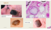
Round, flat, coinlike, waxy plaques that are tan-brown describes what?
Seborrheic Keratoses

Which morphological feature is helpful in distinguishing seborrheic keratoses from melanoma?
Pore-like ostia impacted with keratin
Activating mutation in what are seen in many sporadic seborrheic keratoses?
FGFR3

Small-keratin filled cysts (horn cysts) and invaginations of keratin into the main mass (invagination cysts) are characteristic features of what benign tumor?
Seborrheic keratoses

Benign tumor of skin described as “coin-like” or “stuck-on” is characteristic of what?
Seborrheic keratoses

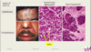
What are the 2 most common associations with acanthosis nigricans?
Obesity and diabetes
Epidermis with underlying enlarged dermal papillae undulating sharply to form numerous peaks and valleys describes the histology of what?
Acanthosis nigricans
Sebaceous adenomas can be associated with internal malignancy in which syndrome?
Muir-Torre syndrome, subset of HNPPC (deficit in DNA mismatch repair)

Cylindromas are appendage tumors with what kind of differentiation and where do they arise?
- Ductal (apocrine or eccrine) differentiation
- Usually arise on forehead and scalp
- With time may produce turban tumor

Characteristic histology of cylindromas?
Islands of cells resembling normal epidermal or adnexal basal cell layer (basaloid cells) which fit together like pieces of a jigsaw puzzle
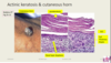
Actinic keratosis can progressively worsen over time finally culminating into what?
Cutaneous SCC

What is the gross morphology of Actinic Keratosis?
- <1cm, tan-brown, red or skin colored, rough sandpaper-like consistency
- Excess keratin prod. —> cutaneous horns

Histologically what is seen with Actinic Keratosis?
- Atypical basal cells w/ pink or red cytoplasm due to dyskeratosis
- Intracellular bridges
- Superficial dermis w/ thickened, blue-gray elastic fibers (elastosis)
- Stratum corneum is thickened, cells retaining their nuclei = Parakeratosis*

Cutaneous SCC and BCC incidence is proportional to what?
Degree of lifetime sun exposure

Immunosuppression such as chemotherapy or organ transplantation ↑ the risk of cutaneous SCC due ↑ risk of infection by what?
HPV subtypes 5 and 8

Which rare AR condition is marked by a high susceptibility to cutaneous SCCs and assoc. with HPV types 5 and8?
Epidermodysplasia verruciformis

Which acquired defect in precursor actinic keratosis has a high incidence in Caucasians and is an early event in development of cutaneous SCC?
TP53 mutations –> improper repair of UV damage

Xeroderma pigmentosum is an inherited mutation in what; leads to very increased susceptibility to SCC?
- Inherited mutation in nucleotide excision repair pathway, required for accurate repair of pyrimidine dimers
- ↑ risk of cutaneous SCC and basal cell carcinoma

How do in situ cutaneous SCC’s appear morphologically?
Sharply defined, red, scaling plaques

How do more advanced, invasive lesions of cutaneous SCC appear?
Nodular, variable keratin production (hyperkeratotic scale) and may ulcerate

How do cutaneous SCC’s differ from actinic keratosis histologically?
Cells with atypical (enlarged and hyperchromatic) nuclei involve ALL levels of the epidermis
Highly anaplastic cells that exhibit only abortive, single-cell keratinization seen with some cutaneous SCC is known as what?
Dyskeratosis

What is typical behavior of basal cell carcinoma and risk of metastases?
Slow growing, rarely metastasize; if caught early –> cured by local excision

Which distinctive locally aggressive cutaneous tumor is associated with mutations that activate the Hedgehog pathways signaling?
Basal Cell Carcinoma
Nevoid basal cell carcioma syndrome aka Gorlin syndrome aka basal cell nevus syndrome is inherited how and what is seen?
- Autosomal dominant
- Development of multiple BCC’s <20 y/o + medulloblastoma and ovarian fibromas; odontogenic keratocysts, and pits of the palms and soles
Nevoid basal cell carcioma syndrome aka Gorlin syndrome is associated with with what germline mutation?
- Loss-of-function in one PTCH allele –> Two-hit hypothesis
- PTCH protein = receptor for sonic hedgehog (SHH)

Which DNA base substitution is considered the hallmark of UV damage and is seen in 1/3 of basal cell carcinomas?
Cytosine (C) –> Thymine (T) = pyrimidine dimers
How do basal cell carcinomas typically present grossly?
Pearly papules w/ prominent dilated subepidermal vessels (telangiectasias)

Why is the term rodent ulcers used with some forms of aggressive basal cell carcinomas?
May ulcerate, and extensive local invasion of bone or facial sinuses may occur after many years of neglect or in unusually aggressive tumors

Which common and important variant of BCC presents as an erythematous, occasionally pigmented plaque that may resemble early forms of melanoma?
Superficial basal cell carcinoma
What are the 2 histological patterns of growth seen with BCC?
- Multifocal growths: originate from epidermis and sometimes extend over several square cm’s or more of skin surface
- Nodular: grow ↓ deeply into dermis as cords/islands of basophilic cells w/ hyperchromatic nuclei in a mucinous matrix w/ fibroblasts and lymphocytes –> cells at periphery arranged radially = palisading

Benign Fibrous Histiocytomas (Dermatofibroma) most often occurs in whom and is seen on which parts of the body?
Adults; often on the legs of young and middle-aged women

Benign Fibrous Histiocytomas (Dermatofibroma) are composed at least partially of what?
Factor XIIIa-positive dermal dendritic cells
Many cases of Benign Fibrous Histiocytomas (Dermatofibroma) are associated with a history of what?
Antecedent trauma, suggesting an abnormal response to injury and inflammation

Dermatofibroma is the most common type of benign fibrous histiocytoma and appears how morphohistologically?
- Benign spindle-shaped cells, well-defined, non-encapsulated mass in the mid dermis
- Overlying epidermal hyperplasia, downward elongation of hyperpigmented rete ridges = pseudoepitheliomatous hyperplasia

Which PE finding is benign fibrous histiocytoma (dermatofibroma) associated with?
Dimple sign: lateral pressure on the skin produces a depression
Dermatofibrosarcoma protuberans (DFSP) is best regarded as what; typical behavior?
Malignant, well-differentiated, primary fibrosarcoma of the skin which are slow-growing, locally aggressive and can recur; rarely metz

What is the molecular hallmark of dermatofibrosarcoma protuberans (DFSP); tumor cell growth is driven through what kind of loop?
- Translocation involving collagen 1A1 (COL1A1) and PDGFB
- Leads to ↑ secretion of PDGFβ
- Tumor cell growth through an autocrine loop

Dermatofibrosarcoma protuberans (DFSP) most often presents how; what is the characteristic morphology seen?
- “Protuberant” nodule, most often on the trunk, within a firm (indurated) plaque that may sometimes ulcerate.
- Closely packed fibroblasts arranged radially in storiform pattern, resembles blades of a pinwheel
- Deep extension into SQ fat produces “honeycomb” pattern

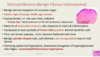
Lesions of mycosis fungoides usually involve truncal areas and include what type of lesions?
- Scaly, red-brown patches
- Raised scaling plaques (can be confused w/ psoriasis)
- Fungating nodules

What is the histologic hallmark of CTCL of the mycosis fungoides type?
- Sezary-Lutzner cells (cerebriform nuclei), forming band-like aggregates with superficial dermis
- Invade epidermis as single cells and small clusters = Pautrier Microabscesses

Prognosis of CTCL of the mycosis fungoides type is related to what?
% of total body surface involved and progression from patch –> plaque —> nodular forms

Which cutaneous form of mastocytosis predominantly affects children and accounts for >50% of all mastocytosis cases?
Urticaria pigmentosa

What can be a clue to the diagnosis of mastocytosis in both pre-menopausal women and in men?
Osteoporosis due to excessive histamine release in the marrow microenviornment

Many cases of mastocytosis have acquired activating point mutation in what?
KIT receptor tyrosine kinase

Which stains can be helpful in identifying mast cells and mastocytomas?
- Metachromatic stains (toluidine blue or Giemsa) for their granules
- Stains for mast cell tryptase and KIT if excessive degranulation has occurred

