Chapter 11: Assembly of the Transcription Initiation Complex Flashcards
Module 8
The two stages in the process that leads from genome to transcriptome.
- Initiation of transcription
- results in assembly upstream of the gene of the complex of proteins
- includes RNA polymerase enzyme and its various accessory proteins
- Synthesis and processing of RNA
- begins when the RNA polymerase leaves the initiation region and starts to make an RNA copy of the gene
- ends after completion of the processing and modification events that convert the initial transcript into a mature RNA molecule
Module 8
The central players in transcription are ___-______ ______ that attach to the genome in order to perform their biochemical functions. Many recognize specific nucleotide sequences and bind predominantly to these target sites, whereas others bind nonspecifically
DNA-binding proteins
Module 8
requires that part of the protein penetrates into the major and/or minor grooves of the helix in order to achieve _____ _____of the sequence. This is usually accompanied by more general interactions with the surface of the DNA molecule, which may
- direct readout
- simply stabilize the DNA–protein complex or which may access the indirect information on nucleotide sequence
Module 8
DNA-binding motifs
- When the structures of sequence-specific DNA-binding proteins are compared, the family as a whole can be divided into a limited number of different groups on the basis of the structure of the segment of the protein that interacts with the DNA molecule
- present in a range of proteins, often from very different organisms, and at least some of them probably evolved more than once
Module 8
helix–turn–helix (HTH) motif

- first DNA-binding structure to be identified
- made up of two a-helices separated by a B-turn
- not a random conformation but a specific structure
- made up of four amino acids
- second one is usually glycine
- This turn, in conjunction with the first a-helix, positions the second a-helix on the surface of the protein in an orientation that enables it to fit inside the major groove of a DNA molecule
- This second a-helix is therefore the recognition helix
- makes the vital contacts which enable the DNA sequence to be read
- structure is usually 20 or so amino acids in length, so its a small part of the protein as a whole
- Some of the other parts of the protein form attachments with the surface of the DNA molecule to help position the recognition helix within the major groove
*
Module 8
helix–turn–helix (HTH) motif
examples
- lactose repressor: in bacteria
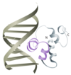
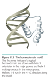
- homeodomain:
- in eukaryotic
- made up of 60 amino acids which form four a-helices, numbers 2 and 3 separated by a b-turn with number 3 acting as the recognition helix and number 1 making contacts within the minor groove

Module 8
Zinc fingers
- rare in prokaryotic proteins but very common in eukaryotes
- 1% of all mammalian genes code for zinc-finger proteins
- at least six different versions of the zinc finger
- versions of the zinc finger differ in the structure of the finger
- some lack the b-sheet component and consisting simply of one or more a-helices
- some differ on the precise way in which the zinc atom is held in place
- multiple copies of the finger are sometimes found on a single protein
- the individual zinc fingers are thought to make independent contacts with the DNA molecule, but in some cases the relationship between different fingers is more complex
Module 8
Zinc fingers
Cys2His2 finger
- first to be studied in detail
- comprises a series of 12 or so amino acids
- includes two cysteines and two histidines, which form a segment of b-sheet followed by an a-helix
- These two structures form the “finger” projecting from the surface of the protein, holding a bound zinc atom, between the two cysteines and two histidines
- a-helix is the part of the motif that makes the critical contacts within the major groove
- b-sheet determines a-helix positioning within the groove and interacts with the sugar–phosphate backbone of the DNA, and the zinc atom
- zinc atom holds the b-sheet and a-helix in the appropriate positions relative to one another
Module 8
Often the first thing that is discovered about a DNA-binding protein is not the identity of the protein itself but the features of the _____ ______ that the protein recognizes because many of the proteins that are involved in genome expression bind to _____ DNA sequences immediately _____ of the genes on which they act. Because of this, a number of methods have been developed for locating protein-binding sites working perfectly well even if the relevant DNA-binding proteins have not been identified.
- DNA sequence
- short
- upstream
Module 8
Gel retardation / gel shit analysis
- technique is carried out with a collection of restriction fragments that span the region thought to contain a protein-binding site
- two nuclear extracts have been mixed with a DNA
restriction digest - a DNA-binding proteing is added to one sample
- DNA-binding protein in the extract attaches to one of the restriction fragments
- this results in a DNA–protein complex
- has a larger molecular mass than the “naked” DNA
- runs more slowly during gel electrophoresis.
- the band for this fragment is retarded
- naked DNA and DNA–protein are run in separate wells
- DNA–protein sample is recognized by comparison with the banding pattern produced by naked DNA which runs faster
- A nuclear extract is used because at this stage of the project the DNA-binding protein has not usually been purified. If, however, the protein is available then the experiment can be carried out just as easily with the pure protein as with a mixed extract
Module 8
Gel retardation / gel shit analysis
drawbacks
- gives a general indication of the location of a protein-binding site in a DNA sequence, but does not pinpoint the site with great accuracy
- no indication of where in the retarded fragment the binding site lies
- if retarded fragment is long then it might contain separate binding sites for several proteins
- if it is quite small then there is the possibility that the binding site also includes nucleotides on adjacent fragments, not forming a stable complex with the protein and so do not lead to gel retardation
- Gel retardation studies are therefore a starting point but other techniques are needed to provide more accurate information.
Module 8
Modification protection assays
- pinpoint binding sites with greater accuracy
- basis of these techniques is that if a DNA molecule carries a bound protein then part of its nucleotide sequence will be protected from modification
- two ways of carrying out the modification
- treatment with a nuclease
- cleaves all phosphodiester bonds except those protected by the bound protein.
- exposure to a methylating agent
- such as dimethyl sulfate which adds methyl groups to G nucleotides
- Any Gs protected by the bound protein will not be methylated
- treatment with a nuclease
- DNA footprinting used with these assays
Module 8
DNA footprinting / Modification protection
- DNA fragment is labeled at one end
- achieved by treating a set of longer restriction fragments with an enzyme that attaches labels at both ends
- cutting these labeled molecules with a second restriction enzyme
- purifying one of the sets of end fragments.
- nuclease treatment is carried out under limiting conditions
- low temps/very little enzyme
- so each copy of the DNA fragment is cleaved at just one position along its length
- all bonds are cleaved except those protected by the bound protein
- carried out in the presence of a manganese salt, which induces the enzyme to make random, double-stranded cuts in the target molecules, leaving blunt-ended fragments
- protein is now removed, the mixture electrophoresed, and the labeled fragments visualized
- all fragments have labels at one end and a cleavage site at the other
- results in a ladder of bands corresponding to fragments that differ in length by one nucleotide, with the ladder broken by a blank area in which no labeled bands occur
- blank area/“footprint,” corresponds to the positions of the protected phosphodiester bonds, of the bound protein, in the starting DNA
Module 8
Modification interference
- identifies nucleotides central to protein binding
- not modification protection
- provides an extra dimension to the study of protein binding
- works on the basis that if a nucleotide critical for protein binding is altered, for example by addition of a methyl group, then binding may be prevented
Module 8
The dimethyl sulfate (DMS)
modification protection assay
- similar to DNase I footprinting
- Instead of DNase I digestion, the fragments are treated with limited amounts of DMS so that a single guanine base is methylated in each fragment
- Guanines that are protected by the bound protein cannot be modified
- Now the binding protein or nuclear extract is added, and the fragments electrophoresed.
- Two bands are seen, one corresponding to the DNA–protein complex and one containing DNA without bound protein
- The latter contains molecules that have been prevented from attaching to the protein because the methylation treatment has modified one or more Gs that are crucial for the binding
- To identify which Gs are modified, the fragment is purified from the gel and treated with piperidine,
- compound cleaves DNA at methylguanine nucleotides
- The result of this treatment is that each fragment is cut into two segments, one of which carries the label
- The length(s) of the labeled segment(s), determined by a second round of electrophoresis, tells us which nucleotide(s) in the original fragment were methylated and hence identifies the positions in the DNA sequence of Gs that participate in the binding reaction
- Equivalent techniques can be used to identify the A, C, and T nucleotides involved in binding
Module 8
It is now recognized that the nucleotide sequence also influences the precise _____ of each region of the helix, and that these conformational features represent a second, less direct way in which the DNA sequence can influence _____ _____
- conformation
- protein binding
Module 8
Direct readout of the nucleotide sequence
- although the nucleotide bases are on the inside of the DNA molecule, they are not entirely buried
- some of the chemical groups attached to the purine and pyrimidine bases are accessible from outside the helix
- Direct readout of the nucleotide sequence is possible without breaking the base pairs and opening up the molecule
Module 8
Direct readout
all 3 DNA forms
- B-form of DNA
- identity and orientation of the exposed parts of the bases within the major groove is such that most sequences can be read unambiguously
- whereas within the minor groove it is possible to identify if each base pair is A–T or G–C but difficult to know which nucleotide of the pair is in which strand of the helix
- A-form
- major groove is deep and narrow and less easily penetrated by any part of a protein molecule
- shallower minor groove is therefore likely to play the main part in direct readout
- Z-DNA
- the major groove is virtually nonexistent and direct readout is possible to a certain extent without moving beyond the surface of the helix.
Module 9
DNA-dependent RNA polymerases
- enzymes responsible for transcription of DNA into RNA
- three different RNA polymerases:
- RNA polymerase I,
- RNA polymerase II
- RNA polymerase III
- Each is a multisubunit protein (8–12 subunits) with a molecular mass in excess of 500 kDa
- all are structurally quite similar, but functionally quite distinct
- Each works on a different set of genes, with no interchangeability
- Each of the three eukaryotic RNA polymerases recognizes a different type of promoter sequence
Module 9
RNA polymerase I
transcribes the multicopy repeat units containing the 28S, 5.8S, and 18S rRNA genes
Module 9
RNA polymerase II,
- most researched
- transcribes
- genes that code for proteins
- snRNAs that are involved in RNA processing
- genes for the miRNAs
Module 9
RNA polymerase III
transcribes other genes for small RNAs, including those for tRNAs
Archaea possess a single RNA polymerase that is structurally very similar to the ______ enzymes
eukaryotic
Module 9
bacterial RNA polymerase
- has five subunits: α2ββ’δ
- 2α subunits
- one each of β and β’
- one of δ
- α2ββ’ are equivalent to the three largest subunits of the eukaryotic RNA polymerases
- δ has its own special properties
mitochondrial RNA polymerase
- consists of a single subunit with a molecular mass of 140 kDa
- more closely related to the RNA polymerases of certain bacteriophages
Module 9
Two ways in which RNA polymerases bind to their promoters
- Bacteria: direct recognition of the promoter by the RNA polymerase
- eukaryotic and archaeal:
- DNA binding protein binds to promoter forming a platform
- RNA polymerase binds to DNA binding protein
Module 9
prokaryotic promoter
- in bacteria, the target sequence for RNA polymerase
- immediately upstream of gene
- binding site for the RNA polymerase
- consists of two consensus sequences
- 6 nucleotides long
- names indicate their positions relative to the point at which transcription begins, starting point is labeled +1
- –35 box: 5’–TTGACA– 3’
- changes to sequence affect ability of RNA polymerase to bind
- –10 box: 5’–TATAAT– 3’
- changes to sequence affect the conversion of the closed promoter complex into the open form
- most are comprised mainly or entirely of A–T base pairs
- space between the 2 very important
- between 20 and 600 nucleotides upstream of the start of the coding region of the gene
- both are on the same face of the double helix

Module 9
Eukaryotic promoters
- more complex than prokaryotes
- describe all the sequences that are important in initiation of transcription of a gene
- numerous and diverse in their functions
- core promoter / basal promoter
- where the initiation complex is assembled
- upstream promoter elements
- lie upstream of the core promote
- Assembly of the initiation complex on the core promoter can usually occur in the absence of the upstream elements

Eukaryotic promoters
RNA polymerase I promoters
- core promoter spanning the transcription start point
- between nucleotides –45 and +20
- has an upstream control element (UCE) about 100 bp further upstream
Module 9
Eukaryotic promoters
RNA polymerase II promoters
- variable and can stretch for several kilobases upstream of the transcription start site
- consists of two main consensus segments
- –25 or TATA box: 5’–TATAWAAR–3’
- W is A or T
- R is A or G
- initiator (Inr) sequence
- mammalian: 5’–YCANTYY–3’
- Y is C or T, and N is any nucleotide
- located around nucleotide +1
- –25 or TATA box: 5’–TATAWAAR–3’
- Some genes have only one of these two components and some have neither, called null genes
- still transcribed
- start position for transcription is more variable
- A few genes have additional sequences
- DPE, downstream promoter element
- located at positions +28 to +32
- has a variable sequence
- binds TFIID, a protein complex that plays a central role in the preinitiation complex
- 7 bp GC-rich
- immediately upstream of the TATA box
- recognized by TFIIB, another component of the preinitiation complex
- PSE, proximal sequence element
- located between positions –45 and –60
- upstream of those snRNA genes that are transcribed by RNA polymerase II
- DPE, downstream promoter element
Eukaryotic promoters
RNA polymerase III promoters
- variable promoters
- at least three categories
- Two are unusual
- important sequences located within genes
- span 50–100 bp
- have two conserved boxes separated by a variable region
- third category
- similar to RNA polymerase II promoters
- have a TATA box and a range of additional promoter elements (sometimes including the PSE mentioned above) located upstream
Assembly of the transcription initiation complex
eukaryotic / prokaryotic
- bacterial polymerase attach directly and the three eukaryotic enzymes attach vi accessory proteins, to their promoter or core promoter sequences
- closed promoter complex is converted into an open promoter complex by breakage of a limited number of base pairs around the transcription initiation site
- RNA polymerase moves away from the promoter
- some attempts by the polymerase to achieve promoter clearance are unsuccessful and lead to truncated transcripts that are soon degraded
- successful initiation is only achieved after polymerase moves away from the promoter region
- correct in outline for all four RNA polymerases, the details are different for each one

Module 9
prokaryotic transcription initiation
- RNA polymerase forms direct contact w/the promoter via the δ subunit (core enzyme)
- δ subunit provides sequence specificity to the polymerase
- δ subunit recognizes –35 box
- RNA polymerase spans some 80 bp from upstream of the –35 box to downstream of the –10 box
- β’ & δ convert closed promoter complex to open form by breaking base pairs /win the –10 box
- Opening of helix involves contacts between the polymerase and the non-template strand
- δ plays a central role
- δ subunit, usually but not always dissociates soon after initiation is complete, converting the holoenzyme (α2ββ’δ) to the core enzyme (α2ββ’)
- elongation phase of transcription starts
- polymerase initial size of core enzyme is 60 bp, after the start of elongation it undergoes a second conformational change, down to just 30–40 bp
Module 9
eukaryotic transcription initiation
Preinitiation complex: Building the complex
- eukaryotic RNA polymerases do not directly recognize their core promoter sequences
-
initial contact is made by the general transcription factor (GTF) TFIID made up of
- TATA-binding protein (TBP)
- sequence-specific protein thant binds to DNA
- makes contact w/the minor groove where the TATA box is
- has a saddle-like shape
- wraps partially around the double helix, forming a platform
- at least 12 TBPassociated factors (TAFs)
- assists TBP to attach to TATA box
- works w/TAF-and-initiator-dependent cofactors (TICs) to recognize Inr sequence, especially those promoters w/out a TATA box
- proposed they can form a DNAbinding structure resembling a nucleosome, acting as the platform for assembly of the initiation complex
- TATA-binding protein (TBP)
- TBP attaches to core promoter
- Preinitiation complex (PIC) is formed w/help of additional GTFs (TFIIA) which stabilize TBP and TAF binding
- TBP binding induces formation of a ~80˚ bend in the DNA, widening the minor groove of the TATA box
-
TFIIB attaches to the complex
- upstream of TATA box
- contacts with TATA box via widened minor and major goove
- ensures correct positioning between RNA polymerase II, and transcription start site
-
TFIIF attaches
- interacts w/non-template strand
- recruits RNA polymerase II
-
TFIIE attaches
- recruits TFIIH
- modulates the various activities of TFIIH
-
TTFIIH attaches
- Helicase activity responsible for the transition from the closed to open promoter complex
- possibly influences promoter clearance by phosphorylation of the C-terminal domain of the largest subunit of RNA polymerase II
-
Activation of initiation complex
- phosphorylation of the C-terminal domain (CTD) of the largest subunit of RNA polymerase II
- Once phosphorylated, polymerase leaves initiation complex and synthesizing RNA
Module 9
eukaryotic transcription initiation
Preinitiation complex: Activation / Phosphorylation can be carried out by _____ or _____
- TFIIH
- mediator
eukaryotic transcription initiation
After departure of the polymerase, at least some of the GTFs detach from the core promoter, but _____, ______, and ______ remain, enabling ______ to occur without the need to rebuild the entire assembly from the beginning. Reinitiation is therefore a more rapid process than primary initiation
- FIID, TFIIA, and TFIIH
- reinitiation
Module 9
controlling transcription initiation in bacteria
Primary and secondary levels of genome regulation
- “primary” regulation
- occurs at the level of transcription initiation
- determines which genes are expressed in a particular cell at a particular time
- sets relative rates of expression of genes that are switched on
- “Secondary” regulation
- all steps in the genome expression pathway after transcription initiation
- modulates the amount of protein that is synthesized
- or changes the nature of the protein in some way (i.e. by chemical modification)
controlling transcription initiation in bacteria
two types
- Constitutive control:
- depends on the structure of the promoter
- intrinsic fitness of a promoter
- differences in the basal rate of transcription initiation
- its sequence, fitness can vary by 1000 fold
- directing 1000 times as many productive initiations as the weakest promoters
- called strong promoters
- Regulatory control:
- depends on the influence of regulatory proteins
- changes the ability of RNAP to initiate at a promoter
Module 9
Efficiency
- number of productive initiations that are promoted per second
- results in RNA polymerase clearing the promoter and beginning synthesis of a fulllength transcript
- controlling transcription initiation in bacteria
- precise sequence of the –35 box would influence recognition by the _____ _____ subunit and hence the rate of attachment of RNA polymerase.
- the ransition from the closed to open promoter complex might be dependent on the sequence of the _____ _____
- frequency of abortive initiations might be influenced by the sequence at, and immediately downstream of, _____ _____
- All this is speculation but it is a sound “working hypothesis.”
- δ subunit
- –10 box
- nucleotide +1
controlling transcription initiation in bacteria
basal rate of transcription initiation for a gene
- is preprogrammed by the sequence of its promoter
- can only be changed by a mutation
controlling transcription initiation in bacteria
The bacterium can, determine which promoter sequences are favored by changing the _____ _____ of its RNA polymerase, resulting in a different set of _____ being recognized. i.e. in E. coli _____ is the standard δ subunit. When it’s heat shock, E. coli alters the structure of its RNA polymerase to use _____ instead which switches on a set of genes with ______ specific to it. these genes help the bacteria deal w/the stress
- δ subunit
- promoters
- δ70
- δ32
- promoters
Regulatory control over bacterial transcription
operator
- region in operon
- usually overlaps the 3′ end of promoter and sometimes 5′ end of first structural gene

Regulatory control over bacterial transcription
inducer
- small molecule that binds to ACTIVE repressor protein altering it so it can’t bind to DNA and inhibit transcription
- stimulates transcription in inducible system
Regulatory control over bacterial transcription
corepressor
- small molecule that binds to INACTIVE repressor protein altering it so it can bind to DNA and inhibit transcription
- inhibits transcription in repressible system
Regulatory control over bacterial transcription
two types of transcriptional control
- negative control
- regulatory protein is a repressor
- binds to DNA and inhibits transcription
- positive control
- regulatory protein is an activator
- stimulates transcription
- it binds to DNA (other than the operator) and stimulates transcription
Regulatory control over bacterial transcription
Inducible operons
- transcription is normally off
- something must happen to induce transcription, or turn it on
Regulatory control over bacterial transcription
Repressible operons
- transcription is normally on
- something must happen to repress transcription, or turn it off
Regulatory control over bacterial transcription
negative inducible operons
- transcription initially turned off
- can be turned on by inducer
- regulator gene encoded an ACTIVE repressor protein that binds to operator & physically blocks binding of RNA polymerase preventing transcription

Regulatory control over bacterial transcription
negative repressible operons

- transcription initially turned on
- can be turned off by corepressor
- regulator gene encoded INACTIVE repressor protein. corepressor binds 2 inactive protein, activates it, it then binds to operator, blocks RNA polymerase & stops transcription

Regulatory control over bacterial transcription
regulator protein that acts on a negative repressible operon is synthesized as:
a. an active activator
b. an inactive activator
c. an active repressor
d. an inactive repressor
d. an inactive repressor
Regulatory control over bacterial transcription
lactose metabolism takes place when …
lactose is present & glucose is missing
Regulatory control over bacterial transcription
lac operon structure

Regulatory control over bacterial transcription
When glucose and lactose are both present, E. coli prefers _____ because it is …
- glucose
- requires less energy to metabolize than other sugars
Regulatory control over bacterial transcription
lac operon
absense of lactose
- repressor synthesized in active form
- attaches to operator
- RNA polymerase cannot bind to promoter

Regulatory control over bacterial transcription
lac operon
presence of lactose
- some lactose is converted to allolactose
- allolactose binds to lac repressor altering its shape & ability to attach to operator and it’s removed from operator
- RNA polymerase attaches and transcription begins
Regulatory control over bacterial transcription
repression never completely shuts down transcription of the lac operon…
- even with active repressor bound to the operator, there is a low level of transcription
- this allows cycle of active transport of lactose in cell & processing of it
Regulatory control over bacterial transcription
In the presence of allolactose, the lac repressor
a. binds to the operator
b. binds to the promoter
c. cannot bind to the operator
d. binds to the regulator gene
C. cannot bind to the operator
Regulatory control over bacterial transcription
catabolite repression
- When glucose is available, genes responsible for metabolism of other sugars are turned off
- results from positive control in response to glucose
Regulatory control over bacterial transcription
catabolite activator protein (CAP)
- functions in catabolite repression
- When bound with cAMP, cAMP-CAP binds upstream to promoters of operons
- This enhances binding of RNA polymerase to promoter increasing transcription of lac genes
Regulatory control over bacterial transcription
- concentration of cAMP is inversely proportional to concentration of _____.
- _____ levels of glucose _____ the amount of cAMP, so little cAMP–CAP complex is available to bind to DNA _____ transcription
- _____ concentrations of glucose _____ the amount of cAMP, resulting in increase cAMP–CAP binding to DNA _____ transcription
- glucose
- high, lower, decreasing
- low, increase, increasing
Regulatory control over bacterial transcription
cap, low level
- ↑ level of cAMP, cAMP binds to CAP
- cAMP-CAP binds to DNA
- cAMP-CAP on DNA increases polymerase binding, increasing transcription

Regulatory control over bacterial transcription
cap, hi level
- low levels of cAMP → less likely to bind to CAP
- polymerase can’t bind as effectively → slower transcription rate

Regulatory control over bacterial transcription
trp Operon
- negative repressible operon
- transcription is usually on
- has five structural genes
- has regulator gene, trpR coding inactive repressor protein w/2 binding sites: for operator & corepressor (tryptophan)
Regulatory control over bacterial transcription
trp Operon
mechanism
- ↓ levels of tryptophan levels cause RNA polymerase to bind to promoter and transcribes the five genes
- ↑ levels of tryptophan cause inhibition of transcription & synthesis of more tryptophan stops

Regulatory control over bacterial transcription
Jacob & Monod - lac operon mutations
regulator gene mutation
- affects regulation of transcription
- Mutations in lacI gene affect synthesis of β-galactosidase & permease (both in same operon)
- Some were constitutive → proteins produced all the time
- some prevented transcription all 2gether
- lacI-: Mutations that block oligomerization, cannot bind DNA
- lacIS: Mutations in inducer. Binding pocket cannot bind inducer, they are non-inducible
- lacI-d: dominant negative
Control of transcription initiation in eukaryotes
The ______ RNA polymerase has a strong affinity for its ______ and the basal rate of transcription initiation is relatively ______ for all but the weakest promoters. With most ______ genes, the reverse is true. The RNA polymerase II and III preinitiation complexes do not assemble efficiently and the basal rate of transcription initiation is therefore very low, regardless of how “strong” the promoter is. Compared with bacteria, eukaryotes use different strategies to control transcription initiation, with ______ playing a much more prominent role than repressor proteins
- bacterial
- promoter
- high
- activators
- eukaryotic
Control of transcription initiation in eukaryotes
The modules for an RNA polymerase II promoter
- The core promoter modules
- TATA box
- Inr sequence
- Basal promoter elements are modules
- present in many RNA polymerase II promoters
- sets the basal level of transcription initiation
- CAAT box recognized by the activators NF-1 and NF-Y
- GC box recognized by the Sp1 activator
- octamer module recognized by Oct-1.
- Response modules
- upstream of various genes
- enable transcription initiation to respond to general signals from outside of the cell
- cyclic AMP response module CRE recognized by the CREB activator
- heat-shock module recognized by Hsp70 and other activators
- serum response module recognized by the serum response factor
- Cell-specific modules
- located in the promoters of genes that are expressed in just one type of tissue
- erythroid module which is the binding site for the GATA-1 activator
- pituitary cell module recognized by Pit-1
- myoblast module recognized by MyoD
- lymphoid cell module, or kB site recognized by NF-kB
- in lymphoid cells, the octamer module is recognized by the tissue-specific Oct-2 activator.
- Modules for developmental regulators
- mediate expression of genes that are active at specific developmental stages
- Bicoid module and the Antennapedia module
Control of transcription initiation in eukaryotes
modules can also be contained within _____, which are 200–300 bp in length and can be located some distance _____ or _____ of their target gene. Silencers are similar to enhancers but, as their name suggests, their modules have a _____ rather than _____ influence on transcription initiation
- enhancers
- upstream
- downstream
- negative
- enhancing
Control of transcription initiation in eukaryotes
alternative promoters
- give rise to different versions of the transcript specified by the gene
- used to generate related versions of some proteins at different stages in development
- enables a single cell to synthesize similar proteins with slightly different biochemical properties
Control of transcription initiation in eukaryotes
activator
- sequence-specific DNA-binding protein that stimulates transcription initiation
- some recognize upstream promoter elements and influence transcription initiation only at the promoter to which these elements are attached
- other activators target sites within enhancers and influence transcription of several genes at once
- stabilizes the preinitiation complex
Control of transcription initiation in eukaryotes
coactivator
- binds nonspecifically to DNA or works via protein–protein interactions
- stimulates transcription initiation
Control of transcription initiation in eukaryotes
enhancers
- can be some distance from their genes
- has insulators at either side of each functional domain
- insulators prevent enhancers within that domain from influencing gene expression in adjacent domains
Control of transcription initiation in eukaryotes
Coactivators
- histone acetylation complexes such as SAGA and nucleosome remodeling complexes such as Swi/Snf
- influence gene expression by introducing bends and other distortions into DNA
- possibly as a prelude to chromatin modification
- possibly to bring together proteins attached to nonadjacent sites, enabling the bound factors to work together in a structure that has been called an enhanceosome
Control of transcription initiation in eukaryotes
Activators have been looked upon as important in initiation by RNA polymerases _____ and _____, but their role at RNA polymerase ______ promoters has been less well defined
- II
- III
- I
Control of transcription initiation in eukaryotes
mediator
- one of the components of the basal transcription apparatus
- interacts with RNA polymerase
- Transcriptional regulator proteins bind to sequences in the regulatory promoter (or enhancer) make contact with the mediator and affect the rate at which transcription is initiated
Control of transcription initiation in eukaryotes
Transcriptional Repressors
- competes w/transcriptional activators for binding sites on the DNA
- binds near activator binding sites preventing activator from contacting basal transcription apparatus
- interferes directly w/assembly of basal transcription apparatus, blocking initiation of transcription


