Chapter 1 - Growth Adaptations, Cellular Injury, and Cell Death Flashcards
What is the mechanism of the increase size in the Left Ventricle?
Hypertrophy
Hyperplasia is not possible in permanent tissues, cardiac muscle, skeletal muscle, and nerves.
What is the purpose of the Ubiquitine-Proteosome pathway?
Degradation of the cytoskeleton.
Important part of atrophy and the mechanism of cell size decrease.
Filaments are tagged by ubiquitine and removed by proteosomes.
What is the mechanism of Hypertrophy?
Gene activation, protein synthesis and organelle production.
What is the mechanism of Hyperplasia?
Generation from Stem cells.
Which Pathologic Hyperplasia does not increase the risk for cancer?
Benign prostatic hyperplasia.
What is the pathology pictured?
Barrett esophagus, the squamous cells of the esophagus undergoes metaplasia and becomes collumnar cells.
Which pathologic metaplasia does not increase the risk of cancer?
Apocrine metaplasia of the breast.
Which vitamin may be involved in Promyelocytic leukemia?
Vitamin A
15’ - 17’ translocation involves the retinoic acid receptor alpha which hinders the maturation of promyelocytes. It responds to ATRA (All-Trans Retinoic Acid) treatment.
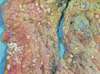
Which deficency causes the metaplasia pictured?
Vitamin A deficency
Vit A deficency leads to metaplasia of the conjunctive (squamous to stratified keratinizing). It is called Keratomalacia.
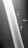
Case: 16 year old male presents with a hard immobile mass in the right upper arm. Looking through his file you see that he had previously visited due to a bike accident three weeks previous where he had hurt his arm, but there had been no fracture on the x-rays. Worried that you may have missed something you order a new x-ray. What does it show?
Myositis ossificans
It is a form of mesenchymal metaplasia which is caused by imflamation of the muscle following trauma. It turns the muscle to bone.
Can be differantiated from osteosarcoma due to the normal viewing of the bone and the fact that the tumor is not connecte to the bone.
How can you distinguish Myositis ossificans from osteosarcome in an x-ray?
- Normal bone
- Distinct separation between the tumour and the bone.
It is a form of mesenchymal metaplasia which is caused by imflamation of the muscle following trauma. It turns the muscle to bone.
What is the two most common mechanisms of dysplasia?
- Longstanding pathologic hyperplasia
- Metaplasia
What are the common causes of Cellular injury?
- Inflammation
- Nutritional deficency
- Nutritional excess
- Hypoxia
- Trauma
- Genetic mutations
What are the common causes of Hypoxia?
- Ischemia
- Hypoxemia
- Decreased O2-carrying capacity
What is Ischemia, and what causes it?
Ischemia is decreased blood flow through an organ. Has three common causes:
- Decreased arterial perfusion (atherosclerosis)
- Decreased venous drainage (e.g., Budd-Chiari, teticular hemorraghic infarction)
- Shock (generalised hypotension)
Case: 22 year old patient presents with intense abdominal pain in the upper right quadrant. The patient has severe nausea and is quite jaundice.
The patient history is mostly unremarkable except a previous diagnosis of polycytemia vera. What illness is the patient presenting with?
Budd-Chiari syndrom
Embolous of the hepatic vein, causes infarction of the liver and may be fatal. Commonly caused by polycytemia vera (increased red bloodcells -> increased blood viscosity) or Lupus anticoagulant (causes hypercoagulant state)
What is Budd-Chiari Syndrome?
Embolous of the hepatic vein, causes infarction of the liver and may be fatal. Commonly caused by polycytemia vera (increased red bloodcells -> increased blood viscosity) or Lupus anticoagulant (causes hypercoagulant state)
What is the mechanism behind testicular hemorraghic infarction?
The testicular cord consists of a among others a thick walled artery and a thin walled vein. During testicular torsion the artery remains open but clamps off the vein. This leads to hypoxia through decreased venous drainage.
What are the 5 types of shock?
- Anaphylactic
- Cardiogenic
- Hypovolemic
- Neurogenic
- Septic
What is Hypoxemia?
It is low partial preassure of oxygen in the blood (PaO2)
What causes hypoxemia?
FiO2 → PAO2 → PaO2 → SaO2
Decreased FiO2: High altitude
Decreased PAO2: Increased PACO2 (hypoventilation, COPD)
Decreased PaO2: Diffusion defect (interstitial pulmonary fibrosis)
What causes low O2-carrying capacity of the blood?
- Anemia (PaO2 normal, SaO2 normal)
- Carbon monoxide poisioning (PaO2 normal, SaO2 low)
- Methemoglobinemia (PaO2 normal, SaO2 low)
Case: You are called out to a house fire together with the paramedics. There is only one person in the building, an elderly lady who had been found sleeping in her bedroom. The firedepartment had been alerted to the fire by a neighbour. The fire had not yet reached the bedroom of the house when the firefighters broke through the window. The woman is upset and shaken, but says she feel fine except for a light headache. You decide to take her to the hospital. What treatment should you give?
If possible the patient should be put in a hyperbaric chamber to combat the carbonmonoxide poisioning she might have developed due to smoke inhalation, while sleeping in a confined room (closed window). Headache is an early sign. It also causes dizziness, confusion, and redness of the skin. If a hyperbaric chamber is not availible she should be put on oxygen therapy.
What are the characteristic symptoms of carbonmonoxide posioning?
Cherry-red apparanca of the skin, this is caused by the tightly bound hemoglobin. It is paradoxical though because the patient is hypoxic.
OBS! Early sign is headache!
What are the characteristic symptoms of Methemoglobinemia?
Cyanosis with Chocolate coloured blood
What is the treatment for Methemoglobinemia?
Methylane blue it turns the Fe3+ back to Fe2+
How does Hypoxia cause cellular injury?
Disruption of the electron transport chain leads to decreased ATP, which affects:
- Na+-K+ pump → cellular swelling
- Ca2+ pump → Increased Ca2+ → enzyme activation
- Aerobic glycosylation → Increased lactic acid → decreased pH → presipitation of proteins and DNA
What are the findings of reversible injury?
Swelling
- Cytosol swelling → loss of microvilli and cellular blebbing
- RER swelling → Dissociation of ribosomes → decreased protein synthesis
What are the findings of irreversible injury?
Membrane damage
- Plasma membrane damage → enzyme leakage (troponines), and Ca2+ influx.
- Mitochondrial membrane damage → Loss of electron transport chain and cytochrome C leakage causing apoptosis
- Lysosome membrane damage → Intracellular enzyme leakage causing intracellular damage (effect increased by increased intracellular Ca2+)
What is the morphological hallmark of cell death?
Loss of nucleus
What is the mechanism of nucleus loss?
- Pyknosis: nuclear condensation
- Karyorrhexis: fragmentation
- Karyolysis: dissolution
What are the characteristics of Necrosis?
- Many cells
- Followed by acute inflammation
- Always pathological
What are the two causes of inflammation?
- Infection
- Necrosis
Which 6 types of necrosis are there?
- Coaglative: Ischemic infarction, pale wedge shaped.
- Liquefactive: Enzymatic lysis
- Gangrenous: Mumifiation, dry or wet
- Caseous: Cottage cheese, granulomatous inflammation
- Fat: Chalky white, saponification
- Fibrinoid: Blood vessels
What is characteristic for the type of necrosis pictured?
Coagulative necrosis
Characteristic: Ischemic infarction of almost all organs. Often wedge shaped (due to branching vessels) and pale.
What is characteristic for the type of necrosis pictured?
Liquefactive necrosis
Enzymatic lysis of cells. Characteristic for brain infarction (proteolytic enzymes from the microglial cells), abcesses (proteolytic enzymes from neutrophils), and Pancreatitis (pancreatic enzymes digests itself. The surrounding fat undergoes fat necrosis)
What is characteristic for the type of necrosis pictured?
Gangrenous necrosis
Resembles mumified tissue. Affects lower limb and GI tract. May be dry or wet. Wet is with a superimposed infection on top.
What is characteristic for the type of necrosis pictured?
Fat necrosis
Chalky white apparance due to calcium deposition. Characteristic of trauma (breast) and pancreatitis (surrpunding tissue)
Saponification (dystrophic calcification of dead or dying tissue) Normal serum calcium and phosphate levels.
Metastatic calcification (not necesarrily cancer) deposition due to High serum calcium and phosphate levels.
What is characteristic for the type of necrosis pictured?
Caseous necrosis
Granulomatous inflammation resembles “chunky” liquefactive necrosis. Associated with fungal infections and tubercolosis.
What is characteristic for the type of necrosis pictured?
Fibrinoid necrosis
Necrosis of blood vessels, causes protein leakage into the vessel wall showing as bright pink on Histology.
Caused by Malignant hypertension, vasculitis, and Pre-eclampsia.
What is the mechanism behind a red infarction?
Blood reenters loose infarcted tissue. (e.g., testicular hemorraghic infarction)
What are the characteristics of apoptosis?
- Few cells
- Genetically programed
- ATP dependent
Which are the three pathways of apoptosis activation?
Intrinsic: DNA damage → inactivation of Bcl2 → destabilization of mitochondria → release of cytochrome C → Caspase activation.
Extrinsic: FAS ligand binds to CD95 (Death receptor) → Caspase activation. OR TNF binds TNF receptor → Caspase activation.
Cytotoxic (CD8+ T-cell mediated): Perforins create pores in the virally infected cell → Granzymes enters pores → Caspase activation
What is the pathway of free radicals production?
What causes pathological production of free radicals?
- Ionizing radiation - .OH
- Inflammation - O2-.
- Metals - .OH
- Drugs and chemicals
What causes free radical damage?
- Peroxidation of lipids
- Oxidation of DNA
Which protective measures against free radicals does the body use?
- Antioxidants (glutathione, and vitamins A, C, and E)
- Metal carrier proteins (e.g., transferrin) hinders metal in reacting in the Fenton reaction.
**- Enzymes**: Superoxide dismutase (mitochondria) O<sub>2</sub><sup>-.</sup> → H<sub>2</sub>O<sub>2</sub>
Glutathione peroxidase (mitochondria) GSH + free radical → GSSH + H2O
Catalase (peroxisomes) H2O2 → O2 + H2O
Case: 50 year old male presents with malaise, abdominal distention and pain, and a slight jaundice to the eyes and skin.
When taking a medical history he reports that his son is also similarily affected. He suffers from hypertension, is a smoker (1 pack a day) and drinks moderatly. He owns a family run dry cleaning store, and has recently started remodeling his home.
Tests shows hypocoagulability and fatty streaks on the liver. What is the cause of his symptoms?
Answer: Free radical injury caused by CCL4 exposure. CCL4 is used in the dry cleaning industry.
CCL4 is converted to CCL3 by the p450 system of the liver. It results in cell injury with cellular swelling. Swelling of the RER reduces protein production leading to hypocoagubility, but most notably decreased apolipoproteins which leads to fatty change.
Why does the cardiac enzymes keep rising after the occlusion, which caused an ischemic episode, is removed?
Reperfusion injury
when blood flow is reestablished it brings with it oxygen and inflammatory cells which together produces free radicals. This increases the area of injury.
What is an amyloid?
A misfolded ß-plated sheet protein which is deposited in the extracellular space and damages the tissue. It often deposits around blood vessels and is stained congo red on histological staining. Turns characteristically apple green under polarised light.
Which amyloid is connected with primary systemic amyloidosis? What type of illness is associated with it?
AL amyloid (immunoglobulin light chain)
Plasma cell dyscrasias (e.g., multiple myeloma)
Which amyloid is connected with secondary systemic amyloidosis? What type of illness is associated with it?
AA amyloid (serum amyloid associated protein)
Acute phase reactant: Chronic inflammatory states (e.g., Rheumatoid arthritis, Lupus, Ulcerative collitis, Chron’s, Osteomyilitis), malignancy, Familial medeterrainian fever.
Case: 33 year old female originally from Spain presents with severe chest pain, feet swelling, fever and proteinuria.
The EKG shows an arrythmia, but cardiac markers are negative. She has an enlarged liver and spleen and her kidney tests indicate damage. You perform a tissue biopsy. Which type of staining should you apply?
Your test confirms your suspision. How should you treat the patient?
Congo red
The patient has famillial medetarainian fever (rare!) a form of secondary amyloidosis. It causes fever and serosal inflammation (pericardium, appendix, etc.) it might threrefore mimic a MI, appendicitis, arthritis etc.
The amyloid affected tissues can not be treated, and must be transplanted.
What are the clinical findings of amyloidosis?
- Neprhotic syndrome
- Restrictive cardiomyopathy or arrythmia
- Tounge enlargment, malabsorption, and hepatosplenomegally
Match the disease and the amyloid:
Disease: Alzheimers disease, Dialysis associated amyloidosis, Familial amyloid cardiomyopathy, Medullary carcinoma of the thyroid, Non-insulin dependent diabetes mellitus, Senile cardiac amyloidosis.
Amyloid: Aß amyloid, Amylin, ß2-microglobulin, Calcitonin, Mutated serum transthyretin, Non-mutated serum transthyretin
Disease - Amyloid
AD - Aß amyloid
DAA - ß2-microglobulin
FAD - MST
MCotT - Calcitonin
NIDDM - Amylin
SCA - NMST