Ch. 1. Anatomy and organisation of the body Flashcards
Types of blood vessels
Arteries – carry blood away from heart Veins – return blood to the heart Capillaries – link arteries and veins
Abnormal immune mechanisms
undesirable responses of the immune system. Allergies as responses to antigens
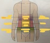
Pelvic girdle:
two innominate bones and the sacrum
Types of blood cells
Erythrocytes – red blood cells – transport O2 and CO2 Leukocytes – white blood cells – protection of the body Platelets (thrombocytes) – cell fragments, play part in blood clotting
Urine
formed by kidneys. Consists of water and waste products (from protein breakdown – urea). Hormones from endocrine system influence kidneys to regulate water balance, blood pH. Bladder stores urine until it is excreted during micturition.
Capillaries
link arteries and veins – tiny blood vessels, very thin walls made of one layer of cells which enables exchange of substances between blood and body tissues
Survival of the species
depends on successful reproduction, involves fusion of male and female sex cell (sexual reproduction). Individuals with most advantageous genetic make-up are most likely to survive (natural selection, survival of the fittest)
Heart
involuntary muscular sac with four chambers, pumps blood round the body and maintains blood pressure. Beats 65-75 times per minute.
Lungs
pulmonary circulation, oxygen absorbed, and CO2 excreted Nitrogen in the air is not used by the body
Lymphatic system organs
Spleen and thymus
Complications
other consequences that might arise if the disease progresses
Cranical cavity
contains the brain. It containes: Anteriorly – 1 frontal bone Laterally – 2 temporal bones Posteriorly – 1 occipital bone Superiorly – 2 parietal bones Inferiorly – 1 shenoid and 1 ethmoid bone and parts of frontal, temporal, and occipital bones
Three body planes
Median plane Frontal (coronal) plane Transverse plane


Thoracic cage functions
Protects thorax, heart, lungs, large blood vessels Forms joints between upper limbs and axial skeleton. The upper part of sternum (manubrium) articulates with the clavicles Intercostal muscle occupies spaces between the ribs Diaphragm is dome-shaped muscle that separates thoracic and abdominal cavities
Anatomy
Study of structure of the body and physical relationships between its parts
Alveoli
are surrounded by tiny capillaries, where the gas exchange between lungs and blood takes place
Special senses
stimulation of specialised receptors in sensory organs gives rise to sensation of sight, hearing, balance, smell, and taste.
Motor or efferent nerves
transmits signals from brain to effector organs (muscles and glands)
Urinary system ageing
Fewer nephrons, lower glomerular filtration rate Less able to regulate fluid balance
Thoracic cavity
in the upper part of the trunk, it containes: Anteriorly – sternum and coastal cartilages of the ribs Laterally – 12 pairs of ribs and intercostal muscles Posteriorly – structures forming root of the neck Inferiorly – diaphragm Main organs contained within: trachea, 2 bonchi, 2 lungs, heart, aorta, superior and inferior venae cavae, oesophagus, lymph vessels, lymph nodes The mediastinum – space between the lungs
Lymph nodes
filter lymph, removing microbes and other materials Site of formation and maturation of lymphocytes (white blood cells)
Special senses - Brain
initiates response with electrical impulses in motor (efferent) nerves to effector organs, muscles, and glands
causes of diseases:
Genetic abnormalities (inherites or acquired) Infection (by bacteria, viruses, microbes, parasites) Chemicals Ionising radiation Physical trauma Degeneration (excessive use or ageing)