Central Nervous System Flashcards

1
Q

What 2 branches can the Nervous System (NS) be divided into?

A

CNS - Central Nervous System

PNS - Peripheral Nervous System

2
Q

What is the CNS composed of?

A

Spinal cord and brain

3
Q

What is the PNS composed of?

A

Composed of nerves (cranial and spinal)

Ganglia outside the brain and spinal cord

4
Q

What are the spinal nerves?

A

Part of the PNS

Nerves that emerge out of the spinal cord and join onto other nerves

5
Q

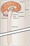
What is the brain composed of? Fill in the labels of this diagram:

A

Forebrain, midbrain, hindbrain

6
Q

Fill in the divisions of the brain:

A

>>

7
Q

What type of section is this of the brain?

Fill in the missing labels of this diagram:

A

Mid-saggital section

The darker green below the hypathalamus is the pituitary gland, and the hypathalamus and pituitary gland communicate with each other for regulation of the body

8
Q

What are the 4 lobes of the brain?

A

Frontal - higher order, coginitive, executive (planning, attention, memory), and motor functions,

Parietal - Sensation, sensory aspects of speech, spatial orientation and self-perception

Temporal - processing auditory information

Occipital - processing visual information

9
Q

Label the 4 different lobes on this diagram:

A

>>

10
Q

What are the different areas circled called?

What are the functions of these areas?

A

Baby pink = central sulcus (divides the frontal and parietal lobes)

Yellow = pre-central gyrus (anterior) and post-central gyrus (posterior) - house the primary cortices of the motor and sensory respectively

Brown = lateral fissure (separated the temporal, parietal and front lobes)

Baby blue = parieto-occipital sulcus (divides the parietal and occipital lobes)

11
Q

What is the lobe called highlighted in green and what is its function?

A

Limbic Lobe - composed of the amygdala, hippocampus, mamillary body, and congulate gyrus

Limbic system involved in learning, memory, motivation (esp. reward systems)

12
Q

What is the lobe that lies deep within the lateral fissure and what is its function?

A

Insular cortex

Functions include - receives viceral sensations from the organs, autonomic control, introception (understanding what is going on inside you), some auditory processing, visual vestibular integration (breakdown causes dizziness / motion sickness)

13
Q

What are the 3 layers of the meninges?

What are the 2 layers of the dura, and what do they form?

What is the space called between the arachnoid and pia?

A

Dure - divides into the periosteal and meningeal layer, both together form the dural sinuses

Arachnoid - thin, transparent, fibrous membrane that forms spiderlike projections (does not go into the folds)

Pia - thin membrane, that follows the folds

Sub-arachnoid space

14
Q

What is CSF, where is it formed, where does it travel and what does it do (i.e. why is it important?

A

Cerebrospinal fluid - made within the ventricles, flows out of the ventricles, pools into the subarachnoid space, reabsorbed into the bloodstream at these sinuses

Cushions the brain within the skull, shock absorber for the CNS, also circulates nutrients, removes waste products from the brain

15
Q

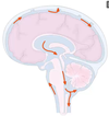
The red arrows indicate the flow of CFS.

Where specifically is CSF produced?

Where does it occupy and how does it get there?

Through what is the CSF reabsorbed?

How much CSF is produced daily?
A

Produced in modified epithelial cells called the choroid plexuses of lateral, 3rd and 4th ventricles

Occupies ventricular system (closed system but greatly expanded within the brain) and sub-arachnoid space (when it emerges from the ventricles at 3 foramen)

Reabsorbed via arachnoid villi (granulations on the SSS) into the superior sagittal sinus (SSS)

~125 ml volume and 500 ml produced each day

16
Q

What is the Ventricular System?

Where are the 4 ventricles and what else are they connected to?

A

Brain ventricles - four cavities located within the brain that contain CSF

Two lateral ventricles - one on each side of the cerebral cortex. The lateral ventricles are continuous with the third ventricle, which is lower in the brain. The third ventricle is continuous with the fourth ventricle, which runs along the brainstem

Ventricles are interconnected with each other, and the central canal of the spinal cord and the subarachnoid space

17
Q

)What are the differences (especially in consistency) between the CSF and blood plasma?

What is CFS used for clinically?

A

Lower pH, less glucose, protein and potassium than plasma

As an indicator something may be wrong / a diagnostic tool - it should not contain many proteins, or any blood

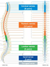
18
Q

What is the spinal cord made up of?

A

Segments

19
Q

How many nerves come out of each segment?

How are the segments named?

A

One from each side of the spinal cord (a pair of spinal nerves)

Corresponds to their position in the back / where it is on the spinal cord

20
Q

What is the difference between the substances that make up the structures at 2 and 4?

A

Grey matter and white matter

21
Q

What are the 2 projections (8 and 6) that go into and out of the grey matter?

What signals are conveyed through them?

A

Dorsal Root (posterior) - sensory / afferent

and Ventral Root (anterior) - motor / efferent

22
Q

What are the smaller versions of the projections (5 and 7)?

A

The Dorsal and Ventral Rootlets

23
Q

The grey matter in the spinal cord can be divided into which 2 parts (1 and 3)?

A

The Dorsal and Ventral Horns (posterior and anterior respectively)

24
Q

What is that part of the nerve (10) and what signal travels though there?

A

Mixed spinal nerve - at this point the afferent nerves have not yet broken away, and the efferent have already joined the nerve

25
Q

What is the slight swelling (9) on the Dorsal Root?

Why is there no swelling on the Ventral Root?

A

Dorsal Root Ganglion - swelling contains the collection of cell bodies of the sensory nerves

Collection of cell bodies for the motor nerves are inside the spinal cord

26
Q

Fill in all the missing labels in this diagram:

A

:)

27
Q

What type of fibres are found in the projection circled in the diagram? (i.e. afferent, efferent or both?)

A

Posterior Ramus - a smaller branch of the mixed nerves, also containing both, afferent and efferent nerves - supplies the muscles and skin down the back of the spine, whilst the main Mixed Spinal Nerve branch supplies the other parts of the body

28
Q

How many segments make up the spinal cord?

Into which 5 sections are the segments divided between?

A

31 segments

Cervical - 8 pairs, Thoracic - 12 pairs, Lumbar - 5 pairs, Sacral - 5 pairs, Coccygeal - 1

29
Q

How do the pair of nerves in each segment of the spinal cord leave to get outside the vertebrae?

A

Through the intervertebral foramina - gap between 2 bones

30
Q

How does the relationship between the nerves and foramina change as you go lower down the spine?

Why is this important clinically?

A

Same nomenclature for the bones as there are for the nerves, but the relationship between them changes between the cervical and thoracic regions

Nerves C1-C7 emerge before the vertebrae, C8-Co1 emerge below the vertebrae

The lower down the spine, the further apart the bones and segments can be found?

31
Q

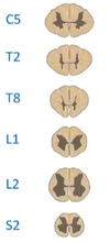
Where are the 2 spinal cord enlargements and why are they enlarged?

A

C5 - Cervical enlargement, huge amount of innervation going out to and coming in from the upper limbs

L2 - Lumbar englargement, huge amount of innervation going out to and coming in from the lower limbs

32
Q

What is the cerebral cortex?

What are gyri?

What are sulci?

A

Outer layer of neural tissue of the brain, separated into the left and right cerebral hemispheres

Ridges on the surface of the brain whih are sepaated by sulci

Fissures / Grooves in the brain

33
Q

What is the major (motor) descending pathway for voluntary movement called?

What is it composed of, and where is it found?

Where does it cross?

A

Corticospinal tract

Composed of 2 motor neurons - one in the primary motor cortex of the brain called the upper motor neuron, and the other in the brainstem / spinal cord called the lower motor neuron

The one that goes down to the brainstem is the corticobulbar tract, the one that goes down to the spinal cord is the corticospinal tract

Medulla

34
Q

What are the major (sensory) ascending pathways for sensation called?

What are they responsible for?

Where do they cross?

A

Dorsal column pathway - fine touch, proprioception, and vibration from the skin and joints; crossing at the medulla

and Spinothalamic tract pathway - pain, temperature, and crude touch from the skin; crossing lower down the spinal cord

35
Q

Both the shaded areas in the diagram occur on both sides of the spinal cord

Label the lateral and ventral corticospinal tract, dorsal columns, and the lateral and ventral spinothalamic tract:

Briefly explain what the ascending (sensory) tracts are responsible for?

A

Spinothalamic goes from the spinal cord, up to the thalamus

36
Q

Through which structure does almost all sensory information entering the body go through?

A

Thalamus - the relay station for vitually anything coming in, via thalamus to the cerebral cortex

37
Q

What is the central sulcus?

What sits in front and behind the central sulcus?

A

Found in the cerebral cortex, seprates the parietal and frontal lobes

Pre-central and post-central gyri

38
Q

Where the upper motor neuron of the corticospinal tract is found

What is found in the pre-central gyrus?

A

Primary motor cortex - this region of the brain projects to muscles

39
Q

How is it known that the area named primary motor cortex is projecting to muscles?

A

Different areas of the brain was stimulated by electrodes

As the strip above the central sulcus (pre-central gyrus) was stimulated along, different parts of the body moved

Penfield did all this systematically - hence technically, a founder of somatotopy

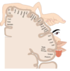
40
Q

What is somatotopy?

A

A region of the body is represented by the brain e.g. for the primary motor cortex - look at the diagram: stimulating at the top causes the movement or our arms / shoulders, stimulating towards the bottom causes facial movement etc.

Broadly similar in all individuals

Diagram = coronal plane

41
Q

To recap, what are the 2 neurons of the corticospinal tract and where are they found?

How many of the fibres of the corticospinal tract cross to the other side of the body and where, and what do they generally control?

What is controlled by the fibres that remain on the same side?

A

Upper motor neuron - primary motor cortex

Lower motor neuron - brainstem / spinal cord

85-90% of the corticospinal tract cross to the other side in the medulla - generally control the limb muscles

The ones that remain on the same side tend to control the trunk muscles

42
Q

Begins with ‘d’ / begins with ‘i’

What is the term used to describe crossing over of the tracts from one side to the other?

What is the term used to describe the fibres or tracts remain on the same side?

A

Decussation e.g. most of the fibres of the corticospinal tract decussate in the medulla

ipsilateral e.g. fribres of the corticospinal tract controlling the trunk muscles descend ipsilaterally

43
Q

What is the corticobulbar tract?

What is the difference between the corticospinal tract and the corticobulbar tract?

A

Bulb = another word for brainstem

Tract that travels from the cortex to the brainstem, primarily controlling the muscles of the face, whereas the corticospinal tract travels from the brain to the spinal cord and primarily controls the movement of the limb muscles and trunk

44
Q

What are the functions of the corticobulbar tract?

Fill in the diagram:

A

Corticobulbar tract that emerges from the:

Oculomotor nucleus (CN III) goes to the extraocular muscles (the medial, inferior, and superior recti, the inferior oblique, and levator palpebrae muscles)

Trochlear nucleus (CN IV) goes to the extraocular muscles of the eyes (the superior oblique)

Trigeminal motor nucleus (CN VI) goes to the mandibular branch going to the muscles of mastication

Absucens nucleus (CN VII) goes out to the extraocular muscles of the eyes (lateral rectus muscles)

Facial nucleus (CN V) goes to the muscles of facial expression

Hypoglassal nucleus (CN XII) goes to the muscles of the tongue

45
Q

What are the extraocular muscles?

A

Six muscles that control movement of the eye and one muscle, levator palpebrae, that controls eyelid elevation

Six muscles include: medial, inferior and superior recti, inferior and superior oblique and lateral rectus muscle

46
Q

How many of the CNs control eyemovement?

Which CN(s)?

Which muscles it / they correspond to?

A

3 CNs

CNs - III, IV, and VI

CN III - the medial, inferior, and superior recti, the inferior oblique, and levator palpebrae muscles

CN IV - the superior oblique muscle

CN VI - the lateral rectus muscle

47
Q

Is there somatotopy as the fibres descend down from the brain?

Why is this important clinically?

A

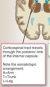
Yes - fibres are bunched up into discrete somatotopic representations as they come from a region that is specific for a function

If there is an infarct or stroke, symptoms will relate to which fibres in that area are affected

Plane of diagram = horizontal / transverse

48
Q

Hint: involuntary tracts beginning with ‘v’, ‘t’, ‘r’, ‘r’

What are some other tracts that have nuclei in the brainstem?

(Covered nect year in more detail, don’t really need to remember them)

A

Involuntary movement - reflex actions, balance, posture:

Vestibulospinal - provides information about head movement and position and mediates postural adjustments

Tectospinal - orientation of the head and neck during eye movements

Reticulospinal - control of breathing and emotional motor function

Rubrospinal - innervate lower motor neurons of the upper limb

49
Q

From which region in the brain do the somatosensory pathways stem? (i.e the dorsal columns and spinothalamic pathway)

A

Post-central gyrus - primary somatosensory cortex

50
Q

What are the 2 tracts the spinothalamic splits into called?

Where are the dorsal columns and spinothalamic tract found in the spine?

What are they responsible for?

A

Dorsal columns - posterior, coveys information about touch: mechanical i.e. fine discriminative touch, pressure, vibration, proprioception

Spinothalamic - anterior, split into two (lateral and ventral / anterior) conveys information about pain and temperature: mechanical, chemical and thermal i.e. crude touch, pain, temperature

51
Q

What are the 3 main neurons / sections of the sensory pathways?

Do all neurons have cell bodies?

A

Both the sensory pathways, dorsal column and spinothalamic tract are composed of 3 neurons - the primary, secondary and tertiary sensory neurons

Yes

52
Q

For both the sensory pathways, are the 3 cell bodies of the 3 sensory neurons that each compose the pathways found in the same place?

Where are the cell bodies of the 3 sensory neurons found?

A

No, the primary and tertiary cell bodies are found in the (sensory receptor? /) dorsal root ganglion and thalamus respectively, however, the secondary cell body is found in different places

For the dorsal column pathway, the secondary cell body is found in the medulla.

For the spinothalamic pathway, the secondary cell body is found in the lower spinal cord

53
Q

For each of the sensory pathways, from where to where do the 3 neurons travel?

What always occurs at the first synapse between the primary and secondary neuron?

A

Primary - sensory receptor to medulla / spinal cord

Secondary - medulla / spinal cord to thalamus

Tertiary - thalamus to cerebral cortex

In the synapse between the primary and secondary neuron, is always where the pathways dessucates (crosses over to the other side)

54
Q

Where do the dorsal column and spinothalamic pathway cross? What determines this?

A

Dorsal column - medulla, where the secondary neuron cell body is found

Spinothalamic pathway - lower spinal cord, where the secondary neuron cell body is found

55
Q

For the dorsal column pathway, where do the fibres enter?

What is found in the dorsal root ganglion?

How is the information conveyed from the lower limbs and body (i.e. via which tract)?

How is the information conveyed from the upper limbs (i.e. via which tract)?

A

Firbres enter via the dorsal horn and enter the ascending dorsal column pathway

Cell bodies

(Below T6) - information travels ipsilaterally along the gracile tract

(Above T6) - information travels ipsilaterally along the cuneate tract

56
Q

Where is the first synapse of the gracile tract?

What is the first synapse of the cuneate tract?

A

Gracile nucleus

Cuneate nucleus

57
Q

Describe this image: (of the dorsal column pathway)

What tract does the secondary neuron form?

A

The red, white and black lines / arrows are the 3 sensory neurons that make it up

First synapse is in the medulla on the ipsilateral side

Then the secondary neuron dessucates, in the caudal medulla, and forms the contralateral medial lemniscus tract

Which then ends at the second synapse in the thalamus

58
Q

What happens after the synapse in the thalamus?

What is the size of the somatotopic area proportional to?

A

The tertiary / 3rd order neuron then projects from the thalamus to the somatosensory cortex in the post-central gyrus (in the cerebral cortex) - the sensory receptor ends in the area resonsible for that body region

The density of sensory receptors in that body region

59
Q

(anterolateral)

What 2 tracts is the spinothalamic tract split into, and which is responsible for what sensations?

Where is the first synapse / beginning of the secondary neuron?

A

Lateral spinothalamic tract - pain and temperature

Anterior spinothalamic tract - crude touch

Lower spinal cord - where the primary sensory axons terminate and second order neurons dessucate immediately to form the spinothalamic tract

60
Q

Describe this diagram: (of the spinothalamic tract)

A

The cell body of the primary neuron is found in the dorsal root ganglion

As it enters the spine, the primary axon ends, after the first synapse the secondary neuron crosses to the other side and splits into 2 tracts. The informatiion travels up on of these 2 tracts, depending on whether it is crude touch, or pain or temprature

61
Q

Where do the 3rd order neurons go to from the thalamus for the spinothalamic tract?

A

Same as dorsal column, project from the thalamus to the somatosensory cortex in the cerebral cortex