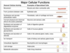
Cell (Part 1) Flashcards
Plasma membrane
- primary function:
- why it’s important
- 2 other functions:
Plasma Membrane:
-primary function: semipermeable barrier that separates the intracellular from extracellular environment (and regulates what goes in/out of cell)
- channels and transporters in the membrane enable the import and export of small molecules
-
important for homeostasis:
- Hydrophobic portions of membrane phospholipids - barrier to most hydrophilic molecules
- Cells must also have a way to allow molecules to enter and exit the cytosol in a controlled fashion
-
important for homeostasis:
-also involved in: cell communication, cell growth and motility
- Receptor proteins in the plasma membrane enable the cell to receive signals from the environment
- flexibility of the membrane and its capacity for expansion allow the cell to grow, change shape, and move.

Nucleus
- contains:
- stains what color in H&E
Nucleus:
- Contains genetic information
- stains purple in H&E (because of nucleic acids that attract hemotoxylin)

Cytoplasm contains: (4)
- color when stained with H&E
1) organelles
2) cytoskeleton
3) cytosol (or cytoplasmic matrix)
4) inclusions
* Cytoplasm is pink when stained with H&E because of protein content (attracts eosin)

Organelles
- what type of function
- location
- 2 formats
Organelles:
- metabolic function
- located in cytoplasm
- can be membranous (membrane bound) or non-membranous

Cytoskeleton
- 3 main functions
- location
Cytoskeleton:
- proteins for transportation, movement, structural support
- located in cytoplasm

Cytosol or cytoplasmic matrix
- definition
- location
Cytosol or cytoplasmic matrix
- aqueous gel containing molecules
- located in cytoplasm

Inclusions
- definition
- location
Inclusions:
- products of metabolic activity (they do not have a metabolic function)
- located in cytoplasm

Homeostasis of plasma membrane
- hydrophobic portions of membrane phospholipids = barrier to most hydrophilic molecules
- cells must also have a way to allow molecutes to enter and exit the cytosol in a controlled fashion
In addition to a plasma membrane, eukaryotic cells also have
-
internal membranes that enclose individual organelles.
- All cell membranes prevent molecules on one side from freely mixing with those on the other

Phospholipid bilayer
- 2 parts & location of each
- overall structure and how it forms
- Hydrophpobic tail = inside
- Hydrophilic head = outside
- Phospholipid bilayers spontaneously close in on themselves to form sealed compartments (circles)
- The closed structure is stable because it avoids the exposure of the hydrophobic hydrocarbon tails to water, which would be energetically unfavorable.

How does cholesterol effect cell membranes?
Cholesterol tends to stiffen cell membranes
- cholesterol fits into the gaps between phospholipid molecules in a lipid bilayer
- non-polar hydrophobic tail of cholesterol is chemically equivalent to the hydrophobic tails of the phospholipids

The fluidity of lipid bilayer depends on (2)
Compositon:
1) cholesterol
2) membrane proteins
Types of membrane proteins (4)
1) Channels and Transporters: allow for movement/tight control of cells homeostasis
2) Anchors: anchor to extracellular matrix or cytoskeletal proteins
3) Receptors: involved in cell signaling
4) Enzymes: involved in cell signaling
* Membrane proteins form a mosaic of particles penetrating the lipids*

The lateral mobility of plasma membrane proteins can be restricted in several ways
a)
b)
c)
d)

Proteins can be tethered to:
(A) intracellular cytoskeleton inside the cell
(B) to extracellular matrix molecules outside the cell
(C) to proteins on the surface of another cell (connecting one cell to another)
(D) Diffusion barriers (shown as black bars) can restrict proteins to a particular membrane domain.

Some of the ___ and ___ in the plasma membrane have ___ attached ____ to them
Some of the lipids and proteins in the plasma membrane have sugars (aka carbohydrates) covalently attached to them
- (aka - the cell surface is coated with carbohydrates)*
-
this carbohydrate-rich layer is made of:
- oligosaccharide side chains attached to membrane glycolipids and glycoproteins,
- polysaccharide chains on membrane proteoglycans
- As shown, glycoproteins that have been secreted by the cell and then adsorbed back onto its surface can also contribute.
- Note that all the carbohydrate is on the external (noncytosolic) surface of the plasma membrane.

What is the distance between the 2 phosphate heads in the plasma membrane bilayer?
- can you see that with a light microscope?
8-10 nm
- cannot see with a light microscope because its resolution is 200 nm (not strong enough)
- need TEM at least to see

Resolution of light microscope
200 nm
TEM appearance of plasma membrane
- Question marks?
- MV?

TEM appearance of plasma membrane = trilaminar structure; two dark bands (e- density) at phosphate heads, sandwiching a light band.
- Question marks: Glycocalyx (sugar chains off phospholipid bilayer)
- “MV” = microvilli - curvy, short finger link projections from the edge of an epithelial cell
(cannot see with plasma membrane light microscope, must use TEM or stronger)
Can you see the plasma membrane indicated by the arrow?

No - this is a light microscope, the resolution isn’t powerful enough
What you are seeing is the proteins associated with the plasma membrane (so can see where the plasma membrane is without actually seeing it itself)

nuclear envelope
-surrounded by what?
- houses nucleus
- surrounded by 2 membranes
-
the outer membrane continuous with the endoplasmic reticulum (ER) membrane
-
The space inside the endoplasmic reticulum (the ER lumen) is colored yellow; it is continuous with the space between the two nuclear membranes.
- The lipid bilayers of the inner and outer nuclear membranes are connected at each nuclear pore.
-
The space inside the endoplasmic reticulum (the ER lumen) is colored yellow; it is continuous with the space between the two nuclear membranes.

nuclear lamina
- definition
- made of what?
- purpose
•Nuclear lamina – meshwork of proteins (brown in image)
- made of intermediate filaments called lamins
- Stabilizes nuclear envelope
- A sheetlike network of intermediate filaments (brown) inside the nucleus forms the nuclear lamina (brown), providing mechanical support for the nuclear envelope.

strategies to protect DNA in nucleus (5)
strategies to protect DNA in nucleus (5)
1) nuclear envelope
* surrounds nucleus, has 2 membranes (inner and outer)
2) nuclear lamina
* meshwork of proteins, layer underneath nuclear envelope (next to its inner membrane), stabilizes nuclear envelope
3) nuclear pore complexes
* forms a gate through which selected macromolecules and larger complexes enter or exit the nucleus
4) chromatin
* DNA packing occurs on several levels in chromosomes
5) RNA copies of DNA sequences (transcription)
* RNA is what leaves nucleus so proteins can be made, DNA stays in nucleus

Nuclear pore complexes
- purpose
- structure
- how it works
nuclear pore complexes: control what can enter and exit the nucleus (keeps DNA protected inside nucleus)
- have fibrils that extend into cytosol and into nucleus (dark red)
- in nucleus: form basket shape
- Proteins with a nuclear localization signal (NLS) will bind to an NLS receptor/nuclear import receptor (ex. importin)
- Help chaperone protein of interest through the pore complex and into the nucleus
- Conformation of the nuclear pore complex changes as the protein passes through
- similar process happens in reverse to chaperone proteins out of nucleus to the cytosol
-
there are many different types of NLS signals and they will bind to and need help from different types of NLS receptors, will somtimes also need help from a third adaptor protein
- Overall: tightly regulated process that has many moving pieces

Nucleus Microscopy (TEM)
A)
B)
C)
-dashed line:

Nucleus TEM Appearance:
-
A - Heterochromatin = Electron dense areas representing tightly packed chromatin fibers. No transcription happening.
- Called “peripheral heterochromatin” if it’s associated with the nuclear membrane.
- B - Euchromatin = Lighter areas with dispersed granular material. Represents loosely arranged chromatin fibers. Transcription occuring.
-
C - Nucleolus = Round area containing both light and dark areas. Represents the site of rRNA synthesis and initial ribosome assembly.
- There may be more than one if the cell produces a lot of proteins
- dashed line: nuclear envelope