Cardiology Peer Teaching Flashcards

1
Q

what sort of thing can a bicuspid aortic valve predispose to

A
  • go undetected initially but later leads to:
    • aortic stenosis
    • aortic regurgitation
  • predisposes to
    • IE
    • aortic dissection
2
Q

what would the treatment be for a bicuspid aortic valve be

A

surgical valve replacement

3
Q

what are the differences between the two types of ASD

A
  • Primum: presents earlier and may involve the AV valves
  • Secundum: asymptomatic until adulthood - affects higher in the septum
4
Q

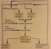
what happens in an ASD and what can this lead to?

A
  • there becomes a L–>R shunt
  • as heart compliance falls with age the shunt increases
  • pulmonary hypertension ensues
  • heart failure and SoB by 40
  • can lead to Eisenmenger’s complex where the shunt is reversed due to the PHTN
  • this leads to cyanosis and organ damage
5
Q

what happens in a VSD

A

there’s a L–>R shunt

there’s no cyanosis as LVP is still greater than RVP

larger holes can cause problems during infancy while smaller ones may be asymptomatic

both increase IE risk

6
Q

what condition would you see a boot shaped heart on x ray

A

teratology of fallot

7
Q

what is coarctation of th aorta

A

it is a narrowing at the site of the ductus arteriosus

8
Q

what happens in mild and severe coarctation of the aorta

A
  • severe:
    • blocks aorta, patient may collapse with heart failure
  • mild
    • raised BP and systolic murmur
      • murmur best heard over left scapula ‘scapula bruit’
  • both cause a radio-femoral delay
    • i.e. BP higher in right arm than left
9
Q

how would you treat mild and severe coarctation of the aorta

A

both need repair: surgically or with a stent

10
Q

which is most common ASD primum or secundum

A

secundom

11
Q

what is eisenmenger’s complex

A

it is a complication of VSD or ASD

reversal of the L–>R shunt due to pulmonary HTN and right sided hypertrophy

causes marked cyanosis, clubbing, heart failure, syncope and polycythaemia

there is very poor prognosis and it can only be cured with a transplant

12
Q

how would VSD present in an infant

A

SOB

poor feeding

failure to thrive

needs fixing before eisenmenger’s syndrom arises

13
Q

name two conditions associated with coarctation of the aorta

A

bicuspid aortic valve and Turner’s syndrome

14
Q

Mother comes to see you. Her two year old has been having episodes where he gets restless and cries for no reason, however as soon as he is allowed to squat down the crying stops. He is a bit underweight for his age and on examination you notice a bit of clubbing.

diagnosis?

A

Teratology of Fallot

15
Q

what are the 4 features of teratology of fallot

A

VSD

Pulmonary stenosis

RV hypertrophy

overriding aorta

16
Q

why do toddlers squat in teratology of fallot

A

it increases TPR so helps to alleviate some of the R->L shunt

17
Q

what happens in teratology of fallot

A

they have the 4 deformities

these cause R->L shunt

then after the DA closes they’ll become progressively more cyanotic as there’s less and less flow to the lungs

18
Q

mortality of teratology of fallot

A

without surgery it’s 95%

with surgery it’s 5-10%

19
Q

what number of live births have teratology of fallot

A

3-6/100,000 live births

commonest cyanotic cardiac disorder

20
Q

how long shoult the PR interval be

A

120-200ms

21
Q

how wide should the QRS be

A

110ms

22
Q

in which leads will the QRS be upright in

A

I and II

23
Q

in which leads will QRS and T waves have the same direction

A

I, II and III

24
Q

what proportion of men and women die from IHD in the UK

A

one in 7 men and one in 11 women

25
what number of deaths does IHD cause in the UK every year
70,000
26
three broad causes of IHD
1. inhibited blood flow 2. increased distal resistance 3. reduced O2 carrying capacity (anaemia) or availability (hypoxia)
27
4 modifiable risk factors for IHD
* smoking * obesity * exercise * diet * cocaine use
28
4 clinical risk factors for IHD
depression hypertension diabetes hyperlipidaemia
29
three non-modifiable risk factors for IHD
age genetics gender M\>F
30
psychosocial risk factors for IHD
high demand, low control jobs low social interaction/support low social class low income
31
9 things included in the QRISK2
BP Age Ethnicity Smoking Cholesterol RA DM Anti-hypertensives BMI
32
what is the gold standard investigation for angina pectoris
Angiogram – Gold standard, shows luminal narrowing
33
what does qrisk tell us
the risk of CV event in the next ten years
34
presentation of angina pectoris
chest pain brought on by exertion but rapidly resolves with rest may radiate to arms, jaw and neck pain can be exacerbated by emotion May also get some dyspnoea, palpitations or syncope
35
what is the most common manifestation of stable IHD
angina pectoris
36
what is the prevalence of angina pectoris
5% among men, 4% among women
37
ECG in angina pectoris
usually normal, may show ST depression and T wave inversion
38
what is the lifestyle advice for angina pectoris
eat less move more stop smoking control diabetes better
39
what investigations would you do on a patient with angina pectoris
ECG Bloods: looking for anaemia CXR - check heart size and pulmonary vessels Angiogram - this is gold standard and will show luminal narrowing of coronary arteries
40
what is the medical treatment of angina pectoris
* control hypertension and diabetes * beta blockers * atenolol * CCB if beta blockers contraindicated e.g. asthma * amlodipine * statin * simvastatin * aspirin * ACE inhibitor * ramipril * if severe try ARB * candesartan
41
surgical treatment of angina pectoris
* PCI (percutaneous coronary intervention) * stenting or ballooning the narrowing * risk of restenosis or thrombosis * less invasive * CABG (coronary artery bypass graft) * good prognosis but longer recovery * not for the frail
42
ACS includes
unstable angina NSTEMI STEMI
43
rare causes of ACS
emboli coronary spasm vasculitis
44
how would ischaemis show on an ECG
ST depression and T wave flattening
45
Ix for ACS
* ECG * Bloods: * FBC * U&E * glucose * lipids * Cardiac enzymes: * Troponin T * CK-MB * myoglobin
46
Acute changes seen on ECG following ACS
Tall T waves, ST elevation, new LBBB
47
* draw the differentiating ACS diagram with the timeframes: * admission * working diagnosis * ECG * Biochemistry * Diagnosis
48
what is the definition of unstable angina
acute coronary syndrome that is defined by the absence of biochemical evidence of myocardial damage
49
what is the outlook for patients with unstable angina
50% will have an MI within 30 days if left untreated
50
Ix for unstable angina §
* FBC = anaemia aggravates it * Cardiac enzymes = excludes infarction * ECG = when in pain shows ST depression * Coronary angiography
51
symptoms of MI
acute central chest pain radiating to jaw or shoulder and lasting \>20 mins, with nausea, SOB and palpitations
52
pulse and BP in MI
can be up or down
53
signs of MI
* clammy and pale * 4th heart sound * pansystolic murmur * may later develop peripheral oedema
54
who are silent MIs most commonly seen in
the elderly or diabetics
55
Acute management of a STEMI
* ABCDE * C- secure IV access * MONA * morphine * oxygen (only if sats \<94%) * nitrates * aspirin (chew) * Refer for PCI immediately
56
actue management of NSTEMI
* MONA * anti-coagulation with fondaprinux (Xa inhibitor) * double up with a second anti-platelet like clopidogrel * give IV nitrate if the pain then continues * glyceryl trinitrate (GTN)
57
Advice points for patient following ACS
1 month no sex drivers, airline pilots must not return to work diet: high in oily fish, fruit and veg, low in sat fats increase exercise stop smoking improve management of diabetes
58
what is the most common cause of pericarditis
cocksackie B virus
59
what 4 drugs for post MI management
aspirin B blocker (CCB like verapamil if CI) ACEi like ramipril statin like simvastatin
60
differential diagnoses of chest pain
* Cardiac: ACS, aortic dissection, pericarditis, myocarditis * Respiratory: PE, pneumonia, PT, pleurisy, ca. lung * Musculoskeletal: rib fracture, chest trauma * Costochondritis * GORD * Oesophageal spasm * Anxiety/panic attack
61
signs and symptoms of peripheral vascular arterial disease
* 6 Ps of limb ischaemia * pain (cramping relieved by rest) * pallor * pulselessness * paresthesis * paralysis * perishing cold * may cause 'punched out' ulcers
62
test for peripheral arterial disease
* Buerger's test * postural colour change * 45 degree elevation of legs when lying down --\> observable colour change * colour returns when hung over bed * one leg at a time to compare
63
Ix for peripheral arterial disease
* exclude: * DM * arteritis * anaemia * ABPI * normal is 1-1.2 * PAD is 0.5-0.9 * colour duplex USS * MR/CT angiography
64
management of peripheral arterial disease
* risk factor modification * quit smoking * treat HTN * lower cholesterol * improve DM control * lower fat diet * exercise improves blood flow * medicatons * clopidogrel as 1st line * PTA if severely stenosed * percutaneous transluminal angioplasty
65
when is PAD critical limb ischaemia
triad of ischaemic rest pain, gangrene and arterial insufficiency ulcers
66
how would the limb appear in critical limb ischaemia
'dusky'
67
management of critical limb ischaemia
surgical embolectomy local thrombolysis if not revascularised in 4-6hrs they'll lose the limb
68
what are the two subdivisions of low output heart failure
* systolic failure * low CO due to insufficient ventricular contraction * EF\<40% * diastolic failure * inability of the ventricle to relax and fill normally leads to increased filling pressures * EF \>50% (heart failure with preserved EF: HFpEF) * caused by restrictive pericarditis, tamponade, ventricular hypertrophy, restricted cardiomyopathy etc
69
what's the socrates for pericarditis
–S = central, retrosternal –O = 3 days ago –C = sharp –R = left shoulder –A = SOB, cough, hiccups fever –T = constant –E = made worse on inspiration, relieved by leaning forwards –S = 7/10
70
causes of pericarditis
* mostly viral * cocksackie B * EBV * mumps * bacterial * staph/strep * pneumonia * Post MI - dressler syndrome * autoimmune * SLE * RA
71
signs of pericarditis
* tachypnoea * tachycardia * fever * SOB * auscultation --\> pericardial friction rub * pericardial effusion may be visible on X-ray
72
7 Ix for pericarditis
* ECG * FBC * CRP * Cardiac enzymes (^ Trop) * Echo * CXR * Pericardial tap / biopsy - for rarer causes
73
what would ECG of pericarditis show
PR depression on all leads
74
management of pericarditis
–Treat the cause! –NSAIDs – to manage pain and inflammation –Corticosteroids – for symptomatic relief –Manage complications
75
what is the clinical definition of heart failure
A state where the heart is unable to pump enough blood to satisfy the needs of metabolising tissues
76
equation for CO
–CO = HR x SV
77
what is the difference between high output and low output HF
* high output: blood requirements of the body are too high * anaemia * pregnancy * Low output: heart is not functioning efficienty * IHD
78
causes of systolic heart failure
IHD MI cardiomyopathy
79
causes of diastolic heart failure
constrictive pericarditis cardiac tamponade restrictive cardiomyopathy
80
causes of high output heart failure
anaemia pregnancy hyperthyroidism
81
pathophysiology of heart failure
* compensatory changes made in response to the beginning of heart failure themselves become pathological * RAAS * Sympathetic stimulation * Cardiac changes
82
how does sympathetic stimulation change in HF
* peripheral vasoconstriction * meant to increase perfusion of organs by increasing BP * but actually increases afterload in heart causing increased hypertrophy and failure
83
how does RAAS change in HF
* RAAS is increased in order to increase perfusion of organs * increased volume and vasoconstriction leads to: * salt and water retention * increases afterload and preload
84
cardiac changes in HF
* –Ventricular dilatation * –Myocyte hypertrophy * both are meant to increase CO * but actually they both reduce EF
85
what happens with pre-load in heart failure
* Increased preload: * Failure of the heart muscle means more blood is left behind in the ventricles after systole * This results in increased preload * This stretches myocardium à which maintains cardiac output up to a point
86
why is there salt and water retention in HF
* Reduced cardiac output leads to diminished renal perfusion, activating RAAS by secretion of renin from the kidney * this increases BP which increases afterload
87
why is there myocyte remodelling in HF
* In response to ischaemia, myocyte damage, etc. * Hypertrophy, loss of myocytes and increased interstitial fibrosis
88
draw the HF diagram
89
Left sided heart failure symptoms
Exertional dyspnoea Fatigue Paroxysmal nocturnal dyspnoea nocturnal cough (pink frothy sputum)
90
left sided heart failure signs (8)
–Cardiomegaly (displaced apex beat) –3rd and 4th heart sounds –Crepitations in lung bases –Weight loss –Reduced BP –Tachycardia –Cool peripheries –Heart murmur
91
symptoms of right sided heart failure
–Peripheral oedema –Ascites –Nausea –Anorexia
92
signs of right sided heart failure
–Raised JVP –Hepatomegaly –Pitting oedema –Ascites –Weight gain (fluid)
93
causes of right sided heart failure
LVF pulmonary stenosis lung disease (cor pulmonale)
94
Ix for heart failure
* Bloods: * B-type Natriuretic Peptide * also raised in PE * FBC, LFTs, U&Es, BNP, TFTs * Cardiac enzymes: * troponin I, troponin T * CXR * ECG * Echo (TTE)
95
what would you see on an HF x ray
* ABCDE * **A**lveolar oedema (bats wings) * Kerley **B** lines (interstitial oedema) * **C**ardiomegaly * **D**ilated prominent upper lobe vessels * Pleural **E**ffusion
96
heart failure management
* Lifestyle advice * symptomatic control * loop diuretic - furosemide * disease altering * ACEi - Ramipril * ARB if cough with ACEi - candesartan * BB - atenolol, propanolol, bisoprolol * not in asthma * aldosterone antagonist - spironolactone
97
chronic heart failure treatment
* ABCDE * •ACE inhibitors (ramapril) / ARB (candesartan, valsartan) * B-blockers (atenolol) * CCB and other vasodilators (amlodipine, hydralazine) * Diuretics and digoxin (furosemide - loop diuretic, spironolactone - aldosterone antagonist)
98
what is supraventricular tachycardia
any tachycardia which arises from the atria or atrioventricular junction
99
what is bradycardia
\<60bpm
100
what is tachycardia
\>100bpm
101
what are some pathological conditions where sinus tachycardia occurs
–Anaemia –Fever –Heart failure –Acute PE –Hypovolaemia
102
what are the 3 types of supraventricular tachycardia
–Atrial fibrillation –Atrial flutter –Atrioventricular nodal re-entry tachycardia (AVNRT)
103
what is the management of sinus tachycardia
–Correction of the cause –Beta blockers to slow sinus rate (e.g. atenolol)
104
what is Atrioventricular nodal re-entry tachycardia (AVNRT)
this is where there's an accessory pathwat from the AV node to the SA node that restarts the cycle before ventricular contraction most common type is wolf parkinson white where the accessory pathway is called the bundle of kent can lead to up to 200bpm
105
what is CHADS2VASc used to calculate
risk of stroke in AF
106
what is the most common arrhythmia
atrial fibrillation
107
what happens in AF
there are many smaller, disorganised contractions in the atria rather than one bigger coordinated contraction this is mechanically ineffective the AVN will conduct a proportion of these impulses with irregular ventricular response
108
what does the pulse feel like in AF
irregularly irregular
109
causes of AF
* any condition that leads to raised atrial pressure * heart failure * hypertension * rheumatic heart disease
110
what is the pathophysiology of atrial fibrillation
* atrial activation 300-600/min * only a proportion of these conducted by AVN * HR 120-180
111
What are the symptoms of atrial fibrillation
## Footnote –Asymptomatic –Palpitations –Fatigue –Heart failure
112
how is the risk of stroke affected with AF
•5 x risk of stroke (embolism due to thrombus formed in atrium)
113
what are the three principles of AF management and give an example of each
•Rate control: –Beta blockers (atenolol) or CCB (verapamil, diltiazem) or Digoxin •Rhythm control: –Electrical DC cardioversion –Amiodarone •Anticoagulation with Warfarin: –Target INR is 2-3
114
how does amiodarone work
prolongs phase 3 of the cardiac action potential, the repolarization phase where there is normally decreased calcium permeability and increased potassium permeability
115
how does AF look on an ECG
* ’Irregularly irregular’ * F waves * No clear P waves * QRS is rapid and irregular
116
whats on chadsvasc and what does the scoring mean
●0 = No anticoagulation ●1 = consider oral anticoagulation or aspirin ●2 = oral anticoagulation (warfarin, rivaroxaban)
117
what would you see on an ECG of atrial flutter
sawtooth flutter waves
118
what is atrial flutter and what is the tretment
often associated with af regularly irregular atrial rate is ~300bpm and you'll have a 2:1 or 3:1 ratio with ventricular rate so here ventricular rate would be 150bpm (based on how it conducts it's a 2:1 if at 300 but don't worry about why) Rx is radiofrequency catheter ablation
119
extrinsic causes of bradycardia
–Drug therapy - BB, digoxin –Hypothyroidism –Hypothermia –Raised intracranial pressure
120
intrinsic causes of bradycardia
– Acute ischaemia – Infarction of SAN – Sick sinus syndrome
121
treatment for intrinsic causes of bradycardia
treat with atropine
122
how does atropine work
competitive, reversible antagonist of the muscarinic acetylcholine receptors so it counters rest and digest
123
what is sick sinus syndrome
sinoatrial block leads to intermittent failure of SAN depolarisation ECG will have severe sinus bradycardia or intermittent long pauses between consecutive p waves if symptomatic needs pacemaker insertion
124
what are teh two places there can be heart block
1. block in AVN or bundle of His = AV block 2. block in lower conduction system = RBBB or LBBB
125
what is the PR interval normally
0.12-0.2 seconds
126
what is the length of the QRS complex normally
0.08-0.1 seconds
127
what is the normal timing of the QT interval
0.4-0.43 seconds
128
what is the RR interval normally
0.6-1.0 seconds
129
what are the causes of heart block
coronary artery disease cardiomyopathy fibrosis of the conducting tissues (in elderly people)
130
what is first degree heart block
delayed AV conduction leads to PR interval being prolonged more than 0.2 seconds its asymptomatic so no treatment
131
what are the two types of seconds degree heart block
Mobits typ I (wenckebach) Mobitz type II
132
describe Mobitz type I
–Progressive PR interval prolongation, until a P wave fails to conduct and a QRS is dropped —\> then the cycle repeats itself –Light headedness, dizziness, syncope
133
describe mobitz type II
same as Mobitz type I except you don't have the progressive lengthening of the PR and the QRS is dropped quite randomly they may have fatigue, chest pain, SOB, syncope and postural hypotension
134
causes of third degree heart block
CHD, infection, HTN
135
Describe third degree heart block
all atrial activity fails to conduct to the ventricles - there is no association between atrial and ventricular activity ventricular contractions are maintained by spontaneous escape rhythm
136
what would the ECG show on 3rd degree heart block
P waves and QRS are totally independent of each other p waves are said to march through
137
treatment for 3rd degree heart block
atropine or permanent pacemaker
138
how will a RBBB appear on ECG
M shape in V1 and W shape in V6
139
causes of right bundle branch block
PE IHD ASD VSD
140
what are the causes of LBBB
IHD Aortic valve disease
141
what would the ECG look like in LBBB
W shape in V1 and M shape in V6
142
equation for blood pressure
CO x peripheral resistance = blood pressure
143
who should you suspect HTN in in clinic and how would you confirm this
\>140/90 in clinic confirm with 24hr ambulatory BP monitor
144
three things you need to Ix in HTN and how to do it
* Eye problems * fundoscopy looking for: * papilloedema * bilateral retinal haemorrhages * Overall CVD risk * QRISK * fasting glucose * cholesterol * End organ damage * urine analysis (looking for proteinuria/haematuria) * 12 lead ECG (looking for past MI/LV hypertrophy)
145
what is essential HTN and how common is it
it's if the HTN has a primary cause this is 95% of HTN
146
name 4 secondary causes of HTN
* Renal * CKD * Endocrine * Conn's * Other * Coarctation of aorta * Pregnancy
147
lifestyle advice for HTN
* Stop smoking * Low-fat diet * Reduce alcohol and salt intake * Increase exercise * Reduce weight if obese
148
what is the management for HTN
149
name the 4 important types of valvular disease
* Aortic Stenosis * Mitral Regurgitation * Aortic Regurgitation * Mitral Stenosis
150
3 causes of aortic stenosis
congenital bicuspid valve degenerative calcificaton rheumatic heart disease
151
3 types of aortic stenosis
Supravalvular, Subvalvular, Valvular
152
when do symptoms occur in aortic stenosis
when valve area is about 1/4 of normal
153
presentation of Aortic stenosis
exertional syncope angina dyspnoea heart failure
154
what is the gold standard for diagnosis of aortic stenosis
echocardiography
155
can you remember the poem for types of heart block
156
management of aortic stenosis
good dental hygeine and IE prophylaxis in dental procedures surgical valve replacement or Transcatheter Aortic Valve Implantation (TAVI)
157
management for mitral valve regurgitation
* vasodilator * ACEi: ramipril * rate control: BB, CCB, Digoxin * diuretics to control symptoms * surgery if deteriorating symptoms
158
causes of valve regurge
bicuspid aortic valve infective endocarditis
159
signs of aortic regurgitation
## Footnote * Collapsing (water hammer) pulse * Wide pulse pressure * Displaced hyperdynamic apex beat * Early diastolic murmur
160
what is Mitral stenosis
•Obstruction of LV inflow that prevents proper filling during diastole it's common with AF
161
management of aortic regurge
surgery to replace valve before LV dysfunction
162
Name 3 causes of mitral stenosis
infective endocarditis RA IE
163
Presentation of mitral valve stenosis
* Dyspnoea * Fatigue * Palpitations * Chest pain…
164
signs of mitral valve stenosis
* Malar flush on cheeks * Low volume pulse * Tapping, non displaced apex beat * Rumbling mid-diastolic murmur * Loud opening S1
165
what is shock
•Circulatory failure resulting in inadequate organ perfusion Low BP below \<90mmHg
166
name 4 types of shock
septic anaphylactic neurogenic hypovolaemic
167
Management of mitral stenosis
anticoagulate with warfarin percutaneous mitral balloon valvotomy
168
describe septic shock
•infection with any organism --\> acute vasodilation from inflammatory cytokines
169
describe anaphylactic shock
•Type-I IgE-mediated hypersensitivity, release of histamine
170
describe neurogenic shock
distributive type of shock resulting in low blood pressure, occasionally with a slowed heart rate, that is attributed to the disruption of the autonomic pathways within the spinal cord. It can occur after damage to the central nervous system, such as spinal cord injury and traumatic brain injury
171
what causes hypovolaemic shock
•Bleeding, trauma, ruptured aortic aneurysm, GI bleed
172
investigation and management of sepsis
* Sepsis 6 (bufalo) should be achieved in first hr * Blood cultures * Urine output measured * Fluid given * Antibiotics given * Lactate measured * Serum lactate \>2mmol/l is indicative of tissue hypoperfusion and severe sepsis * Oxygen given if sats low
173
what should your first approach to management of shock be
* ABCDE * secure airway * assess breathing * assess circulation (BP and capillary refil time) and establish IV access * Disability * Environment
174
what is the ECG of pericarditis
widespread concave ST elevation and PR depression
175
55yo Caucasian Man with HTN develops renal impairment. Which of the following medications should be stopped a. Ramipril b. Verapamil c. Losartan d. Spironolactone e. Amlodipine
ramipril
176
example of thiazide like diuretic
indapamide