Cardiac Exam Lecture 1 Flashcards
(30 cards)
Explain blood’s pathway through the heart
Blood enters the heart from the body through the superior and inferior vena cava and into the right atrium. Blood then goes from the RA to the RV via the tricuspid valve. From the Right Ventricle, blood goes through the pulmonary valve and into the pulmonary artery to reach the lungs. From the lungs, oxygenated blood then goes into the pulmonary vein and into the Left Atrium. From the LA, blood goes through the mitral/bicuspid valve into the Left Ventricle. From the Left Ventricle, blood gets pumped out of the aortic valve, into the aorta and then to the rest of the body.
Explain the electrical conduction pathway of the heart.
Pathway is SA node (which then sends a wave of depolarization through the atria) to the AV node > Bundle of His > Bundle Branches > Perkinje Fibers
Which ventricle wall is thicker? What are the pressure differences between the RV and LV?
The left ventricle wall is much thicker.
Left Ventricle: 125 mmHg
Right Ventricle: 25mMHg
What are the medical names of the following valves:
Right AV valve
Left AV valve
Semilunar Valves
Right AV valve: Tricuspid
Left AV valve: Mitral/Bicuspid
Semilunar valves: Pulmonary and Aortic
How much blood does the ventricle hold and how much does it actually eject during contraction?
Ventricle holds 150 mL. Ejects around 80mL per contraction.
Explain the function of Purkinje fibers
Purkinje fibers are VERY FAST… they need to activate all of the cells of the ventricles at once
Allows coordinated ejection of blood instead of just sloshing around
Explain the function of Papillary Muscles
What happens when a heart attack causes an infarction of the papillary muscles
Papillary muscles: contract to support the valve leaflets connected by cordinae tendinae
Sometimes heart attacks cause an infarction of the papillary muscles: valve will then blow backwards like an umbrella in the wind- leaks blood, exposes atrium to high pressure.
Draw the standard depiction of an EKG (lead two) and showcase which sections depict the following:
Atrial Activation
Ventricle Activation
Ventrical Recovery

Explain the role gap junctions play in cardiac tissue
- Gap junctions are intercalcalated discs that connect cardiac myocytes to each other.
- Low electrical resistance connections btwn cells
- cell membranes are very close
- primary determinant of internal resistance in cardiac tissue
Remember, to get a fast conduction velocity you want low internal resistance. (which increases the space constant and makes conduction a lot better)
That is what these gap junctions provide to cardiac tissue
Explain the following about each type of structure:
Diameter size, how many gap junctions and how many myofibrils.
Then relate the above to the function of each structure:
- SA node and AV node
- Atrial and Ventricular Muscle
- His bundle, bundle branches, Purkinke fibers

Explain the roles of the following:
Na/K Pump
Na-Ca exchanger
- Na/K Pump:
- maintains Na/K gradients across membrane
- electrogenic - net outward current
- requires metabolic energy in form of ATP
- specifically inhibited by digitalis
- Na-Ca exchanger
- exchanges 3 sodium going into the cell for one calcium going outside the cell
- electrogenic, net inward current
- forward direction: extrudes intracellular calcium to maintain low Ca inside cell
- driven by the Na+ gradient across the membrane…therefore indirectly affected by alterations in the Na/K pump activity
K+ in the heart affects its own ________
As K+ is reduced outside, K+ permeability _____
Explain inward rectification
K+ in the heart affects its own permeability
As K+ is reduced outside, K+ permeability is decreased,
Less K+ leaking out means less negative
This behavior is called inward rectification, it is a way for cells to conserve K+, it limits how much K+ leaks out, and it also keeps the membrane protention from getting too negative.
The K+ channel also turns off when the heart _______.
As the gradient is streghtened (aka as K+ decreases extracellularly), it is balanced by a ______
The K+ channel also turns off when the heart depolarizes, so it stops fighting the upstroke of the AP.
As the gradient is stregthened (aka as K+ decreases outside of the cell), it is balanced by a decrease in K+ permeability. So decreasing K+ has less of an effect on RMP than you might expect
Draw the cardiac action potentials for the following: Ventricles, SA node
Explain the following phases: 0, 1, 2, 3, 4
Phases:
0 : Na+ channels activate/open, membrane potential approaches Ena
1: Na+ channels inactivate/close, and Ito (K+ channels that transiently open) open
2: Calcium channels activate/open and background K conductance (Ik1) decreases [inward rectification]
3: delayed actification of K+ channels called Ik and background Ik1 conductance increases again (reversal of inward rectification)
4: background K+ conductance is high (Ik1), delayed Ik channels closed, calcium channels closed and sodium channels recover from inactivation but still closed.

Explain’s calcium’s role in the cardiac AP
Calcium is the cause AND the reason for the long plateau…. Ca depolarizes the membrane and is the signal for contraction and contraction takes time.
Ca channels are a lot like sodium channels but WAY SLOWER at activation and inactivation
Explain how the cardiac AP gets repolarized
Repolarization happens when the delayed rectifier K+ channel kicks in and starts bringing the membrane potential back toward the K Nernst potential.
Also Ca is decreasing at this time because Ca channels are becoming inactivated
Explain the SA node AP
the SA node has NO fast depolarization, no plateau phase and no “resting membrane potential”
Note: the SA node has no sodium channels, only has L type calcium channels
Na channels are blocked by what kind of toxin?
Draw/explain the effect this has on the AP
Na channels are blocked by TTX, turning the fast reponse into a slow response.
Phase O gets blocked
Explain the differences in Slow Response vs Fast Response in the various categories:
Tissue type, Phases, Membrane Potential, Threshold, Upstroke, Duration, Conduction Velocity

What are the two factors that determine cardiac conduction. Explain them in further detail.
- Space/Length Constant
- Rm is inversely related to K permeability
- Ri is inversely related to number of gap junctions and cell diameter (more gap junctions means lower Ra which increases space constant)
- Rate of rise AND amplitude of action potential
- slow vs fast response
- premature responses initiated during relative refractory period
- level of RMP (fast response only)
Explain the electrical conduction through the AV node
AV Node Conduction:
- normally conduction delay permits optimal ventricular filling
- action potential is slow response due to slow inward calcium current
- long refractory period
- AV nodal conduction time is clinically determined by P-R interval on EKG
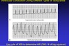
What do the P-R interval and the QRS complex represent on the EKG?
P-R interval: conduction time from atria to ventricular muscle/ AV nodal conduction time
QRS complex: intra-ventriular conduction time
Explain the following: 1st, 2nd, and 3rd degree heart block
1st Degree Block: Abnormal prolongation of P-R interval
2nd Degree Block: Some atrial impulses fail to activate ventricles; not all P waves are followed by QRS complexes
3rd degree heart block: complete AV nodal block, no consistent P-R interval

Explain what the following mean on an EKG:
Slurred QRS complex
Notched QRS complex
Slurred QRS complex: slowed intraventricular conduction (hyperkalemia, ischemia, ventricular tachycardia)
Notched QRS complex: asynchronous electrical activation of left and right ventricles (bundle branch blocks)